Arquivos Brasileiros de Cardiologia - Volume 86, Nº 5, May 2006
Case Report
Case Report
Mailing Address: Helena Furtado Martino • Rua das Laranjeiras, 441/501 – 22240-002 – Rio de Janeiro, RJ - Brazil
E-mail: [email protected] Received on 05/03/05 • Accepted on 09/30/05
Cell Therapy in Dilated Cardiomyopathy
Helena Furtado Martino, Paulo Sérgio Oliveira, Edmilson Assunção, Rita Villela, Miriam Gaze,
Patrícia C. dos Santos Costa, Fernando César Castro Souza, Luiz Henrique Weitzel,
Ana Paula R. Velloso, Amarino Oliveira Júnior, Antônio C. Campos de Carvalho
Instituto Nacional de Cardiologia Laranjeiras, Hospital Prócardíaco - Rio de Janeiro, RJ - Brazil
A forty-one-year-old male with systolic heart failure, FC-III NYHA, clinical stage C due to dilated cardiomyopathy was submitted to an autologous transplant of the mononuclear fraction of bone marrow via coronary artery system through heart catheterism. Two months after the procedure, there was a decrease in plasma BNP and cardiac area reduction at the thorax X-ray and nuclear magnetic resonance. The echocardiogram showed decrease of the secondary regurgitation and mitral ring dilatation. There was a better performance at the ergospirometry, with increase of the maximum oxygen consumption and consequent reduction in drug therapy. The absence of adverse events characterized by clinical/hemodynamic instability, enzymatic alteration or electrocardiogram demonstrate the safety and feasibility of this procedure carried out and described with pioneering spirit in dilated cardiomyopathy.
I
NTRODUCTIONDespite the progresses made regarding the drug therapy of chronic systolic heart failure, which is the evolution stage of several cardiomyopathies such as dilated cardiomyopathy, the number of individuals who present the syndrome increases worldwide, constituting a serious public health problem. For a signifi cant number of individuals, the syndrome progresses even with drug therapy. Thus, heart failure Functional Class (FC) III and IV of the New York Heart Association has been one of the main causes of hospital admissions, using a large part of healthcare resources with a high mortality rate (40%/year). The development of new, low-cost and low-risk therapeutic procedures, such as the myocardial implant of stem cells obtained from bone-marrow aspirate from the individual’s own bone marrow, constitutes a promising therapeutic option for these advanced cases. From the 90s on, several experimental studies in animal models of cardiac lesion showed the capacity of bone marrow-derived cells to improve heart performance when injected directly into the myocardium or in the systemic circulation. In the beginning of the new millennium, the knowledge generated by experimental models started to
be utilized in clinics. In a pioneering work, Menasché and cols. performed a transplant of skeletal muscle satellite cells to the heart of an elderly patient with refractory heart failure, in France1. After that, two groups in Germany and one in Hong Kong utilized bone marrow mononuclear cells in the treatment of patients with ischemic disease2-4. In Brazil, within the context of the project developed at the Instituto do Milenio de Bioengenharia Tecidual (Tissue Bioengineering Millennium Institute), the same cells were used to treat patients with post-ischemic heart failure5 and, more recently, in patients with chronic Chagas disease cardiopathy6
C
ASER
EPORTArquivos Brasileiros de Cardiologia - Volume 86, Nº 5, May 2006
For the cell implant to the heart, 50 mL of the patient’s bone marrow was aspirated from the iliac crests. The mononuclear fraction was separated after pre-fi ltering (bone marrow fi ltering system - Washington University – USA) and centrifuged through a Ficoll gradient. After resuspension in 20 mL of autologus serum solution (5%), 108 cells were injected in the coronary artery system (10 mL in the DA, 4 mL in the CX, 6 mL in the CD) with no occurrence of pain, arrhythmia or hemodynamic instability during the procedure and in the immediate post-procedure period.
Electrocardiographic and enzymatic monitoring was carried out during the fi rst 24 hours post-procedure in an intensive care unit, with no alterations of these parameters (Table 1).
The patient was released from the hospital seventy-two hours post-procedure, and returned every two weeks for follow-up; the exams described above were repeated after two months. At physical examination, the patient’s weight was unaltered (66.5 kg), BP was 100/60 mmHg, CF was 60 bpm, RF was 18 rpm, with regular cardiac rhythm, but no B3, and reduction of the holosystolic murmur, now 1+/6+ and no irradiation.
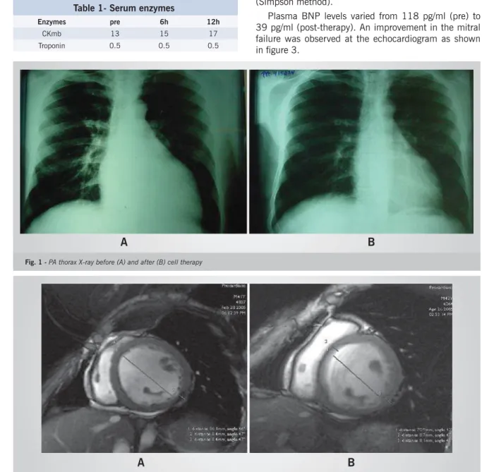
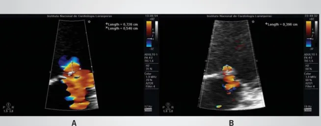
The initial MLWHFQ score was 63, being reduced to 39 after the cell therapy. Thorax X-ray showed a pre-treatment thoracic cardiac index of 0.55, and post-treatment of 0.48 (Fig 1). The magnetic resonance also showed a decrease in the systolic and diastolic volumes of the left ventricle, going from 302 mL (FSV) and 371 (FDV) pre-treatment, to 217 and 263 mL, respectively, after cell therapy (Fig 2). However, there was no alteration in the ejection fraction, which remained around 17% (Simpson method).
Plasma BNP levels varied from 118 pg/ml (pre) to 39 pg/ml (post-therapy). An improvement in the mitral failure was observed at the echocardiogram as shown in fi gure 3.
Table 1- Serum enzymes
Enzymes pre 6h 12h
CKmb 13 15 17 Troponin 0.5 0.5 0.5
Fig. 1 - PA thorax X-ray before (A) and after (B) cell therapy
A
B
A
B
Fig. 2 - Nuclear magnetic resonance (A) before and (B) 2 months after therapy
Arquivos Brasileiros de Cardiologia - Volume 86, Nº 5, May 2006 The improvement observed in the mentioned
varia-bles was followed by a better performance at the ergospirometry as shown in Table 2. It is noteworthy that the patient was classifi ed as being in functional group III, by ergospirometry, before therapy, and improved to being group II after treatment. The favorable clinical evolution of the patient allowed us to decrease drug therapy with the reduction of the daily dose of furosemide (40 mg) and spironolactone (12.5 mg).
Fig. 3 - Bidimensional echocardiograms: (A) before and (B) after therapy, showing a reduction of the mitral regurgitation volume from 42 ml with PISA of 0.726 (A) to 13 ml with PISA of 0.395 (B). PISA= proximal isovelocity surface area
A
B
Table 2 - Ergospirometry - Ramp Protocol
Variable Pre Post
Duration 5:49 11:40 Distance (miles) 0.15 0.38 Max. CF* 100 102 Max. Pot. (W) 55.9 130 Max. VO2 mL(Kg.min) 15.61 18.64
Functional group III II T ½** (s) >120 <90 O2 pulse (ml/beat) 10.3 12.1 LV/VCO2 29.7 23.8
*CF: cardiac frequency; **T1/2: time required for peak VO2 to
reach 1/2 the value at maximum stress
D
ISCUSSIONCell therapy has been applied to patients with acute and chronic ischemic cardiopathy through the intracoronary release of bone marrow cells or intramyocardial injection1-5. The action mechanism of bone marrow mononuclear cells (BMMC) remains to be clarifi ed, and there is a great deal of controversy in literature on their capacity of regenerating cardiomyocytes7-9. Still, in all animal models of cardiopathy and clinical assays carried out to date, these cells promoted an improvement in cardiac function, although these studies were performed with the main objective of testing the safety and feasibility of this new procedure.
In patients with chronic Chagas cardiopathy with severe systolic dysfunction, the intracoronary release of BMMC also showed to be safe and resulted in functional improvement6. Based on these results, we utilized the intracoronary BMMC release model in a patient with dilated cardiomyopathy. In this case report, we observed that the procedure was carried out with no adverse events, suggesting the method is safe and viable. This observation, together with the improvement in the patient’s clinical parameters and complementary exams leads us to propose cell therapy as an alternative to be investigated for the treatment of severe dilated cardiomyopathy.
1. Menasché P, Hagege AA, Scorsin M, et al. Myoblast Transplantation for heart failure. Lancet. 2001;357:279–80.
2. Strauer BE, Brehm M, Zeus T, et al. Repair of infarcted myocardium by autologous intracoronar y mononuclear bone marrow cell transplantation in humans. Circulation. 2002;106:1913-18.
3. Assmus B, Schächinger V, Teupe C, et al. Transplantation of progenitor cells and regeneration enhancement in acute myocardial infarction (TOPCARE-AMI). Circulation. 2002;106:3009–17.
4. Tse HF, Kwong YL, Chan JKF, et al. Angiogenesis in ischaemic myocardium by intramyocardial autologous bone marrow mononuclear cell implantation. Lancet. 2003;361:47–9.
5. Perin EC, Dohmann HF, Borojevic R, et al .Transendocardial, Autologous Bone Marrow Cell Transplantation for Severe, Chronic
R
EFERENCESIschemic Heart Failure. Circulation. 2003;107:r75-r83.
6. Vilas-Boas F, Feitosa GS, Soares MPB, et al. Transplante de Células de Medula Óssea para o Miocárdio em Paciente com Insufi ciência Cardíaca Secundária a Doença de Chagas. Arq Bras Cardiol. 2004; 82 (2):181-4.
7. Orlic D, Kajstura J, Chimenti S, et al. Bone Marow cells regenerate infarcted myocardium. Nature.2001; 410: 701-5.
8. Balsam LB, Wagers AJ, Christensen JL, et al Haematopoietic stem cells adopt mature haematopoietic fates in ischaemic myocardium. Nature. 2004; 428: 668-73.
9. Murry CE, Soonpaa MH, Reinecke H, et al. Haematopoietic stem cells do not transdifferentiate into cardiac myocytes in myocardial infarcts. Nature. 2004; 428: 664-8.