Instituto de Cardiologia do Rio Grande do Sul/Fundação Universitária de Cardiologia – Porto Alegre
Mailing address: Marne de Freitas Gomes - Instituto de Cardiologia do RS – Av. Princesa Isabel, 395 - 90620-001 - Porto Alegre, RS - Brazil
Objective - To test the hypothesis that left ventricular hypertrophy (LVH) reduces the electrocardiographic and functional effects of right coronary artery occlusion.
Methods - We analysed 215 patients (166 males and 49 women,age of 58.9±10.6 years), with occlusion of the right coronary artery without other associated lesions. There was no significant difference (p>0.05) in age and gender distribution between the 78 patients with LVH (left ventricular mass >100g/m2) (Group A) when compared with the 137 patients without LVH (left ventricular mass <100g/m2) (Group B).
Results - The electrocardiographic finding of transmural necrosis was more often found in group B patients than in group A patients (56.9% and 30.8%, respectively; p<0.05). The left ventricular function parameters of group A were better than those of group B: the ratio end-diastolic pressure/systolic pressure (EDP/SP) (A: 0.108±0.036; B: 0.121±0.050; p<0.05); the end-diastolic volume index (A: 75.9±31.3ml/m2; B: 88.0±31.0ml/m2; p<0.01); the end-systolic volume index (A: 16.0±10.0ml/m2; B: 27.0 ±20.0ml/m2; p<0.001); the ejection fraction (A 78.6±10.8%; B 67.7±17.9%; p<0.001); the anteroinferior shortening (A: 43.9±10.3%; B: 35.1±12.8%; p<0.001). A higher degree of coronary tortuosity was observed in group A than in group B (78.2% and 24.1%; p<0.001) and also a more frequent absent or minimal dia-phragmatic hypokinetic area (A: 80.8%; B: 54.0%; p<0.05).
Conclusion - LVH reduces the effects of myocardial sequela and protects LV function when right coronary occlusion develops.
Key words: left ventricular hypertrophy, acute myocar-dial infarction, ischemic cardiopathy
Arq Bras Cardiol, volume 72 (nº 2), 166-170, 1999
Marne de Freitas Gomes, Carlos Antônio Mascia Gottschall
Porto Alegre, RS - Brazil
Protective Effect of Left Ventricular Hypertrophy in Right
Coronary Artery Occlusions
Left ventricular hypertrophy (LVH) is one of the most frequent findings in adult patients with heart disease. Its main cause is high blood pressure (HBP) 1-3. Considering that arterial hypertension is one of the major risk factors for acute myocardial infarction (AMI) 4 and that the identifica-tion of LVH on the electrocardiogram (ECG) is also consi-dered a risk factor 5, it is easy to understand the frequent occurrence of AMI in patients with LVH.
In 1949, Harrison and Wood 6 demonstrated that coro-nary artery caliber varied directly with heart weight. Howe-ver, several studies 7,8 suggest that the increase in the de-mand for myocardial oxygen due to LVH can exceed the ca-pacity of the coronary arteries to provide oxygen. Coronary arteries become tortuous 9, even without obstruction, and this leads to ischemia, lesion, infarction, and heart failure 10. On the other hand, even though in LVH secondary to HBP the coronary flow increases, there is a double increase of the coronary arterial resistance 11 and decrease in the extraction of lactate. In patients with aortic stenosis, during tachycar-dia due to atrial stimulation 7 lactate production can occur suggesting poor myocardial perfusion. However, despite the evidence already documented of the relation between hypertrophy and ischemia, several studies 12-17 have sug-gested a certain protective effect of myocardial hypertro-phy, restricting the expansion of the infarcted area, when an acute coronary artery occlusion develops.
patients with hypertrophy of the non-infarcted myocardium showed improvement of the rest and exercise ejection fraction (EF), but this did not happen to those who did not develop LVH. Therefore, the post-infarction LVH repre-sented a beneficial myocardial adaptation. The group of Ginzton 16, studying AMI due to proximal occlusion of the anterior descending coronary artery in dogs, reported that after three and a half months the recovery of the post-infarction EF depends on a correlated increase of the left ventricular mass.
The objective of our study was to clarify possible cli-nical, electrocardiographic, hemodynamic, and cineangio-cardiographic differences between patients with isolated occlusion of the right coronary artery, with or without LVH.
Methods
This is a retrospective cohort study on 215 patients (166 males and 49 females, mean age 58.9±10.6 years) with proximal occlusion of the right coronary artery. These patients underwent a first coronary angiocardiographic study in the catheterization and Interventional Cardiology Laboratory in 1992 and 1993. This cohort study is part of broader research on the prognosis of ischemic heart disease, which is being developed. Patients with surgical or percutaneous myocardial revascularization, with obstruc-tive lesions >50% in the left coronary tree, and those with associated valvular lesions were excluded from the analysis. All patients underwent a left heart catheterization and coronary angiography more than one month after AMI, usually for risk stratification or due to post-infarction angi-na. The patients were divided into two groups as follows: group A – patients with LVH (left ventricular mass >100g/ m2); group B – patients without LVH (left ventricular mass <100g/m2). The left ventricular mass was measured using the ventriculogram.
The ECGs of all patients were analysed and the follo-wing findings were considered evidence of LVH: R wave >26mm in V5 or V6; R wave >20mm in DI or DII; R wave >12mm in aVL; R wave in V5 or V6 + S wave in V1 >35mm. The necrosis was arbitrarily classified into three degrees, according to the possible transmural characteristic of the infarction estimated through Q wave amplitude in aVF lead, as follows: degree 1- Q wave ≤ 0.03 s, amplitude > 20% of R wave; degree II – Q wave >0.03s, amplitude <50% of R wave; degree III – Q wave >0.03s, amplitude ≥ 50% of R wave.
The measurements of the ventricular pressures and the left ventriculogram were performed at the beginning of the examination, prior to coronary angiography, using conventional techniques, and a Besi recorder and the Philips Polydiagnostic DCI for coronary angiography. For the calculation of ventricular volumes, 40mL of ionic con-trast medium (Meglumine) was injected into the LV in right anterior oblique view at 35°. The following parameters of LV were determined in all patients: systolic pressure (SP) and
end-diastolic pressure (EDP) in mmHg; end-diastolic and end-systolic volumes (EDV and ESV, respectively) in mL; EF of LV and anteroinferior shortening (AIS) in %; left ventricular mass in g/m 2. The measurement of the ventri-cular volumes was performed through the delimitation of the silhouette of the LV by planimetry and applying the formula of Dodge corrected by Kennedy 18. For determining the left ventricular mass, the method of Rackley 19 was used, and a perfect visualization of the cardiac silhouette was conside-red essential. Nineteen out of the 234 original patients were excluded from the analysis because their delimitations of the cardiac silhouette were not considered precise. In addition to the usual coronary angiographic analysis, coronary tortuosity was evaluated through the tortuosity index (TI) using curvimetry 9 and was classified into two degrees, as follows: degree I – TI ≤ 1.1; degree II – TI >1.1. The repercus-sion in the LV of the right coronary artery occlurepercus-sion was eva-luated through the area of contractile abnormality obtained by planimetry after remodeling or normalization of the systolic silhouette 17. According to this area, the abnormalities were classified as follows: absence of or minimum hypokinesia (asynergic area ≤ 1.5cm2, or 5% of the diastolic area); moderate or severe hypokinesia (asynergic area > 1.5cm2), with or without akinesia, dyskinesia, or aneurysm.
In regard to the statistical analysis, the database was formed using blind data, and the analysis was performed through the Statgraphics 5.0 program. The quantitative variables were summarized through their means and standard deviation, and the Student’s t test was used for comparison of the means in two independent series. The analysis of the qualitative data, expressed by their frequen-cies and percentages, was performed by comparison of the proportions in the two groups of patients through the chi-square test or, when convenient, through the Z test for comparison of percentages. The probabilistic values <5% (p<0.05) were considered significant.
Results
The values of left ventricular mass, which was the criterium for placement of patients in groups A or B, are shown on table I.
The distributions of age and gender of the patients in groups A and B were not statistically different (p>0.05), nor was the prior diagnosis of AMI. In group A, the body surface and the incidence of SAH were significantly higher (table II).
The characterization of LVH by means of ECG, accor-ding to the criteria proposed, occurred in 26 patients (33.3%)
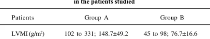
Table I – Values of left ventricular mass index (LVMI) in the patients studied
Patients Group A Group B
LVMI (g/m2) 102 to 331; 148.7±49.2 45 to 98; 76.7±16.6
in group A, and in none of the patients in group B. Findings of necrosis through the relation Q/R in aVF occurred in 133 patients (61.8% of the total) and was more severe in those in group B (p<0.05), where the occurrences of absence of necrosis or minimum necrosis (degree I) and severe necro-sis (degree III) (p<0.05) were compared. The moderate necrosis (degree II) had a similar incidence in both groups (p>0.05) (table III).
Table IV shows that ventricular SP and EDP were not statistically different (p>0.05) in groups A and B. The different values (p<0.05 to <0.001) were the following: the relation EDP/SP, the indexes of end-diastolic volume and end-systolic volume, the EF and the anteroinferior shorte-ning of the LV.
In group A, the percentage of patients with normal or decreased coronary tortuosity (degree I) was significantly smaller than the percentage of patients with moderate to se-vere tortuosity (degree II), but in group B it was the opposite (p<0.001) (table V). There was no significant difference in the presence of collateral flow from the left coronary tree to the right one (88.4% in group A and 83.2% in group B; p>0.05).
Table VI shows that group A had a higher proportion of absence of hypokinetic area or minimum hypokinetic area than group B (p<0.05). The opposite occurred with moderate or severe hypokinesia and dyskinesia (p<0.05).
Figures 1 and 2 present the relations between left ven-tricular mass, necrosis, asynergy in the infarcted area, and behavior of left ventricular function.
Discussion
The presence of LVH in patients with coronary occlu-sion or AMI is a frequent possibility, not always remembe-red, because one can lead to the other and SAH can be com-mon to both. The criterium of division into groups accor-ding to ventricular mass (> or <100g/m2) allowed the separa-tion of two populasepara-tions with normal distribusepara-tion and aleatory degrees of left ventricular mass (table I).
Gender distribution and mean age of the patients (table II) were similar (p>0.05) in the groups with and without LVH. Other studies report older age in patients with LVH 13. Diagnosis of SAH occurred in 69.2% of patients with LVH and in 46.7% of patients without LVH, confirming that SAH is prevalent (p<0.05) in both pathophysiological situations (LVH and AMI).
Table II – Clinical data of the patients studied and statistical significance of the differences
Characteristics Patients Group A Group B p
Gender Males n (%) 58 (74.4%) 108 (78.8%) NS Females n (%) 20 (25.6%) 29 (21.2%) NS Age (years) M ± DP 58,6±12.9 59.1±9.3 NS BS (m2) M ± DP 1,89±0.20 1.83±0.17 <0.05 SAH n (%) 54 (69.2%) 64 (46.7%) <0.05 AMI n (%) 42 (53.8%) 91 (66.4%) NS
BS- body surface; SAH- systemic arterial hypertension; AMI- acute myo-cardial infarction; M- mean; sd- standard deviation; NS- not significant.
Table III – Degrees of necrosis on the electrocardiogram in the patients studied
Degrees I* II III* Total
n % n % n % n %
Group A 38 56.0 16 23.5 14 20.5 68 100 Group B 26 40.0 15 23.0 24 37.0 65 100
Total 64 31 38 133
*p<0.05; degree I- Q wave in aVF ≤ 0.03s, amplitude >20% of the R wave; degree II- Q wave in aVF >0.03s, amplitude <50% of the R wave; degree III- Q wave in aVF >0.03s, amplitude ≥ 50% of the R wave.
Table IV – Hemodynamic data and statistical significance of the differences
Measures Group A Group B p
SP (mmHg) 151.0±35.0 143.0±28.0 NS EDP (mmHg) 15.9±5.3 17.0±6.8 NS EDP/SP (%) 10.8±3.6 12.1±5.0 <0.05 EDVI (ml/m2) 75.9±31.3 88.0±31.0 <0.01 ESVI (ml/m2) 16.0±10.0 27.0±20.0 < 0.001 EF (%) 78.6±10.8 67.7±17.9 <0.001 AIS (%) 43.9±10.3 35.1±12.8 <0.001
Results expressed in mean ± sd; SP- systolic pressure; EDP- end-diastolic pressure; EDVI- end-diastolic volume index; ESVI- end-systolic volume in-dex; EF- ejection fraction; AIS- anteroinferior shortening; NS- not significant.
Table V – Coronary tortuosity degrees
Degrees I* II* Total
n % n % n %
Group A 17 21.8 61 78.2 78 100
Group B 104 75.9 33 24.1 137 100
Total 121 94 215
*p<0.001; I- coronary tortuosity by curvimetry up to 1.1; II- coronary tortuosity by curvimetry >1.1.
Table VI – Degrees of the left ventricle asynergy
Degrees Hypokinesia Hypokinesia Dyskinesia Total
Abs/Min* Mod/Sev* n % n %
N % n %
Group A 63 80.8 14 17.9 1 1.3 78 100
Group B 74 54.0 58 42.4 5 3.6 137 100
Total 137 72 6 215
LVH identified through ECG in patients with AMI has a low prevalence 13,14, less than 25%, but this has been re-vealed to be around twice this value in pathological 20, in ventriculographis 19 and in echocardiographis 21 studies. In our study, 36.2% of the patients had LVH diagnosed by cineventriculography (group A), but ECG identified only 24 patients (30.7% of the patients with LVH or 11.1% of the to-tal number of patients). In the chronic stage, non-Q infarc-tions are characterized by a greater contractile capacity of the affected area in relation to Q infarctions 22. The Q-wave size has been used to characterize the significance of the transmural quality 23 and its relation with R-wave to characterize the size of the infarction 24. Awan et al 25 showed a strong correlation between the number of patho-logical Q-waves and the extension of the asynergy (r=0.84) through ventriculography. The most frequent finding of absence of necrosis or minimal necrosis in patients with LVH (group A) can be an indication of the “protection” promoted by LVH to the myocardium if an infarction happens. As the time gap between the infarction and the angiography was distributed at random between the two groups, it is correct to suppose that the ventricular remode-ling of infarcted and non-infarcted areas was not influenced by time.
Although LV SP and EDP were not significantly diffe-rent (p>0.05) in the two groups, the EDP/SP ratio, which better expresses LV functionality than does isolated EDP 26, was significantly smaller (p<0.05) in group A, indicating once again a better functional capacity. The smaller values of diastolic volume and mainly of end-systolic volume 27 in group A than in group B (p<0.001) also confirm this. This behavior is responsible for an increased EF in group A (p<0.001). It is still a matter of speculation as to what degree this functional behavior can influence a prognosis and clinical behavior.
One can admit that the larger caliber and tortuosity of the coronary arteries represent an expansion of the tree to better nourish the increased ventricular mass 6,9,10. In our series of patients, this fact is confirmed because coronary tortuosity was significantly more frequent (p<0.001) in group A than in group B. The smaller regional motion abnormality we observed due to right coronary occlusion in patients with LVH is not emphasized in the literature. The only reference was that hypertrophy is proportional to the degree of necrosis caused by the obstruction 15. It is worth supposing that the better function in these cases can represent a favorable short- and long-term prognosis.
Several authors 12-17 postulate that the justification for the beneficial role of LVH could rest in the larger muscle mass adjacent to the necrotic process, capable of confining the necrosis to the subendocardium and median myocar-dium. The smallest size of the infarction is demonstrated by the following: smaller rate of enzymatic elevation in patients with AMI and LVH, in comparison to those with AMI without LVH 13; greater percentage of patients with LVH among those with non-Q wave infarction than in the general infarcted population 14.
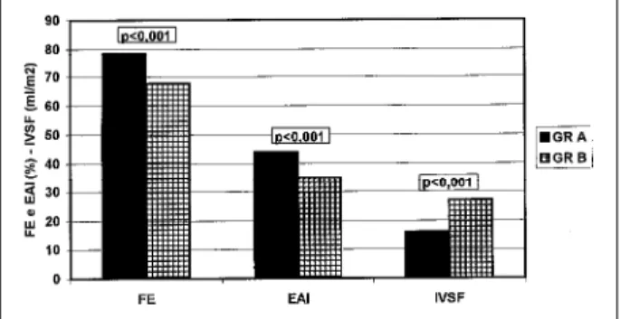
The explanation for a tissular structure, which has in-creased oxygen consumption and dein-creased coronary reser-ve, to develop a smaller infarcted area when after a coronary occlusion has been attributed to the development of an effective intra- and intercoronary collateral flow, according to studies carried out in animals 28,29 and humans 30-32. Since in our groups the majority of the cases presented collateral flow, this was not a differentiating factor. However, the significant difference can rest not only in the presence but in the degree 32, in the microvasculature 29,30, and/or in the moment when the AMI occurs 31,32, and not more than one month thereafter. It is speculated that the harmful effects attributed to hypertrophy are not directly dependent on its presence, but on the disorders causing the circulatory stress established by variegated stimuli such as neuronal, humoral and metabolic stimuli. These effects can also contribute to facilitating phenomena of cardiac events, among which LVH would be a mere marker 33. A confirmed mechanism of overload and risk for coronary events is the increase in heart rate, which frequently is part of the same disorder of adrenergic hyperactivity. This hyperactivity is present in the majority of the hypertensive patients and has also been recognized as a factor that might facilitate the Fig. 2 - In the patients with left ventricular hypertrophy (group A), the greatest left
ventricular mass index (149±49g/m2) is associated with a higher ejection fraction (FE), a higher anteroinferior shortening (EAI) and a smaller end-systolic volume index (IVSF) of the left ventricle (VE). All parameters indicate a better contractile function than in the patients without LV hypertrophy (group B), with a smaller left ventricular mass (77±17g/m2).
References
1. Kannel WB - Prevalence and natural history of electrocardiographic left ventricular hypertrophy. Am J Med 1985; 75: 4-18.
2. Savage DD, Garrison RJ, Kannel WB, et al - The spectrum of left ventricular hypertrophy in a general population sample: The Framingham Study. Circula-tion 1987; 75(suppl): 26-33.
3. Levy D, Anderson KA, Savage DD, Balkus SA, Kannel WB, Castelli WP - Risk of ventricular arrhythmias in left ventricular hypertrophy: The Framingham Heart Study. Arrhythmias and Conduction Disturbances. Am J Cardiol 1987; 60: 560-5. 4. Kannel WB - Role of blood pressure in cardiovascular morbidity and mortality.
Prog Cardiovasc Dis 1974; 17: 5-24.
5. Kannel WF, Gordon T, Castelli WP, Margolis JR - Electrocardiographic left ventricular hypertrophy and risk of coronary heart disease. The Framingham Study. Ann Intern Med 1970; 72: 813-21.
6. Harrison CV, Wood P - Hypertensive and ischemic heart disease. A comparative clinical and pathological study. Br Heart J 1949; 11: 205-13.
7. Trenouth RS, Phelps NC, Neill WA - Determinants of left ventricular hypertrophy and oxygen supply in chronic aortic valve disease. Circulation 1976; 53: 644-50. 8. Bache RJ - Effects of hypertrophy on the coronary circulation. Prog Cardiovasc
Dis 1988; 31: 403-40.
9. Macruz R, Toriano N, Ariê S, et al - Síndrome da insuficiência coronária não obs-trutiva. Tortuosidade das artérias coronárias. Arq Bras Cardiol 1976; 29: 255-62. 10. Carvalho V - Afecções coronárias não obstrutivas. In: Carvalho V, Macruz R (ed)
- Cardiopatia Isquêmica. São Paulo: Sarvier, 1989: 255-80.
11. Strauer BE - Left ventricular dynamic energetics and coronary hemodynamics in hypertrophied heart disease. Eur Heart J 1983; 4(suppl A): 137-42. 12. Pirolo JS, Hutchins GM, Moore W - Morphologic studies. Infarct expansion:
Pathologic analysis of 204 patients with a single myocardial infarct. J Am Coll Cardiol 1986; 7: 349-54.
13. Boden WE, Kleiger RE, Schechtman KB, et al - Clinical significance and prognostic importance of left ventricular hypertrophy non-Q wave acute myocardial infarction. Am J Cardiol 1988; 62: 1000-4.
14. Behar S, Reicher-Reiss H, Abinader E, et al - Long-term prognosis after acute myocardial infarction in patients with left ventricular hypertrophy on the electrocardiogram. Am J Cardiol 1992; 69: 985-90.
15. Ginzton zle, Conant R, Rodrigues DM, Laks MM - Functional significance of hypertrophy of the noninfarcted myocardium after myocardial infarction in humans. Circulation 1989; 80: 816-22.
16. Ginzton LE, Rodrigues D, Garner D, Laks MM - Functional significance of post-myocardial infarction left ventricular hypertrophy: A beneficial response. Am Heart J 1992; 123: 628-35.
17. Gomes MF - Oclusão da artéria coronária direita em pacientes com hipertrofia ventricular esquerda: análise do padrão hemodinâmico e angiográfico. Tese de Mestrado, IC/FUC, Porto Alegre, RS, 1994.
18. Kennedy JW, Trenholme SE, Kasser IS - Left ventricular volume and mass from
single plane cineangiocardiograms: a comparison of anteroposterior and right anterior oblique methods. Am Heart J 1970; 80: 343-52.
19. Rackley CE, Dodge HT, Coble YD, et al - A method for determining left ventricular mass in man. Circulation 1964; 29: 666-71.
20. Ellis LV, Allison RB, Rodriguez FL, et al - Relation of the degree of coronary artery disease and of myocardial infarctions to cardiac hypertrophy and chronic congestive heart failure. N Engl J Med 1962; 266: 525-30.
21. Murray JA, Johnston W, Reid JM - Echocardiographic determination of left ven-tricular dimensions, volumes and performance. Am J Cardiol 1972; 30: 252-7. 22. Horan LG, Flowers NC, Johnson JC - Significance of the diagnostic Q wave of
myocardial infarction. Circulation 1971; 18: 428-36.
23. Williams RA, Cohn PF, Vokonas PS, Young E, Herman MV, Gorlin R - Electro-cardiographic, arteriographic and ventriculographic correlations in transmural myocardial infarction. Am J Cardiol 1973; 51: 595-9.
24. Maroko PR, Hillis LD, Muller JE, et al - Favorable effects of hyaluronidase on electrocardiographic evidence of necrosis in patients with acute myocardial infarction. N Engl J Med 1977; 296: 898-903.
25. Awan NA, Miller RR, Vera Z, Janzen DA, Amsterdam EA, Mason DT - Noninva-sive assessment of cardiac function and ventricular dyssynergy by precordial Q wave mapping in anterior myocardial infarction. Circulation 1977; 55: 833-8. 26. Gottschall C, Miler V, Rodrigues R - Pressão diastólica final como índice
funcional do ventrículo esquerdo. Arq Bras Cardiol 1977; 30: 201-7. 27. White HD, Norris RM, Brown MA, Brandt PW, Whitlock RM, Wild CJ - Left
ventricular end-systolic volume as the major determinant of survival after recovery from myocardial infarction. Circulation 1978; 76: 44-51.
28. Cohen MV - The functional value of coronary collaterals in myocardial ischemic and therapeutic approach to enhance collateral flow. Am Heart J 1978; 95: 396-404. 29. Reimer K, Jennings R, Cobb F, et al - Animal models for protecting ischemic myocardium: Results of the NHLBI cooperative study: comparison of uncons-cious and consuncons-cious dog models. Circ Res 1985; 56: 651-65.
30. Zoll PM, Wessler S, Schlesinger MJ - Interarterial coronary anastomoses in the human heart with particular reference to anemia and cardiac anoxia. Circulation 1951; 4: 797-815.
31. Habib GB, Heibig J, Forman AS, et al - Influence of coronary collateral vessels on myocardial infarct size in humans. Circulation 1991; 83: 739-46.
32. Gottschall C - Angioplastia directa e trombólise no infarto agudo do miocárdio. Rev Port Cardiol 1997; 16: 165-74.
33. Frohlich ED, Tarazi RC - Is arterial pressure the sole factor responsible for hypertensive cardiac hypertrophy? Am J Cardiol 1979; 44: 959-63. 34. Gillman MW, Kannel WB, Belanger A, D’Agostino RB - Influence of heart rate
on mortality among persons with hypertension: The Framingham Study. Am Heart J 1993; 125: 1148-53.
35. Gottschall C - Diástole normal e anormal. In: Gottschall C – Função Cardíaca: da Normalidade à Insuficiência. São Paulo: Fundo Editorial Byk, 1995; 101-10. occurrence of coronary events 34. On the other hand,
pronounced degrees of LVH can determine significant changes in ventricular compliance and contribute to the development of heart failure and arrhythmias 35.
To conclude, we can say: 1) the absence of a previous clinical diagnosis of AMI is a common finding in right coronary artery occlusion, and it is more frequent in patients with LVH; 2) the localized sequela is significantly smaller in patients with LVH than in those without LVH; 3) patients