232
Left ventricular hypertrophy was identified as a risk factor of cardiovascular morbidity and mortality in the Framingham Study 1-3. Patients with heart failure due to ventricular dysfunction undergo cardiac anatomical changes, which are included under the concept of cardiac remodeling 4. Remodeling occurs in different clinical circumstances and in different patients in heterogeneous ways.
From the clinical point of view, great differences are observed in the left ventricular mass estimated on physical examination, on electrocardiography, or by use of the dimension of the cardiac image on chest radiography. Among patients with heart failure of the same etiology, some with a similar functional condition were observed to have different magnitudes of the cardiac image on chest radiography.
From the echocardiographic point of view, greater mortality and hospitalization rates due to cardiovascular diseases were ob-served in patients with left ventricular dysfunction and left ventricular hypertrophy. The echocardiographic estimate of left ventricular mass added prognostic information to other cardiovascular risk factors. However, assessment of left ventricular hypertrophy, as compared with other clinical variables, suggested independence between left ventricular hypertrophy and left ventricular ejection fraction 5, a different finding from that of our initial study. The correlation tests of left ventricular mass with age, duration of symptoms, left ventricular end-diastolic pressure, and pulmonary artery systolic and occlusion pressures did not show statistically significant values. However, the correlation with the left ventricular ejection fraction calculated on echocardiography was significant. The stepwise regression analysis showed a negative correlation between left ventricular mass and left ventricular ejection fraction6. On autopsy study, patients with hypertensive, ischemic, and idio-pathic cardiomyopathies had similar weights, which were greater than those of patients with cardiomyopathy due to Chagas’ disease7. Therefore, the clinical observation, echocardiographic data, and autopsy studies allow the hypothesis that the left ventricular mass may be a relevant variable for the prognosis of patients with heart failure. In this context, we formulated the hypothesis that left ventricular mass may also provide prognostic information about patients with symptomatic heart failure.
This study aimed at assessing, in a large case series of patients with heart failure being followed up for more than 10 years, the dis-tribution of left ventricular mass on echocardiography, its correlations with other clinical variables, and its potential prognostic influence.
Methods
A cross-sectional study 8 was carried out for assessing left ventricular mass in a cohort of patients with heart failure being followed up on an outpatient basis.
Original Article
Left Ventricular Mass in Patients with Heart Failure
Marcello Ricardo Paulista Markus, Humberto Felício Gonçalves de Freitas, Paulo Roberto
Chizzola, Gisela Tunes da Silva, Antonio Carlos Pedroso de Lima, Alfredo José Mansur
São Paulo, SP - Brazil
Heart Institute (InCor) University of São Paulo Medical School, Brazil Mailing address: Marcello Ricardo Paulista Markus - Av. Dr. Eneas Carvalho Aguiar, 44 - Cep 05403-000 - São Paulo, SP, Brazil E-mail: [email protected]
Received for publication: 11/19/02 Accepted for publication: 3/17/04 English version by Stela Maris Costalonga
Objective
To assess left ventricular mass in patients with heart failure and its correlations with other clinical variables and prognosis.
Methods
The study comprised 587 patients aged from 13.8 years to 68.9 years, 461 (78.5%) being males and 126 (21.5%) females. Left ventricular mass was estimated by using M-mode echocar-diography and was indexed by height.
Results
The left ventricular mass index ranged from 35.3 g/m to 333.5 g/m and increased with age. The left ventricular mass index was greater in males (mean, 175.7 g/m) than in females (mean, 165.7 g/m). The left ventricular mass index was greater in patients with hypertensive cardiomyopathy (mean of 188.1 g/m), with idiopathic dilated cardiomyopathy (mean, 177.7 g/m) and with cardiomyopathies of other etiologies (mean, 175.1 g/m) than in patients with chagasic (mean, 164.3 g/m) or ischemic (mean, 162 g/m) cardiomyopathy. The left ventricular mass index in patients with heart failure showed a correlation with age, sex, etiology, and left atrial diameter. The correlation with left ventri-cular ejection fraction was negative: the increase in the left ventricular mass index was associated with a reduction in ejec-tion fracejec-tion. The relative risk of death was 1.22 for each 50-g/m increase in the left ventricular mass index.
Conclusions
The estimate of left ventricular mass may contribute to the prognostic assessment of patients with heart failure.
Key words
233
The diagnosis of heart failure was established based on the Fra-mingham criteria 9, and the diagnosis of the etiology of heart failure was based on previously established criteria 10 and on the International Classification of Diseases 1993, 10th review (ICD 10).
Patients aged < 75 years diagnosed with symptomatic heart failure due to systolic ventricular dysfunction were included in the study.
Patients with the following characteristics were excluded from the study: heart failure due to cardiomyopathies indicated for surgical treatment (myocardial revascularization, aneurysmectomy, valvuloplasty, or valvular replacement); hypertrophic cardiopathy; chronic obstructive pulmonary disease; recent acute myo-cardial infarction; and unstable angina. Patients with the following characteristics were also excluded from the study: creatinine clea-rance lower than 30 mL/kg/min; liver failure; peripheral arterial disease; cerebrovascular disease; type I diabetes mellitus; recent infection; neoplasias; or active peptic ulcer disease.
This study comprised 587 patients with heart failure, who were followed up from April 1991 to December 2000. Their ages ranged from 13.8 to 68.9 years (mean of 45.6 years, standard deviation of 10.3 years), 461 (78.5%) were males and 126 (21.5%) were females.
The time elapsed between symptom onset and entrance into the study ranged from 6.9 days to 243.5 months (mean, 50.1 months; standard deviation, 49 months).
Heart failure was attributed to ischemic cardiomyopathy in 114 (19.4%) patients, chagasic cardiomyopathy in 97 (16.5%), hypertensive cardiomyopathy in 82 (14%), and other etiologies in 73 (12.4%). In 221 (37.7%) patients, heart failure was attributed to idiopathic dilated cardiomyopathy.
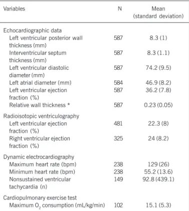
Echocardiographic, radioisotopic, electrocardiographic, and functional characteristics of the population studied are shown in table 1.
The patients were followed up on an outpatient care basis. The clinical treatment included dietary guidance, instruction about the general principles of treatment, and prescription of medications adjusted to their needs and tolerance. The medications included angiotensin-converting-enzyme inhibitors, diuretics, and digitalis. Beta-blockers were gradually introduced in the treatment from 1997 onwards.
Data on evolution were supplemented by a review of hospital records, telephone contact by the researchers, and research in the ProAim (Programa de Aprimoramento de Informações de Mor-talidade do Município da Cidade de São Paulo – Program on im-provement of the information on mortality in the city of São Paulo). Echocardiographic measurements were taken according to the criteria recommended by the American Society of Echocardiography11. Left ventricular mass was estimated by use of the following
formula 12: LVM (grams) = 0.8 X {1.04 X [(LVID + IST + PWT)3–
(LVID)3]} + 0.6, where: LVM = left ventricular mass; PWT = left
ventricular posterior wall thickness; IST = interventricular septum thickness; LVID = left ventricular inner diameter.
The left ventricular mass was indexed by the patient’s height (g/m) 12, and the term “ventricular mass index” began to be used. For comparisons with the left ventricular mass index, this indexation was also performed for other echocardiographic variables.
The left ventricular mass index was studied in regard to age, sex, duration of symptoms, left and right ventricular ejection fraction
on radioisotopic ventriculography, maximum O2 consumption on
cardiopulmonary exercise testing, maximum and minimum heart rates, and the presence of nonsustained ventricular tachycardia on 24-hour dynamic electrocardiography.
The demographic and functional variables of the population studied and the left ventricular mass index were initially examined by using exploratory descriptive analysis, with identification of the minimum, maximum, and mean values, median, and standard deviation of the variables studied. Then the left ventricular mass index was studied in regard to the probability of survival by using
the Kaplan Meier method 13. Death was considered an event; the
surgical interventions, including transplantation, were considered censored data. The comparisons were performed by means of the log-rank and Breslow tests 13.
To assess the relations of the left ventricular mass index with the demographic and functional variables, multivariate linear re-gression was used.
The relative risk of death was estimated by using the Cox proportional hazards regression model 14. An analysis of residues was performed to assess whether the suppositions performed when using the Cox model were satisfied. The results are shown as relative risk, P value, and respective 95% confidence intervals.
The statistical significance level adopted was P < 0.05. The calculations were performed by using SPSS software, version 10.0, and SAS software, version 8.2.
The protocol was approved by the Institutional Committee of Research in Human Beings.
Results
The left ventricular mass index ranged from 35.3 g/m to 333.5 g/m (mean, 173.5 g/m; standard deviation, 44 g/m) and increased
Table I - Laboratory characteristics of the population studied
Variables N Mean
(standard deviation)
Echocardiographic data
Left ventricular posterior wall 587 8.3 (1) thickness (mm)
Interventricular septum 587 8.3 (1.1) thickness (mm)
Left ventricular diastolic 587 74.2 (9.5) diameter (mm)
Left atrial diameter (mm) 584 46.9 (8.2) Left ventricular ejection 587 36.2 (7.8) fraction (%)
Relative wall thickness * 587 0.23 (0.05) Radioisotopic ventriculography
Left ventricular ejection 481 22.3 (8) fraction (%)
Right ventricular ejection 325 24 (8.2) fraction (%)
Dynamic electrocardiography
Maximum heart rate (bpm) 238 129 (26) Minimum heart rate (bpm) 238 55.2 (13.6) Nonsustained ventricular 149 92.8 (439.1) tachycardia (n)
Cardiopulmonary exercise test
Maximum O2 consumption (mL/kg/min) 102 15.1 (5.3)
234
Left ventricular mass index (g/m)
400
Age
< 39.3 years 300
200
100
0
N= 148 144 146 147 39.3 - 46.4 years 46.4 - 52.9 years > 52.9 years
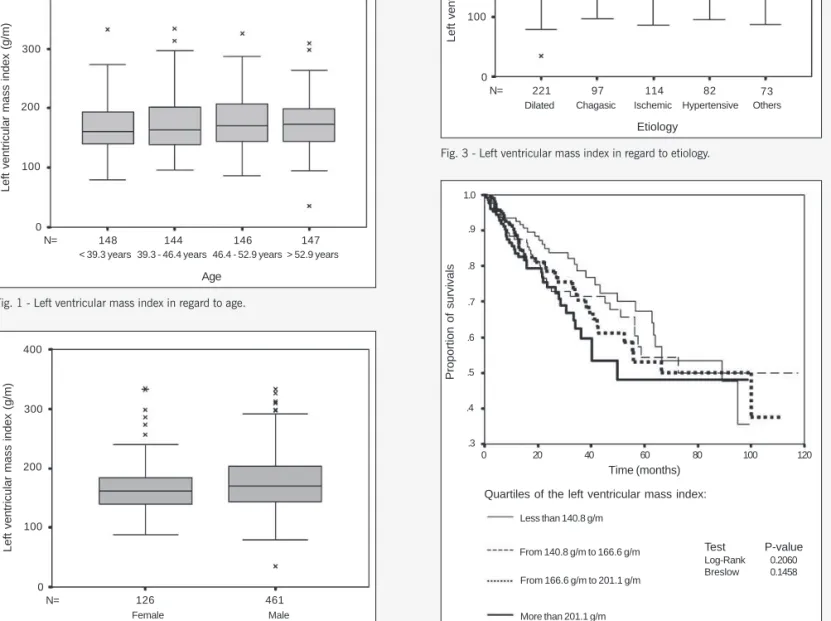
Fig. 1 - Left ventricular mass index in regard to age.
myopathy; from 88.2 g/m to 333.5 g/m (mean, 175.1 g/m; stan-dard deviation, 50.3 g/m) in patients with cardiomyopathy of other etiologies; from 97.5 g/m to 239.6 g/m (mean, 164.3 g/m; stan-dard deviation, 33.7 g/m) in patients with chagasic cardiomyopathy; and from 86.5 g/m to 264.2 g/m (mean, 162 g/m; standard deviation, 39.5 g/m) in patients with ischemic cardiomyopathy.
The left ventricular mass index categorized into quartiles showed no significant difference in regard to the probability of survival (fig. 4). The left ventricular mass index increased 0.39 g/m for each increase in year of the patient’s age, and the other variables (sex, etiology, left ventricular ejection fraction on radioisotopic ventri-culography, left atrial diameter) remained constant (tab. II). The left ventricular mass index in male patients was 11.2 g/m greater than that in female patients.
The left ventricular mass index in patients with hypertensive cardiomyopathy, compared with that in patients with ischemic car-according to age categorized in quartiles (fig. 1). The left
ventricular mass index ranges according to the age groups were as follows: from 79.7 g/m to 332.6 g/m (mean, 168.5 g/m; standard deviation, 41.4 g/m) in patients aged < 39.3 years; from 96.2 g/m to 333.5 g/m (mean, 173.2 g/m; standard deviation, 46.8 g/m) in patients aged from 39.3 years to 46.4 years; from 86.5 g/m to 325.5 g/m (mean, 175.3 g/m; standard deviation, 44.4 g/m) in patients aged from 46.4 to 52.9 years; and from 35.3 g/m to 309.3 g/m (mean, 176.1 g/m; standard deviation, 42.6 g/m) in patients aged > 52.9 years.
The left ventricular mass index was greater among males [range, 35.3 g/m to 333.5 g/m (mean, 175.7 g/m; standard deviation, 44.3 g/m)] than among females [range, 88.2 g/m to 332.6 g/m (mean, 165.7 g/m; standard deviation, 42.3 g/m)] (fig. 2).
Left ventricular mass index was lower in patients with chagasic and ischemic cardiomyopathies than in those with hypertensive cardiomyopathy, idiopathic dilated cardiomyopathy, and cardiomyo-pathies of other etiologies (fig. 3). The left ventricular mass index ranges according to the etiologies of cardiomyopathy were as follows: from 96.2 g/m to 309.3 g/m (mean, 188.1 g/m; stan-dard deviation, 44.6 g/m) in patients with hypertensive cardio-myopathy; from 35.3 g/m to 332.6 g/m (mean, 177.7 g/m; stan-dard deviation, 45.9 g/m) in patients with idiopathic dilated
cardio-Left ventricular mass index (g/m)
400
Etiology
Dilated 300
200
100
0
N= 221 97 114 82 Chagasic Ischemic Hypertensive
Fig. 3 - Left ventricular mass index in regard to etiology.
73 Others
Proportion of survivals
Time (months)
Quartiles of the left ventricular mass index:
Less than 140.8 g/m From 140.8 g/m to 166.6 g/m From 166.6 g/m to 201.1 g/m More than 201.1 g/m
Test P-value
Log-Rank 0.2060 Breslow 0.1458 1.0
.9
.8 .7 .6 .5 .4 .3
0 20 40 60 80 100 120
Fig. 4 - Left ventricular mass index in regard to the chance of survival. Fig. 2 - Left ventricular mass index in regard to sex.
Left ventricular mass index (g/m)
Sex
300
Female 200
100
0
235
diomyopathy, was 27.4 g/m greater. The left ventricular mass indi-ces of patients with cardiomyopathy of unknown etiology and with cardiomyopathy of other etiologies were 16.7 g/m and 12.9 g/m greater, respectively, and that of patients with chagasic cardiomyopathy showed no statistically significant difference when compared with that in patients with ischemic cardiomyopathy.
The left ventricular mass index increased 6.96 g/m for each 5-mm/m increase in the left atrial diameter.
The left ventricular mass index on radioisotopic ventriculography had a negative relation with left ventricular ejection fraction. The left ventricular mass index decreased 1.2 g/m for each 1-unit increase in ejection fraction.
For each 1-g/m increase in the left ventricular mass index, the relative risk of death increased 0.4% (P = 0.0418) (95% CI: 0 to 1%). Because the left ventricular mass index in our case series ranged from 35.35 to 333.52 g/m, the relative risk of death was 1.22 (95% CI: 1 to 1.49) for each 50-g/m increase.
Discussion
This study comprised a large cohort of patients with sympto-matic heart failure of different etiologies, including Chagas’ heart disease, who were followed up on an outpatient care basis at a single institution for 10 years. Patients with cardiomyopathy of unknown etiology (idiopathic, 37.7%) were the most frequent, followed by patients with ischemic cardiomyopathy (19.4%), cha-gasic cardiomyopathy (16.5%), and hypertensive cardiomyopathy (14%). This etiologic distribution differs from that of other case series, in which ischemic cardiomyopathy (34% to 60% of the
cases)15-17, idiopathic cardiomyopathy (18.2% to 59% of the
cases)16,17, and hypertensive cardiomyopathy (3.8% to 23.6% of
cases)16 predominated. Therefore, our results were assessed
ac-cording to these characteristics, including the etiologic distribution. M-mode echocardiography was used to assess left ventricular mass. Alterations in ventricular dimension and geometry may indu-ce inaccuracies in the estimate of left ventricular mass index. The left ventricular mass was indexed by height because patients with heart failure may vary in weight due to fluid retention or loss. This indexation was validated in the literature in a study with extreme methodological strictness, which assessed 864 individuals and found an association between left ventricular mass and height (r=0.39, P < 0.001 in males; r=0.23, P < 0.001 in females) 12. In addition, height is strongly associated with lean body mass, which is an excellent predictor of left ventricular mass 18. Despite of restrictions,
Table II - Relations of the left ventricular mass index with the demographic and functional variables
Varible Estimate Standard error P value
Age 0.3925 0.1696 0.0210
Sex (male) 11.2047 4.2188 0.0081 Hypertensive etiology 27.3629 5.8058 0.0261 Idiopathic etiology 16.6845 4.0165 <0.0001 Other etiologies 12.9527 5.3999 <0.0001 Left atrial inner 13.9206 3.5392 <0.0001 diameter
Left ventricular -1.2411 0.2194 <0.0001 ejection fraction
Ejection fraction obtained on radioisotopic ventriculography.
M-mode echocardiography has been used in large population studies, including those showing the important relation between left ventri-cular mass and cardiovasventri-cular morbidity and mortality 12,19.
Age influenced left ventricular mass index in an independent way. The 1-year increase in age was associated with a 0.39-g/m increase in left ventricular mass index. This observation differs from the previous population studies of individuals with no cardiomyo-pathy, in which age had no influence on left ventricular mass 20,21. On the other hand, the Framingham study showed a relation between age and left ventricular mass in patients with cardiomyopa-thy, but this relation was not shown in patients without cardiomyo-pathy 22. Therefore, the appearance of heart failure may represent a modulatory factor of the relations between left ventricular mass and age.
Sex influenced the left ventricular mass index adjusted for height in an independent way; the left ventricular mass index was 11.2 g/m greater in males as compared with that in females. This finding confirms data of previous epidemiologic studies inclu-ding hypertensive patients 12,21,23,24. Therefore, in regard to sex and with adjustment of the other variables of comparison, patients with heart failure maintain the difference in left ventricular mass. The left ventricular mass index was higher in patients with hypertensive cardiomyopathy, followed by those with idiopathic dilated cardiomyopathy. The left ventricular mass index in patients with ischemic cardiomyopathy and chagasic cardiomyopathy did not show a statistically significant difference. A study of patients with aortic stenosis showed that the increase in left ventricular mass resulted from a combination of hypertrophy and hyperplasia of myocytes 25. Therefore, the mechanisms acting on the increase in ventricular mass may act differently according to the etiology of the cardiomyopathy that causes heart failure.
The relation between the left atrial diameter on echocardio-graphy and the left ventricular mass index was assessed. The left ventricular mass index increased 6.96 g for each 5-mm increase in the left atrial diameter. In this study, the numerical estimate of this relation is noteworthy. One hypothesis is that the same va-riables that influence ventricular mass may also influence left atrial diameter 26,27. On the other hand, the increase in left ventricular mass could contribute to an increase in the atrial dimensions, due to both hemodynamic and biochemical factors. Although the hy-pothesis of the left atrial enlargement secondary to left ventricular diastolic dysfunction may be attractive, a study in patients with arterial hypertension by use of Doppler diastolic indices did not
show this occurrence 28. On the other hand, this same study
showed that the left atrial size in hypertensive patients with left ventricular hypertrophy on electrocardiography did not depend on left ventricular mass 28. Therefore, a relation between left atrial diameter and left ventricular mass in patients with heart failure exists, but not in myocardial hypertrophy of patients with arterial hypertension.
236
Although the comparison of the probabilities of survival of pa-tients, whose left ventricular mass indices were categorized in quarti-les, showed no statistically significant difference, the Cox proportional hazards regression model revealed that for each 1-g/m increase in left ventricular mass index, the relative risk of death increased 0.4%. Because the left ventricular mass index in our case series ranged from 35.35 g/m to 333.52 g/m, the relative risk of death was 1.22 for each 50-g/m increase in the left ventricular mass index. In the Framingham study with patients without cardiomyo-pathy, for each 50-g/m increase in the left ventricular mass, a relative risk of death due to heart diseases of 1.73 was observed in males and of 2.12 in females 30. In a case series of elderly patients without cardiomyopathy, whose ages ranged from 59 years to 90
years, the incidence of coronary events for each 50-g/m increase in
left ventricular mass was 1.67 in males and 1.60 in females 31.
Therefore, the increase in left ventricular mass index is an unfavorable prognostic factor.
It is worth noting that the case series studied comprises sympto-matic patients in an advanced phase of the disease. These observa-tions, however, may not be applicable to the general population or patients with heart failure in another phase of clinical evolution.
In conclusion, the influence of left ventricular mass was not very strong, but could contribute to the prognostic assessment of patients with heart failure. Therefore, the relations with other va-riables, including the negative correlation with left ventricular ejec-tion fracejec-tion on radioisotopic ventriculography, require further studies.
1. Kannel WB, Dawber TR, Kaga A, Revotskie N, Stokes J. Factors of risk in the de-velopment of coronary heart disease-six year follow-up experience: the Fra-mingham Study. Ann Intern Med 1961; 55: 33-50.
2. Kannel WB, Gordon T, Offutt D. Left ventricular hypertrophy by electrocardiogram. Prevalence, incidence, and mortality in the Framingham Study. Ann Intern Med 1969; 71: 89-105.
3. Kannel WB. Prevalence and natural history of electrocardiographic left ventricular hypertrophy. Am J Med 1983; 75: 4-11.
4. Francis GS. Pathophysiology of chronic heart failure. Am J Med 2001; 110 Suppl 7A:37S-46S.
5. Quinones MA, Weiner DH, Shelton BJ, Greenberg BH, Limacher MC, Koilipillai C et al. For the SOLVD Investigators. Echocardiographic predictors of one-year cli-nical outcome in study of left ventricular dysfunction (SOLVD) Trial and Registry: an analysis of 1172 patients (abstract). Circulation 1993; 88 Suppl I:304. 6. Nastari L, Mansur AJ, Freitas HFG et al. Massa miocárdica em portadores de
in-suficiência cardíaca. Soc Cardiol Estado de São Paulo 1994; 4 Suppl B:27. 7. Simão Filho C. Remodelamento ventricular esquerdo em cardiomiopatias de
dife-rentes etiologias na sua forma dilatada. Estudo morfológico comparativo em peças anatômicas. Tese (Doutorado). São Paulo (SP): Faculdade de Medicina, Univer-sidade de São Paulo; 1998.
8. Rouquaryol MZ, Almeida Filho N. Desenhos de pesquisa em epidemiologia. In: Epi-demiologia e saúde. 5ª ed. Rio de Janeiro (RJ): Medsi; 1999. p. 149-70. 9. Mckee PA, Castelli WP, McNamara PM, Kannel WB. The natural history of
conges-tive heart failure: the Framingham study. N Engl J Med 1971; 285: 1441-6. 10. Report of the WHO/ISFC task force on the definition and classification of
cardio-myopathies. Br Heart J 1980; 44: 672-3.
11. Sahn DJ, De Maria A, Kisslo J, Weyman A. Recommendations regarding quanti-tation in M–mode echocardiography: results of a survey of echocardiographic measurements. Circulation 1978; 58: 1072-83.
12. Levy D, Savage DD, Garrison RJ, Anderson KM, Kannel WB, Castelli WP. Echocar-diographic criteria for left ventricular hypertrophy: the Framingham Heart Study. Am J Cardiol 1987; 59: 956-60.
13. Kleinbaum DG. Survival analysis: a self-learning text. New York, Springer-Verlag, 1996. 14. Cox DR. Regression models and life tables. J R Stat Soc 1972; 34 Suppl B: 187-220. 15. Likoff MJ, Chandler SL, Kay HR. Clinical determinants of mortality in chronic con-gestive heart failure secondary to idiopathic dilated or to ischemic cardiomyopa-thy. Am J Cardiol 1987; 59: 634-8.
16. Teerlink JR, Goldhaber SZ, Pfeffer MA. An overview of contemporary etiologies of congestive heart failure. Am Heart J 1991: 121: 1852-3.
17. Myers J, Gullestad L, Vagelos R, Do D, Bellin D, Ross H et al. Clinical, homodyna-mic, and cardiopulmonary exercise test determinants of survival in patients referred for evaluation of heart failure. Ann Intern Med 1998; 129: 286-93.
18. Devereux RB, Lutas EM, Casale PN et al. Standardization of M-Mode
echcar-References
diographic assessment of left ventricular hypertrophy: comparison to necropsy fin-dings. Am J Cardiol 1986; 57: 450-8.
19. Casale PN, Devereux RB, Milner M, Zullo G, Harshfield GA, Pickering TG et al. Value of echocardiographic measurement of left ventricular mass in predicting cardiovas-cular morbid events in hypertensive men. Ann Intern Med 1986; 105: 173-8. 20. Liebson PR, Grandits G, Prineas R, Dianzumba S, Flack JM, Cutler JA et al.
Echo-cardiographic correlates of left ventricular structure among 844 mildly hypertensive men and women in the Treatment of Mild Hypertension Study (TOMHS). Circula-tion 1993; 87: 476-86.
21. Gardin JM, Siscovick D, Anton–Culver H, Lynch JC, Smith VE, Klopfenstein HS et al. Sex, age, and disease affect echocardiographic left ventricular mass and systolic function in the free–living elderly. The Cardiovascular Health Study. Circulation 1995; 91: 1739-48.
22. Dannenberg AL, Levy D, Garrinson J. Impact of age on echocardiographic left ven-tricular mass in a healthy population (the Framingham Study). Am J Cardiol 1989; 64: 1066-8.
23. Vasan RS, Larson MG, Levy D, Evans JC, Benjamin EJ. Distribuition and catego-rization of echocardiographic measurements in relation to reference limits. The Framingham Heart Study: formulation of a height and sex specific classification and its prospective validation. Circulation 1997; 96: 1863-73.
24. Zabalgoitia M, Rahman NU, Haley WE, Mercado R, Yunis C, Lucas C et al. Com-parison in systemic hypertension of left ventricular mass and geometry with systolic and diastolic function in patients < 65 to ≥ 65 years of age. Am J Cardiol 1998; 82: 604-8.
25. Urbanek K, Quaini F, Tasca G, Torella D, Castaldo C, Ginard BN et al. Intense myo-cyte formation from cardiac stem cells in human cardiac hypertrophy. PNAS 2003; 100: 10440-5.
26. Gottdiener JS, Reda DJ, Williams DW, Materson BJ. Left atrial size of hypertensi-ve men: influence of obesity, race and age. J Am Coll Cardiol 1997; 29: 651-8. 27. Gottdiener JS, Domenic J, Reda DJ, Williams DW, Materson BJ, Cushman W et al.
Effect of single-drug therapy on reduction of left atrial size in mild to moderate hy-pertension. Circulation 1998; 98: 140-8.
28. Gerdts E, Oikarinen L, Palmieri V, Otterstad JE, Wachtell K, Boman K et al. Corre-lates of left atrial size in hypertensive patients with left ventricular hypertrophy. The losartan intervention for endpoint reduction in hypertension (LIFE) study. Hyper-tension 2002; 39: 739-43.
29. Parameshwar J, Keegan J, Sparrow J, Sutton GC, Poole–Wilson PA. Predictors of prognosis in severe chronic heart failure. Am Heart J 1992; 123: 421-6. 30. Levy D, Garrison RJ, Savage DD, Kannel WB, Castelli WP. Prognostic implications
of echocardiographically determined left ventricular mass in the Framingham Heart Study. N Engl J Med 1990; 322: 1561-6.