The Medical College of Ohio - Toledo, Ohio, USA
Mailing address – Sergio do Carmo Jorge Rua Tuim 523/51 04514102 -Sao Paulo - Brazil - Email: [email protected]
Blair P. Grubb, Sérgio do Carmo Jorge
Toledo, Ohio – São Paulo, SP - Brazil
A Review of the Classification, Diagnosis, and Management of
Autonomic Dysfunction Syndromes Associated with
Orthostatic Intolerance
Syncope, the transient loss of consciousness and postural tone, is one of the oldest of recorded medical pro-blems. Indeed, Hippocrates, the father of medicine, recor-ded the first description of syncope, and it is from the Greek that we derive the medical term for fainting (syncoptein - to cut short). Both a sign and a syndrome, syncope may result from a wide variety of causes. Over the last decade, conside-rable attention has been focused on one particular cause of syncope, the phenomenon previously called vasovagal syncope. Research into the nature of this disorder has de-monstrated that it is only one aspect of a much broader group of disturbances of the autonomic nervous system that may lead to hypotension, orthostatic intolerance, and ultimately syncope. Indeed, recent discoveries have caused us to reevaluate our entire classification of autonomic di-sorders, and to develop a new system that better reflects our current knowledge. Because it is the cardiologist and the clinical cardiac electrophysiologist who are now fre-quently called upon to recognize and treat these disorders, this review is designed to acquaint the reader with these conditions, their diagnosis, and management.
The autonomic nervous system
Because these nervous disorders all result from a dis-turbance in normal autonomic function, it would seem ap-propriate to briefly review some aspects of the structure and operation of the nervous system.
The human nervous system has two basic compo-nents: the central nervous system, made up of the brain and the spinal cord 1, and the peripheral nervous system, which
is comprises groups of neurons called ganglia, and of peripheral nerves that lie outside the brain and spinal cord. Although anatomically separate, the two systems are func-tionally interconnected. The peripheral nervous system is further divided into somatic and autonomic divisions. The
somatic division is principally concerned with sensory in-formation about the environment outside the body as well as muscle and limb position. The autonomic division (usually called the autonomic nervous system or ANS) is the motor system for the viscera, the smooth muscles of the body (especially those of the vasculature), and the exocrine glands. It is composed of three distinct parts: the sympa-thetic, parasympasympa-thetic, and enteric nervous systems. The sympathetic nervous system helps control the reaction of the body to stress, and the parasympathetic system works to conserve the body’s resources and to restore equilibrium to the resting state. The enteric system controls the functi-on of the gut. The organ systems governed by the ANS are, for the most part, independent of volitional control (althou-gh they sometimes can be affected by volitional or emotio-nal inputs) and include the cardiorespiratory organs, the gastrointestinal and genitourinary tracts. The autonomic system is vital to the maintenance of internal homeostasis and achieves this by mechanisms that regulate blood pres-sure, fluid and electrolyte balance, and body temperature.
Although representative of one of the defining as-pects of the evolution of Homo sapiens, the adoption of upright posture presented a novel challenge to a blood pressure control system that developed to meet the require-ments of an animal in the dorsal position. Indeed, the organ that defines our humanity, the brain, was placed in a so-mewhat precarious position with regards to vascular perfu-sion and oxygenation. It is the ANS that governs both the short- and medium-term blood pressure responses to posi-tional change 2. Normally, around 25% of the circulating
hu-mans, the venous HIP is around the level of the diaphragm and the arterial HIP is at the left ventricle. The venous HIP is somewhat dynamic in that it can be altered by changes in venous compliance brought on by muscular activity 2.
Following standing, the healthy subject achieves orthosta-tic stabilization in one minute or less. It should be noted that the exact circulatory responses brought on by standing (an active process) are somewhat different from those brought on by head up tilt (a passive process). In the moments follo-wing assumption of upright posture, a slow decline in arte-rial pressure and cardiac filling occurs. This causes activa-tion of the high-pressure receptors of the carotid sinus and aortic arch, as well as the low-pressure receptors of the heart and lungs. The mechanoreceptors that are within the heart are linked by unmyelinated vagal afferents in both the atria and ventricles 1-4. These fibers have been found to cause
continuous inhibitory actions on the cardiovascular areas of the medulla (the nucleus tractus solitarii) 1. The fall in
venous return that results from upright posture produces less stretch on these receptors, then discharge rates de-crease, and the change in input to the brain stem causes an increase in sympathetic outflow resulting in systemic vaso-constriction. At the same time, the fall in arterial pressure while upright activates the high-pressure receptors in the carotid sinus, which stimulates an increase in heart rate. These early steady state adaptations to upright posture therefore result in a 10 to 15 beat per minute increase in heart rate, a diastolic pressure increase of 10 mm Hg, and little or no change in systolic blood pressure. Once these adjust-ments are complete, as compared with the supine state, du-ring upright stance the thoracic blood volume is 30% less as is the total cardiac output, and the mean heart rate is 10-15 beats/minute higher. Furthermore, detailed descriptions of this process are available to the interested reader 2.
As a person continues to stand, activation of neurohu-moral responses occurs, the amount of which is dependent on the subject’s volume status. As a rule, the lower the volu-me, the higher the degree of the renin-angiotensin-aldoste-rone system involvement 2. The inability of any of these
pro-cesses to function adequately (or in a coordinated manner) can potentially result in a failure in the normal responses to sudden shifts in posture (or their maintenance) with resultant hypotension that may be sufficiently great as to result in ce-rebral hypoperfusion, hypoxia, and loss of consciousness.
Disorders of orthostatic control
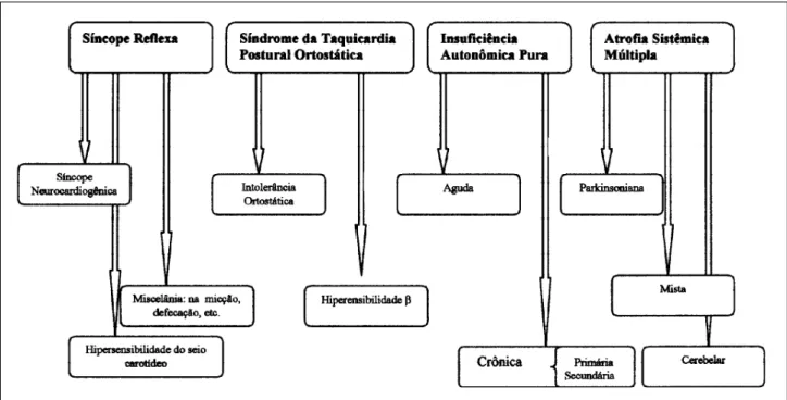
A number of different disorders of orthostatic control have been identified that, although sharing certain charac-teristics, are in many ways unique. It should be remembered that when we observe nature, we see what we want to see according to what we believe we know about it at the time. At one point, supraventricular tachycardia was felt to be a single entity, only later was it found to be composed of mul-tiple subtypes. In order to make some sense of the apparent chaos of nature, we try to classify it into a coherent system that conforms to our expectations. Thus, any system of clas-sification is in some ways arbitrary and open to debate. The following system conforms to that developed by the Ameri-can Autonomic Society in 1996. A basic somewhat simpli-fied outline of the system is provided in figure 1. Many in-vestigators like to divide these disorders into primary and secondary forms. The primary forms tend to be idiopathic and are divided into acute and chronic forms. The seconda-ry types are usually seen in association with a particular di-sease or are known to arise secondarily to a known bioche-mical abnormality.
Reflex syncopes
Physicians tend to be most familiar with this form of syncope and as such, our discussion of it will be limited 3.
First described by both Gower and Sir Thomas Lewis as vasovagal syncope, it is better known today as either neu-rocardiogenic or neurally mediated syncope. Although di-verse in presentation, it most frequently occurs in younger people and is characterized by a distinct prodrome of variable duration followed by an abrupt loss of conscious-ness. Recovery is rapid and is usually not accompanied by a postictal state. These episodes are felt to represent a “hy-persensitive” autonomic system that overresponds to various stimuli. Most commonly, this is prolonged orthos-tatic stress, which is felt to increase venous pooling to the point where venous return to the right ventricle falls so pre-cipitously that an increase in ventricular inotropy causes activation of mechanoreceptors that would normally fire only during stretch 4. This sudden surge in neural traffic to
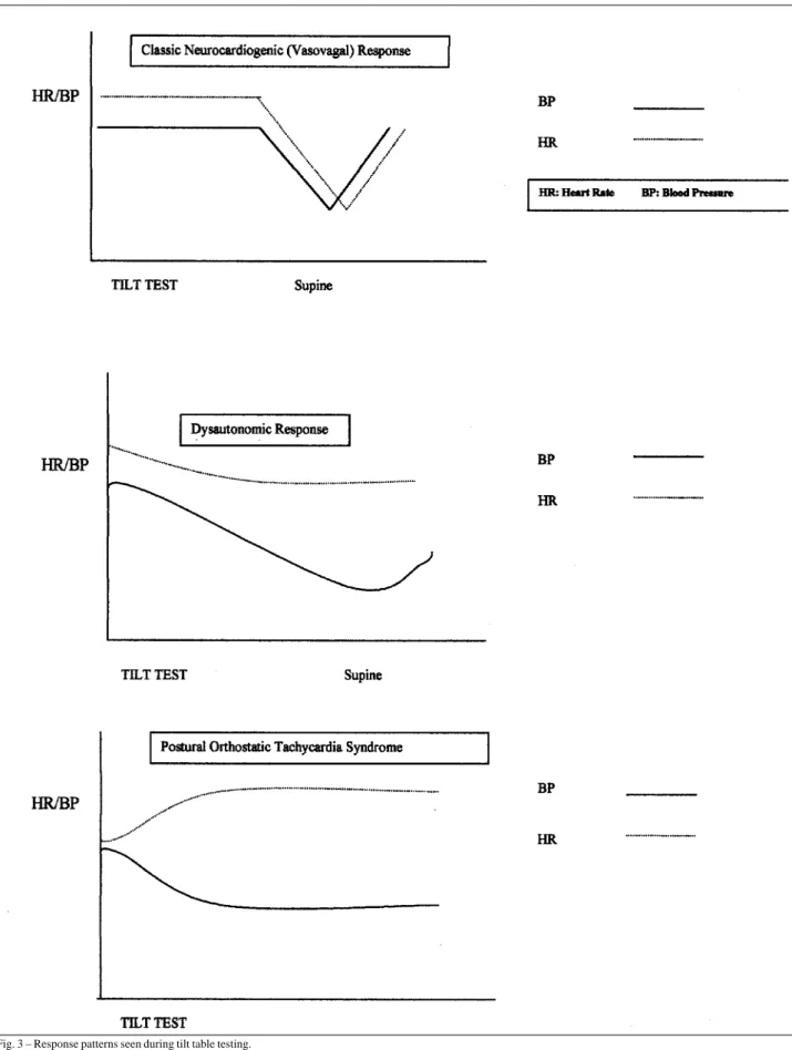
the brain stem mimics the conditions seen in hypertension, thus provoking an apparently “paradoxic” sympathetic withdrawal with resultant hypotension, bradycardia, and syncope. It is important to remember, however, that other stimuli such as strong emotion or epileptic discharge can provoke identical responses, thus suggesting that these in-dividuals have an inherent increase in sensitivity to such stimuli. During head upright tilt table testing, these indivi-duals have a sudden profound fall in blood pressure, that is closely followed by a fall in heart rate (sometimes to the point of asystole).
Sutton has made the insightful observation that the responses seen during neurocardiogenic syncope and ca-rotid sinus hypersensitivity are quite similar and may be dif-ferent aspects of the same disorder 5. Indeed, in such a
pre-disposed individual, rapid mechanoreceptor activation from any site (blood, bladder, cough) could elicit similar respon-ses. More detailed descriptions of these disorders can be found elsewhere 6. What seems to distinguish these
disor-ders from the remainder of those discussed herein, is that in between episodes these patients are quite normal and re-port few if any other symptoms. Indeed, their autonomic systems appear to function normally despite their “hyper-sensitive” nature, as opposed to other conditions where the autonomic system seems to “fail”.
Primary disorders of autonomic failure
Chronic disorders - The physician is more likely to
encounter the chronic forms of autonomic failure than their acute counterparts. The first report of chronic autonomic failure was the groundbreaking report by Bradbury and Eg-gleston in 1925, where they labeled the condition “idiopa-thic orthostatic hypotension” due to an apparent lack of other neurologic features 7. However, since then, it has
be-come quite apparent that in these patients there exists a ge-neralized state of autonomic dysfunction as manifested by orthostatic hypotension and syncope, as well as
disturban-ces in bowel, bladder, thermoregulatory, sudomotor, and se-xual function. The American Autonomic Society has named this disorder Pure Autonomic Failure (or PAF) 8. Although
the cause of PAF remains unknown, several investigators have postulated that a degeneration of the peripheral post-ganglionic autonomic neurons occurs. Although the condi-tion is more commonly seen in older adults, it can occur in almost any age group (including children).
Another type of autonomic failure was reported in 1960 in a landmark paper by Shy and Drager 9. In contrast to
Pure Autonomic Failure, this more severe condition is mani-fested by severe orthostatic hypotension, progressive uri-nary and rectal incontinence, loss of sweating, iris atrophy, external ocular palsy, impotence, rigidity, and tremors. Both muscle fasciculations and distal muscle wasting may be seen late in the disorder. To better identify this complex mul-tisystem disorder, the American Autonomic Society has na-med this disease Multiple System Atrophy (MSA) and has divided it into three major subtypes 10. The first type
pre-sents as tremor that is surprisingly similar to Parkinson’s di-sease (some authors prefer to refer to this type as presen-ting as striatonigral degeneration). A second type presents as mainly cerebellar or pyramidal symptoms, or both (some investigators have termed this the olivopontocerebellar atrophy/degeneration form). A third type presents as cha-racteristics of both of these types. As was alluded to pre-viously, MSA can appear surprisingly similar to Parkin-son’s disease. An autopsy study recently found that so-mewhere between 7 and 22% of people thought to have Parkinson’s disease actually had neuropathologic findings diagnostic for MSA. Although the vast majority of MSA patients do not present with symptoms until somewhere between the 5th and 7th decade of life, some unfortunate in-dividuals do begin to have symptoms in their late thirties.
Recently, a considerable amount of attention has been focused on a milder form of chronic autonomic failure that is referred to as the Postural Orthostatic Tachycardia Syn-drome (or POTS) 11. The hallmark of the syndrome is a
per-sistent tachycardia while upright (that sometimes reaches rates of 160 beats/minute or more) that is associated with se-vere fatigue near syncope, exercise intolerance, and li-ghtheadedness or dizziness. Many also complain of always being cold, while at the same time unable to tolerate extreme heat. During head upright tilt, these patients experience a sudden increase in heart rate of greater than 30 beats/minu-te within the first five minubeats/minu-tes, or achieve a maximum heart rate of 120 beats/minute associated with only mildly reduced blood pressures.
inappropriate sinus tachycardia and who had undergone radio frequency modification of the sinus atrial node (at other centers). After the apparently successful elimination of their “sinus tachycardia”, they were left with profound orthostatic hypotension. These patients may be misdiagno-sed as suffering from Chronic Fatigue Syndrome. Several recent reports have suggested that a great deal of overlap may exist between these two disorders 12.
Acute autonomic dysfunction
Even though these syndromes are rare, the acute auto-nomic neuropathies that produce hypotension and synco-pe are frequently dramatic in presentation 13. These
disor-ders are acute in most and demonstrate severe and wides-pread failure of both the sympathetic and parasympathetic systems while leaving the somatic fibers unaffected. Many of these patients tend to be young and, prior to the illness, were very healthy. The development of the illness is surpri-singly rapid and patients can frequently relate the exact day symptoms first began. One interesting observation has been that a large number of these individuals report having had a febrile illness (presumed to be viral) prior to the onset of symptoms, giving rise to the notion that an autoimmune component to the disorder may exist.
Function of the sympathetic nervous system is often so severely disrupted that orthostatic hypotension occurs of such a degree that the patient cannot even sit upright in bed without fainting. Patients often totally lose their ability to sweat, and suffer bowel and bladder dysfunctions. These patients frequently complain of bloating, nausea, vomiting, and abdominal pain. Constipation is frequent, and someti-mes will alternate with diarrhea. One fascinating finding is that the heart rate will often be at a fixed rate of 40 to 50 beats per minute, associated with complete chronotropic incom-petence. The pupils are often dilated and poorly reactive to light. Patients may experience several syncopal episodes daily. The long-term prognosis of these patients is quite variable, with some enjoying complete recoveries, and others suffering a chronic debilitating course. Patients are often left with significant residual defects.
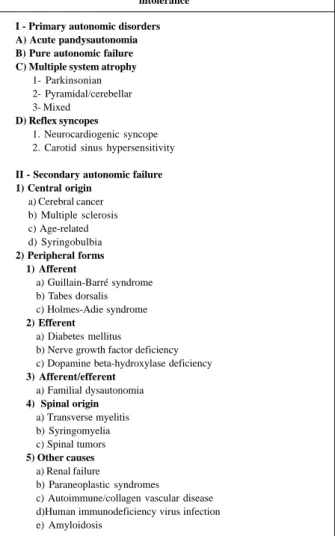
Secondary causes of autonomic dysfunction
A wide variety of disorders may cause varying degrees of autonomic disturbance. A list of some of these disorders is found in table I. It is important for the phy-sician to be able to recognize when autonomic dysfunction is but part of a greater disorder. In occasional patients, several conditions may coexist that produce synergistic detrimental effects on autonomic function. Over the last decade, a number of enzymatic abnormalities have been identified that can result in autonomic disruption. Princi-pal among these is isolated dopamine beta hydroxylase (DBH) deficiency syndrome, a condition that is now easily treated by replacement therapy. Additional deficiency syndromes involving nerve growth factor, monamine
oxidase, aromatic L-amino decarboxylase, and some sensory neuropeptides may all result in autonomic failure and hypotension. Diffuse systemic illnesses, such as re-nal failure, cancer, or acquired immune deficiency syn-drome (AIDS), may all cause hypotension and syncope. Studies have also demonstrated a link between orthostatic hypotension and Alzheimer’s disease 14.
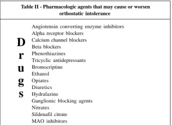
Perhaps one of the most important things to remember is that a vast number of pharmacologic agents are available that may either cause or worsen orthostatic hypotension (table II). Chief among these are the peripherally acting vaso-dilatory agents, such as the angiotensin converting enzyme (ACE) inhibitors, prazosin, hydralazine, guanethidine. Beta-blocking agents may also worsen syncope in some patients. Lately we have observed an increased frequency of dysau-tonomic syncope in patients suffering from congestive heart failure. In this group, the combination of a low cardiac output and volume depletion due to diuretics and vasodila-tor therapy serve to interfere with the body’s aforementio-ned mechanisms for adapting to upright posture. Centrally acting agents, such as the tricyclic antidepressants, reser-pine and methyldopa, may also exacerbate otherwise mild hypotension.
Table I - Autonomic disorders associated with orthostatic intolerance
I - Primary autonomic disorders A) Acute pandysautonomia B) Pure autonomic failure C) Multiple system atrophy
1- Parkinsonian 2- Pyramidal/cerebellar 3- Mixed
D) Reflex syncopes
1. Neurocardiogenic syncope 2. Carotid sinus hypersensitivity
II - Secondary autonomic failure 1) Central origin
a) Cerebral cancer b) Multiple sclerosis c) Age-related d) Syringobulbia
2) Peripheral forms 1) Afferent
a) Guillain-Barré syndrome b) Tabes dorsalis c) Holmes-Adie syndrome
2) Efferent
a) Diabetes mellitus
b) Nerve growth factor deficiency c) Dopamine beta-hydroxylase deficiency
3) Afferent/efferent
a) Familial dysautonomia
4) Spinal origin
a) Transverse myelitis b) Syringomyelia c) Spinal tumors
5) Other causes
a) Renal failure
b) Paraneoplastic syndromes
Clinical features
The principal feature that all of these conditions share is that normal cardiovascular regulation is disturbed resul-ting in postural hypotension. Although orthostatic hypo-tension was once defined as a greater than 20 mm Hg fall in systolic blood pressure over a three minute period after standing upright, a smaller drop in blood pressure asso-ciated with symptoms can be just as important. A large per-centage of these patients display a slow steady fall in blood pressure over a longer time frame (around 10-15 minutes) that can be quite symptomatic. Whether the patient expe-riences symptoms is as much dependent on the rate of fall in pressure as it is upon the absolute degree of change. The loss of consciousness in the dysautonomic tends to be slow and gradual, usually when the patient is walking or stan-ding. However, many older patients do not seem to perceive this decline in pressure and therefore report little or no pro-drome prior to syncope and describe these episodes as “drop attacks”. Those who do experience prodromes des-cribe a wide variety of symptoms, such as dizziness, blur-ring of vision, “seeing stars”, and tunnel vision. A dis-tinguishing feature between neurocardiogenic and dysau-tonomic syncope is that in the latter, bradycardia and dia-phoresis are uncommon during an episode. Dysautonomic syncope tends to be more common in the early morning hours. Any factor that enhances peripheral venous pooling such as extreme heat, fatigue, or alcohol ingestion will exa-cerbate hypotension. As time goes on, some patients may develop a relatively fixed heart rate that shows little respon-se to either postural change or exercirespon-se. In addition, some patients develop a syndrome of supine hypertension that alternates with upright hypotension, presumably due to a failure to vasodilate when prone. Patients suffering from this combination of supine hypertension and upright hypo-tension can be quite difficult to treat. Sometimes distin-guishing between these disorders can be difficult as a con-siderable degree of overlap may exist between them (a situa-tion not dissimilar to that seen with the various forms of chronic obstructive lung disease) (fig. 2).
Evaluation of patients
The cornerstone of evaluation is a detailed history and physical examination. When do syncopal or near syn-copal episodes occur and when did they begin? How often? Is there a pattern to the events or any known precipitating factors? What are episodes like to the patient and how do they appear to bystanders? What other organ systems are involved? Other than syncope, what symptom bothers the patient most? A careful and concise history and physical (which must include a concise neurologic examination) will have a far greater diagnostic yield than the mindless ordering of multiple tests. Laboratory examinations should be ob-tained in a careful and directed manner, based upon history and physical findings, to confirm one’s clinical impressions. It is far beyond the scope of this paper to review every autonomic disorder and the various tests used in evalua-tion. The interested reader is directed to several excellent texts on the subject 8, 15-18. One point that needs to be
em-phasized, is that any drugs the patient is taking that could produce hypotension should be identified. (table II) This includes not only proscriptions but also over-the-counter medications and herbal remedies as well. Sadly, in the mo-dern era, when a young person presents with symptoms of autonomic dysfunction, the potential use of illicit drugs or alcohol should be considered. In women, symptoms may vary with the menstrual cycle or an otherwise mild tendency toward autonomic dysfunction may be exacerbated by the onset of menopause.
Because the autonomic areas of the brain are not ac-cessible to direct measurement, one must measure the res-ponses of various organ systems to various physiologic or pharmacologic challenges. In addition, recent advances have allowed for the determination of serum urine and cere-brospinal fluid levels of some autonomic neuromodulators and neurotransmitters. Foremost, however, is the determi-nation of blood pressure and heart rate response to positio-nal change, with measurements taken while the patient is
Reflex Syncope
Pure Autonomic Failure
Multiple System Atrophy
Postural Orthostatic Tachicardia
Syndrome (STOP)
Fig. 2 – Interelationship between autonomic disorders.
D
r
u
g
s
Table II - Pharmacologic agents that may cause or worsen orthostatic intolerance
Angiotensin converting enzyme inhibitors Alpha receptor blockers
Calcium channel blockers Beta blockers
Phenothiazines Tricyclic antidepressants Bromocriptine Ethanol Opiates Diuretics Hydralazine
Ganglionic blocking agents Nitrates
supine, sitting, and standing. The exact change in pressure considered to be significant is still under discussion, but is usually felt to be between 20-30mmHg systolic and 10-15mmHg diastolic. Remember that when standing, pressure determination should be performed with the arm extended horizontally (to avoid the possible hydrostatic effects of the fluid column of the arm). Because the body’s respon-ses to active standing differ from those of passive tilting, we also frequently perform tilt table testing on these pati-ents, the details of which are given elsewhere 19 (fig. 3). A
number of other autonomic tests are also available and are quite useful in selected patients 15-18.
Therapeutic options
A complete discussion of the treatment options avai-lable is beyond the scope of this review; however, some basic principals will be briefly outlined (more complete dis-cussions can be found elsewhere). One of the physician’s most important tasks is to identify whether hypotensive syncope is primary or secondary in nature, and to determi-ne if there are any potentially reversible causes (i.e., drugs, anemia, volume depletion).
It is equally important to educate patients and their fa-milies as to the nature of the problem. Teaching the patient to avoid aggravating factors (such as extreme heat, dehy-dration, and alcohol consumption) as well as recognizing any prodromal symptoms and assuming a recumbent posi-tion at their onset are extremely helpful measures.
Nonpharmacologic therapies that are useful include sleeping with the head of the bed upright (around 6-12 in-ches), and elastic support hose (at least 30-40 mm Hg ankle counter-pressure). Biofeedback has also proven useful in select patients.
Pharmacotherapy should be used cautiously, and should be tailored to fit the needs of the patient based on the type of autonomic disorder being treated, as well as coexisting symptoms and conditions. It should also be re-membered that virtually any drug used in treatment can oc-casionally worsen symptoms (a “prosyncopal” effect).
In neurocardiogenic syncope, a number of reports have found that beta-blocker therapy is effective presuma-bly because its negative inotropic effects lessen the degree of cardiac mechanoreceptor activation associated with abrupt falls in venous return. The increase in peripheral vascular resistance that accompanies unopposed beta blo-ckade may also contribute to its therapeutic effects. We have not found beta blockade as useful in other forms of reflex syncope, and they may be detrimental in the nomic syndromes. A useful agent in patients with dysauto-nomic syncope (and in younger patients with neurocardio-genic syncope) is the mineral corticoid agent, fludrocorti-sone. It results not only in fluid and sodium retention, but it also appears to raise pressure via an indirect vasoconstric-tive effect resulting from sensitization of peripheral alpha re-ceptors. Because the drug may cause hypokalemia and
hy-pomagnesemia, serum potassium and magnesium levels need to be periodically monitored.
Because failure to properly vasoconstrict the peripheral vessels is common to all of these disorders, vasoconstrictive substances can be employed. Initially, we employed the amphetamine-like agent, methylphenidate with excellent results 20. However, the fact that it is a
controlled substance with potent CNS stimulating activity tended to limit our use of the drug. An excellent alternative is the new alpha stimulating agent, midodrine. It has almost no CNS effects or cardiac stimulation, but provides identical degrees of peripheral alpha- receptor stimulation. Several studies have demonstrated midodrine’s efficacy in both neurocardiogenic and dysautonomic disorders 21,22.
It has been found that the alpha-2 receptor blocking agent, clonidine, can actually elevate blood pressure in dy-sautonomic patients where hypotension is secondary to a se-vere postganglionic sympathetic lesion 23. In patients with
severe autonomic failure, the post- junctional vascular alpha-2 receptors (that are plentiful in the venous system) are actually hypersensitive. Although, in healthy individuals, clonidine acts on the central nervous system to lessen sympathetic output and with it blood pressure; in autonomic failure, some patients exhibit little or no sympathetic output thus permitting its peripheral actions to become manifest.
Interestingly, a number of patients with autonomic failure are anemic. A brilliant study by Hoeldtke and Stre-eten demonstrated that subcutaneous injections of erythro-poietin while raising blood count also produce dramatic in-creases in blood pressure 24. This pressure effect seems to
occur independently of the red cell effect 25.
A series of both animal and human studies have de-monstrated that the neurotransmitter serotonin (5-hydroxy-tryptamine) plays an essential role in the central regulation of blood pressure and heart rate. It has been postulated that some patients with autonomic disorders may have distur-bances in central serotonin production or regulation 26. In
support of this concept has been the observation that the serotonin reuptake inhibitors can be effective in both the treatment of neurocardiogenic syncope and orthostatic hy-potension 27.
The exact role of pacemaker therapy in the treatment of these disorders remains controversial, and is beyond the scope of this discussion; however, a number of investiga-tors have found that in select patients pacemaker therapy can be effective in reducing symptoms and may sometimes eliminate syncope altogether 28.
1. Bernarroch E. The central autonomic network: Functional organization, dysfunc-tion and perspective. Mayo Clinic Proc 1993; 68: 988-1001.
2. Wieling W, Lieshout J. Maintenance of postural normotension in humans. In: Low P. Clinical Autonomic Disorders. (ed). Little Brown Co., 1993: 69-73. 3. Grubb BP. Neurocardiogenic syncope. In: Grubb BP, Olshansky B (eds).
Synco-pe: Mechanisms and Management. Armonk, NY: Futura Publishing, 1998 (in press).
4. Kosinski D, Grubb BP, Temesy-Armos P. Pathophysiological aspects of neuro-cardiogenic syncope. PACE 1995; 18: 716-21.
5. Sutton R, Petersen M. The clinical spectrum of neurocardiogenic syncope. J Car-diovasc Eletrophysiol 1995; 6: 569-76.
6. Kosinski D. Miscellaneous causes of syncope. In: Grubb BP, Olshansky B (eds). Syncope: Mechanisms and Management. Armonk, NY: Futura Publishing, 1998 (in press).
7. Bradbury S, Eggleston C. Postural hypotension: A report of three cases. J Am Heart J 1925; 1: 73-86.
8. Robertson D, Pllinsky E. A Primer on the Autonomic Nervous System (eds). San Diego: Academic Press, 1996.
9. Shy GM, Drager GA. A neurologic syndrome associated with orthostatic hypo-tension. Arch Neurol 1960; 3: 511-27.
10. Mathias CJ. The classification and nomenclature of autonomic disorders: ending chaos, restoring conflict, and hopefully achieving clarity. Clin Auton Res 1995; 5: 307-10. 11. Grubb BP, Kosinski D, Boehm K, Kip K. The postural orthostatic tachycardia
syndrome: A neurocardiogenic variant identified during head up tilt testing. PACE (in press).
12. Bou-Holaigh I, Rowe P, Kan J, Calkins H. The relationship between neurally me-diated hypotension and chronic fatigue syndrome. JAMA 1995; 274: 961-7. 13. Grubb BP, Kosinski D. Acute pandysautonomic syncope. Eur J Cardiac Pacing
Electrophysiol 1997; 7: 10-14.
14. Passant V, Warkentin S, Karlson, et al. Orthostatic hypotension in organic
de-mentia: Relationship between blood pressure, cortical blood flow, and sympto-ms. Clin Auton Res 1996; 6: 29-36.
15. Bannister R, Mathias C. Autonomic Failure: A Textbook of Clinical Disorders of the Autonomic Nervous System (eds). Oxford: Oxford Medical Publications, 1992. 16. Low P (ed). Clinical Autonomic Disorders. Boston: Little Brown Co., 1993. 17. Grubb BP, Olshansky B (eds). Syncope: Mechanisms and Management.
Ar-monk, NY: Futura Publishing, 1997.
18. Robertson D, Biaggioni T. Disorders of the Autonomic Nervous System. (eds) London: Harwood Academic Publishers, 1995.
19. Grubb BP, Kosinski D. Tilt table testing: Concepts and limitations. PACE 1997; 20(PTII): 781-7.
20. Grubb BP, Kosinski D, Mouhaffel A, Pothoulakis A. The use of methylphenidate in the treatment of refractory neurocardiogenic syncope. PACE 1996; 19: 836-40. 21. Low P, Gilden J, Freeman R, et al. Efficacy of midodrine vs placebo in
neurocar-diogenic orthostatic hypotension. JAMA 1997; 277: 1046-51.
22. Sra J, Maglio C, Biehl M, et al. Efficacy of midodrine hydrochloride in neurocar-diogenic syncope refractory to standard therapy. J Cardiovasc Eletrophysiol 1997; 8: 42-6.
23. Robertson D, Davis TL. Recent advances in the treatment of orthostatic hypoten-sion. Neurologic 1995; 5: 526-32.
24. Hoeldtke RD, Streeten DH. Treatment of orthostatic hypotension with erythro-poietin. N Engl J Med 1993; 329: 611-15.
25. Grubb BP, Lachant N, Kosinski D. Erythropoietin as a therapy for severe refrac-tory orthostatic hypotension. Clin Auton Res 1994; 4: 212.
26. Grubb BP, Kosinski D. Serotonin and syncope: An emerging connection? Eur J Cardiac Pacing Eletrophysiol 1996; 5: 306-14.
27. Grubb BP, Samoil D, Kosinski D, et al. Fluoxetine hydrochloride for the treat-ment of severe refractory orthostatic hypotension. PACE 1993; 16: 801-05. 28. Benditt D, Petersen ME, Luriek , et al. Cardiac pacing for prevention of recurrent
vasovagal syncope. Ann Int Med 1995; 122: 204-09.