Session editor: Alfredo José Mansur
Associate editors: Desiderio Favarato Vera Demarchi Aiello
Case 3/ 2000 – Right-sided heart failure in a 37-year-old man with systemic lupus erythematosus (Instituto do Coração of Hospital das Clínicas - FMUSP - São Paulo)
Clinicopathologic Session
The patient was a 37-year-old male who sought medical assistance due to dyspnea even at rest, edema of the lower limbs, and fever.
Five years earlier, after a period of fever and arthralgia, the diagnosis of systemic lupus erythematosus was esta-blished and for five years the patient had been using a spe-cial formulation containing 100mg of chloroquine, 6mg of deflazacort, 130mg of benzobromarone, 30mg of indome-thacin, and 20mg of famotidine.
After three years, appeared dyspnea on strenuous exertion , evolving to dyspnea on medium exertion, and, one year later, edema of the lower limbs and dry nocturnal cough appeared not accompanied by orthopnea. Since that time, the patient had been using 0.25mg of digoxin, 40mg of furosemide, 5mg of enalapril, and 200mg of theophylline.
On physical examination (4/16/97), the patient’s pulse was 80bpm, his blood pressure was 170/130mmHg in the right upper limb and 180/120mmHg in the right lower limb. The pulmonary examination was normal. The heart examina-tion showed an increased intensity of the second cardiac sound in the pulmonary area. The liver was palpated 2cm from the right costal margin. Edema (+/4+) of the lower limbs was evident. Laboratory tests are shown in table I.
An electrocardiogram (4/24/97) showed sinus rhythm, a heart rate of 87bpm, amplitude of the P wave of 0.3 mV, P axis +80° forward, QRS axis +120° backwards, sugestive of right atrial and ventricular hypertrophy (fig. 1).
An echocardiogram (4/24/97) showed an increased thi-ckness of the interventricular septum and posterior wall of the left ventricle, normal left ventricular diameters, and nor-mal left ventricular ejection fraction with no alteration in segmentary contractility. The diameter of the aorta was 35mm, the left atrial diameter was 36mm, and the right ventri-cle was dilated and hypokinetic with a systolic pressure of 110mmHg evaluated through Doppler. Mild mitral and mode-rate tricuspid regurgitations, and a mild pericardial effusion were evident.
A few days later (5/7/97), the patient sought medical assistance because of worsening of the edema. On physical examination, the patient’s pulse was 78bpm and his blood pressure was 140/100mmHg. The pulmonary examination was normal. A cardiac examination showed increased
intensity of the second heart sound in the pulmonary area. The abdomen showed no change. The edema (+++/4+) in the lower limbs persisted. The dose of furosemide was in-creased to 160mg/day.
On May 9, 1997, the patient again sought medical assistance because of worsening of the dyspnea and the appearance of cough and fever (37.8 °C). On physical exa-mination (5/9/97), the patient was in regular condition, his pulse was 108bpm, and his blood pressure was inaudible. The pulmonary examination was within the normal range as was the heart examination. Ascites and edema (++++/4+) in the lower limbs existed. The results of laboratory tests are shown in table I.
The electrocardiogram at that time showed alterations similar to those found on the electrocardiogram of April 1997, except for the generalized reduction in the voltages of the waves (fig. 2).
A chest X-ray showed reduction in transparency in the right pulmonary base, compatible with an alveolar pneu-monic process.
With dobutamine use, the patient’s blood pressure re-mained 106mmHg.
Mixed (cardiogenic and septic) shock with a pulmo-nary infective focus was diagnosed. Ceftriaxone and vanco-mycin were started at daily doses of 2g and 1g, respectively, in addition to dobutamine, sodium nitroprusside, and 40mg of furosemide, by intravenous route.
The patient remained in shock, and in the next morning suffered a irreversible cardiac arrest in asystole (5/10/97).
Discussion
Clinical features – The patient was a young male with
a confirmed diagnosis of systemic lupus erythematosus for 5 years. Despite the reports of fever and arthralgias at the time of the diagnosis, we do not have serologic data for that time. The patient, however, had evidence of renal, pericar-dial, and articular involvement, meeting 3 of the American Rheumatism Association criteria, which are used for classi-fication of systemic lupus erythematosus 1. It is important to
emphasize that the presence of 4 or more of the 11 criteria described is not necessary for the diagnosis of the disease. The criteria have been proposed for classification and inclu-sion in clinical trials.
right-sided heart failure. The increased intensity of the second he-art sound with no third hehe-art sound, the signs of pulmona-ry hypertension and right ventricular hypertrophy obser-ved on the echocardiogram and electrocardiogram indicate
pulmonary hypertension and cor pulmonale as compli-cations of the systemic disease experienced by the patient. Pulmonary hypertension is not one of the most fre-quent complications of systemic lupus erythematosus. More recent series have shown an increase in the incidence of pulmonary hypertension, ranging from 5% to 14% 2.
Among the rheumatic diseases, it is more often a complica-tion of scleroderma and mixed connective-tissue disease. Even considering the diagnosis of systemic lupus ery-thematosus, clinical data, such as erosive arthritis (sugges-ting rheumatoid arthritis), involvement of the skin (sclero-derma) or muscle (polymyositis), and presence of anti-ribonucleoprotein antibodies (anti-RNP) would favor mixed connective-tissue disease as the cause of pulmonary hyper-tension. Even though pulmonary hypertension is not a fre-quent direct complication of lupus, it affects more often young women and is usually associated with Raynaud’s phenomenon, anti-RNP antibodies, nephritis, and lupus an-ticoagulant 3. The search for lupus anticoagulant and
anti-cardiolipin antibody is important because both are associa-ted with the antiphospholipid antibody syndrome, one of the major causes of pulmonary hypertension in systemic lupus erythematosus. These antibodies may interact with the reagents used in determining the activated partial thromboplastin time (APTT) and falsely increase it despite the thrombotic diathesis. In this patient, despite the mild al-teration in the APTT, knowing the antibody dosage would be important for assessment of the patient’s condition. In spite of the considerations about the causes of pulmonary hypertension (whether associated with thromboembolic phenomena, vasculitis, or general vasoconstriction), its cli-nical presentation in systemic lupus erythematosus cannot be distinguished from primary pulmonary hypertension. Even more important is the prognostic implication of this finding, which foretells a mean survival of 2 years after its diagnosis 3.
The evolution of this patient with worsening of the dyspnea and edema suggests progression of the cor pulmo-nale, which is in accordance with the natural history of the disease. In addition, systemic hypertension with left ventri-cular hypertrophy and pericardial effusion were observed. In decompensation of heart failure, no clinical signs of cardiac tamponade, such as paradoxal pulse or decrease in the intensity of the cardiac sounds, occurred. The reduc-Fig. 1 - Electrocardiogram – Hypertrophy of the right chambers.
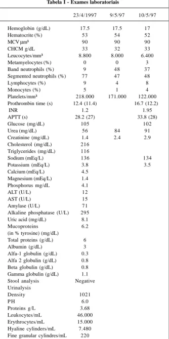
Tabela I - Exames laboratoriais
23/4/1997 9/5/97 10/5/97 Hemoglobin (g/dL) 17.5 17.5 17
Hematocrite (%) 53 54 52
MCV µm³ 90 90 90
CHCM g/dL 33 32 33
Leucocytes/mm³ 8.800 8.000 6.400
Metamyelocytes (%) 0 0 3
Band neutrophils (%) 9 48 37
Segmented neutrophils (%) 77 47 48
Lymphocytes (%) 9 4 8
Monocytes (%) 5 1 4
Platelets/mm³ 218.000 171.000 122.000 Prothrombin time (s) 12.4 (11.4) 16.7 (12.2)
INR 1.2 1.95
APTT (s) 28.2 (27) 33.8 (28)
Glucose (mg/dL) 105 102
Urea (mg/dL) 56 84 91
Creatinine (mg/dL) 1.4 2.4 2.9 Cholesterol (mg/dL) 216
Triglycerides (mg/dL) 116
Sodium (mEq/L) 136 134
Potassium (mEq/L) 3.8 3.5
Calcium (mEq/L) 4.5 Magnesium (mEq/L) 1.4 Phosphorus mg/dL 4.1
ALT (U/L) 12
AST (U/L) 15
Amylase (U/L) 71
Alkaline phosphatase (U/L) 295 Uric acid (mg/dL) 8.1
Mucoproteins 6.2
(in % tyrosine) (mg/dL)
Total proteins (g/dL) 6
Albumin (g/dL) 3
Alfa-1 globulin (g/dL) 0.3 Alfa 2 globulin (g/dL) 0.8 Beta globulin (g/dL) 0.8 Gamma globulin (g/dL) 1.1 Stool analysis Negative Urinalysis
Density 1021
P H 6.0
Proteins g/L 3.68 Leukocytes/mL 46.000 Erythrocytes/mL 15.000 Hyaline cylinders/mL 7.480 Fine granular cylindres/mL 220
atherosclerosis or inflammatory impairment of the coronary arteries. Electrocardiographic or enzymatic changes sug-gesting these complications were not reported in the pa-tient. However, finding coronary artery obstructions at au-topsy, even though not directly related to the cause of death, would not be a surprise.
Finally, it is worth stressing that infections along with cardiovascular and renal complications are the major cause of death in systemic lupus erythematosus. I think that in our patient, respiratory infection played a major role in the development of refractory septic shock and in the patient’s death.
(Dr. Alessandro Alves Fagundes)
Diagnostic hypotheses – systemic lupus
erythemato-sus, pulmonary hypertension, and, in the final event, septic shock secondary to respiratory infection.
Autopsy
At autopsy, 250mL of pericardial effusion and 1000mL of tion in voltage detected on the electrocardiogram may be
associated with edema, diffuse cardiomyopathy or cardiac tamponade; against this latter diagnosis, no electrical alter-nation was observed on the electrocardiogram.
The cardiac complications of systemic lupus erythema-tosus are the major causes of morbidity and mortality in the periods both before and after corticosteroids came into use 4.
In addition to the dilatation of the right chambers, the patient had pericardial effusion that was mild in the echo-cardiographic assessment of April 1997. In clinical evolu-tion no evidence of pericardial tamponade occurred; howe-ver, as already mentioned, the effusion may perhaps have increased during the progression of the disease. Pericardi-tis is the most common cardiac manifestation in systemic lu-pus erythematosus. It may be symptomatic with thoracic pain, fever, and pericardial friction, or asymptomatic with only pericardial effusion revealed by echocardiogram or peri-cardial impairment, as an autopsy finding. Depending on the criteria used in clinical echocardiographic or autopsy studies, pericarditis is found in 40% to 100% of the patients 5,6.
Despite its frequency, pericarditis rarely has a major clinical significance. Cardiac tamponade happens in less than 1% of cases 5 and, occasionally, in cases already treated with
corticosteroids, it may become complicated by infectious pericarditis.
In this patient, we did not observe valvar involvement directly related to systemic lupus erythematosus. Modera-te tricuspid regurgitation seems to be associaModera-ted with pul-monary hypertension. Involvement of left valves, an event first described by Libman and Sacks in 1927, may result in mitral and aortic regurgitation and, less commonly, in valvar stenoses. This type of involvement, also known as verru-cous endocarditis, was found in up to 50% of cases at autop-sy during the period of time before corticosteroids came into use7. This pattern of involvement has decreased in
frequency and severity with the introduction of corticoste-roid therapy 8.
In the case presented, we may also observe hyperten-sion and left ventricular hypertrophy. Even though syste-mic hypertension seems not to have significantly influen-ced the final evolution of the disease, it is a major cause of morbidity and mortality in systemic lupus erythematosus. Corticosteroid therapy has been pointed out as a cause of the increased incidence of systemic hypertension, hyper-cholesterolemia, and probably coronary artery disease 7.
Evidence of respiratory infection complicated with he-modynamic instability occurred as a final event. Hemody-namic support with vasoactive drugs and antibiotics was started. Even though invasive hemodynamic monitoring has not been used, a diagnostic suspicion of mixed shock existed. The patient already had marked functional limitation due to pulmonary hypertension. Additional myocardial dysfunction, however, may occur as a consequence of lupic myocarditis, myocardial ischemia, or septic cardiomyopa-thy. The lupic myocarditis is not a frequent cause of heart failure or cardiogenic shock, being clinically diagnosed in 10% of cases 9. Myocardial ischemia may result from
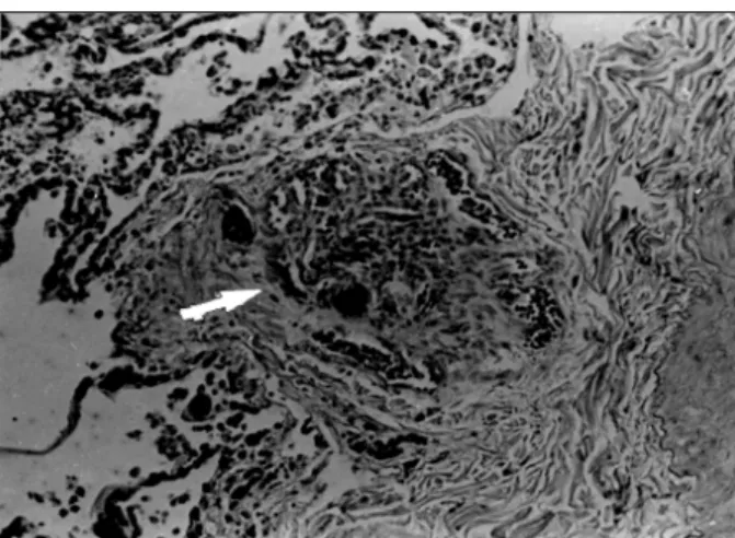
Fig. 4 - Microphotograph of the lung. Severe congestion and vascular hypertrophy (arrows) (hematoxylin and eosin, 4X).
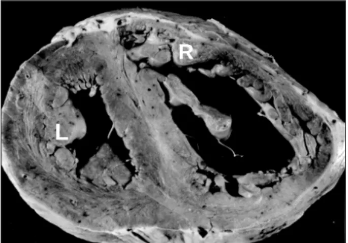
Fig. 3 - Cross section of the heart. Dilation and hypertrophy of the right ventricle. R – right ventricle; L – left ventricle.
L
Fig. 5 – Microphotograph of the lung. Plexiform vascular lesion characterizing Heath-Edwards grade IV pulmonary hypertension (arrow) (hematoxylin and eosin, 10X).
ascites fluid were found, both straw-colored. The pericardium had normal thickness and appearance. The heart weighed 500g, showed mild hypertrophy of the right and left ventricles and dilation of the right ventricle (fig. 3). The cardiac valves showed no alterations. The microscopic examination of the coronary arteries showed mild fibrointimal thickening in the proximal third of the anterior interventricular artery.
The right and left lungs weighed 350g and 300g, res-pectively. Microscopically, both showed vascular altera-tions compatible with severe Heath-Edwards grade IV pul-monary hypertension (figs. 4 and 5) and mild alveolar he-morrhage in the bases. The kidneys were enlarged and weighed 250g each. Histologically, diffuse membranous glomerulonephritis and interstitial nephritis were observed. In the right kidney, a medullary adenoma with a diameter of 3mm was also found.
The spleen was mildly enlarged, weighed 250g, and had fibrous thickening of the capsule.
The liver weighed 1850g and showed chronic passive congestion.
For a better assessment of the impairment caused by sys-temic lupus erythematosus, the search for immunoglobulins, fibrinogen, and complement was performed through im-munofluorescence in fragments of skin, lungs, kidneys, and heart. Deposition of immunoglobulins (IgM, IgG, IgA), complement, and fibrinogen occurred in the walls of the pulmo-nary vessels and renal glomeruli. Deposits of IgA in the tubules and fibrinogen in the interstitium and in vascular walls also occurred in the kidneys. Immunofluorescence was negative for all of these markers in the fragments of skin and heart. The immunofluorescence findings confirmed active lupus erythematosus involving lungs and kidneys, and this involvement was morphologically characterized by pulmonary hypertension and diffuse membranous glomerulonephritis.
No alteration suggesting an infectious process was found at autopsy.
(Dr. Patrícia Maluf Cury)
Anatomopathological diagnoses – active systemic
lu-pus erythematosus; grade IV pulmonary hypertension se-condary to lupus erythematosus; cor pulmonale; cardio-genic shock (final event).
Comments
Cardiac decompensation secondary to pulmonary hy-pertension resulting from lupus erythematosus is rare in male patients. Systemic lupus erythematosus is an uncommon autoimmune disease that is much more frequent in females, with a female:male ratio of 9:1. Pulmonary hypertension secondary to lupus is an even rarer finding, occurring in about 2% of the patients with the disease 10. The
etio-pathogeny of pulmonary hypertension in lupus is still controversial 4, 11, but seems not to be related to the severity
of the disease. Some of the proposed mechanisms are: 1) loss of pulmonary vessels due to interstitial disease; 2) narrowing of the vessels due to vasculitis or thromboembolism; 3) vasoconstriction. Anti-phospholipid antibodies may play an important role because they lead to a state of hypercoagula-bility and thrombosis, and also to a reduction in the synthesis of endothelial prostacyclin, causing an increase in vasoreac-tivity 4. These vascular alterations, mainly
thromboembo-lisms and fibrous intimal thickening, may also occur in the coronary arteries. This could explain the greater coronary involvement in patients with lupus as compared with patients with the same number of traditional risk factors for coronary artery disease 12. Cardiac involvement in systemic lupus
erythematosus may be generalized, even though not always clinically diagnosed 4. In addition to the coronary arteries, the
pericardium, myocardium, endocardium, and cardiac valves may be affected. Pericarditis is the most common finding associated with pericardial effusion, but it does not always cause clinical manifestations. The same is observed with the electrocardiogram that is altered in 50% of the patients with lupus. The most frequent electrocardiographic alteration is sinus tachycardia followed by nonspecific changes in the ST-T segment. The noninfectious lupic endocarditis, Liebman-Sacks endocarditis, was a very common finding in autopsies of patients with lupus before the development of corticosteroids, but is currently rarer. On the other hand, corticosteroid therapy accelerates atherosclerosis and hy-pertension secondary to renal involvement in these patients. In the present patient, pericarditis with pericardial effusion was found, as was mild alteration in coronary arteries. The electrocardiogram showed only right hyper-trophy secondary to pulmonary involvement. The atheros-clerosis was mild, comprising mainly the abdominal portion of the aorta. Therefore, the cardiac alterations here found cannot be attributed exclusively to systemic lupus erythe-matosus, even more with the negative immunofluorescence of the heart. Pericarditis and pericardial effusion might be secondary to right-sided heart failure and the mild vascular alterations could be attributed to age.
1. Tan EM, Cohen AS, Fries JF, et al. The 1982 revised criteria for the classification of systemic lupus eritematosos. Arthritis Rheum 1982, 25: 1271- 7.
2. Murin S, Wiedemann HP, Matthay RA. Pulmonary manifestations of systemic lupus erythematosus. Clin Chest Med 1998, 19: 641-65.
3. Ter Borg EJ, Groen H, Horst G, Limburg PC, Wouda AA, Kallenberg CG. Clinical associations of antiribonucleoprotein antibodies in patients with systemic lupus erythematosus. Semin Arthritis Rheum 1990; 20: 169-78.
4. Roberts WC, High ST. The heart in systemic lupus erythematosus. Curr Probl Cardiol 1999, 24: 6-51.
5. Collins RL, Turner RA, Nomeir AM, et al. Cardiopulmonary manifestations of systemic lupus erythematosus. J Rheumatol 1978; 5: 299-306.
6. Doherty NE, Siegel RJ. Cardiovascular manifestations of systemic lupus erythe-matosus. Am Heart J 1985; 110: 1257-65.
References
7. Ansari A, Larson PH, Bates HD. Cardiovascular manifestation of systemic lupus eritematosus. Prog Cardiovasc Dis 1985, 27: 421-34.
8. Bulkley BH, Roberts WC. The heart in systemic lupus eritematosus and the changes induced in it by corticosteroid therapy. Am J Med 1975, 58: 243-64. 9. Borenstein DG, Fye WB, Arnett FC, Stevens M. The myocarditis of systemic
lu-pus eritematosus association with myositis. Ann Intern Med 1978, 89: 619-24. 10. Mochizuki T, Aotsuka S, Satoh T. Clinical and laboratory features of lupus
pati-ents with complicating pulmonary disease. Respir Med 1999; 93: 95-101. 11. Yokoi T, Tomita Y, Fukaya M, Ichihara S, Kakudo K, Takahashi Y. Pulmonary
hy-pertension associated with systemic lupus erithematosus. Arch Pathol Lab Med 1998; 122: 467-70.