Arq Bras Cardiol 2001; 77: 545-8.
Barretto, ACP Reversibility of ventricular dysfunction
5 4 5
Instituto do Coração do Hospital das Clínicas - FMUSP
Mailing address: Antonio Carlos Pereira Barretto - Rua Piave, 103 - 05620-010 São Paulo, SP - Brasil - E-mail: [email protected]
Objective - To describe clinical observations of
mar-ked improvement in ventricular dysfunction in a medical office environment under circumstances differing from those in study protocols and multicenter studies performed in hospital or with outpatient cohorts.
Methods - Eleven cardiac failure patients with marked
ventricular dysfunction receiving treatment at a doctors office between 1994 and 1999 were studied. Their ages ranged from 20 and 66 years (mean 39.42±14.05 years); 7 patients were men, 4 were women. Cardiopathic etiologies were arterial hy-pertension in 5 patients, peripartum cardiomyopathy in 2, non-defined myocarditis in 2, and alcoholic cardiomyopathy in 4. Initial echocardiograms revealed left ventricular dilatation (average diastolic diameter, 69.45±8.15mm), reduced left ven-tricular ejection fraction (0.38±0.08) and left atrial dilatation (43.36±5.16mm). The therapeutic approach followed consis-ted of patient orientation, elimination of etiological or causal factors of cardiac failure, and prescription of digitalis, diu-retics, and angiotensinconverting enzyme inhibitors.
Results - Following treatment, left ventricular
ejec-tion fracejec-tion changed to 0.63±0.09; left ventricular diame-ters changed to 57.18±8.13mm, and left atrium diamediame-ters changed to 37.27±8.05mm. Maximum improvement was noted after 16.9±8.63 (6 to 36) months.
Conclusion – Patients with serious cardiac failure
and ventricular dysfunction caused by hypertension, alcoholism, or myocarditis can experience marked im-provement in ventricular dysfunction after undergoing appropriate therapy within the venue of the doctor’s office.
Key words: ventricular remodeling, dilated cardiomyopa-thy ventricular dysfunction, cardiac failure
Arq Bras Cardiol, volume 77 (nº 6), 545-8, 2001
Antonio Carlos Pereira Barretto
São Paulo, SP - Brazil
Reversibility of Ventricular Dysfunction. Clinical Experience
in a Medical Office
Original Article
Morbidity and mortality associated with cardiac failu-re afailu-re mofailu-re intense the gfailu-reater the ventricular dysfunction experienced by the patient 1,2. However, patients with a
mar-ked degree of ventricular dysfunction after following appro-priate therapeutic intervention have significant clinical im-provement, achieving survival in good condition 3,4. Such
survival is in many cases surprising in the face of the clini-cal picture presented at the initial evaluation. This effect has also been described in multicenter studies where a large number of cases were analyzed 3-8.
Some doubts exist about the frequency at which such improvement occurs in clinical practice and whether reduc-tion in ventricular dysfuncreduc-tion is observed in any patient with cardiac failure and whether it is more frequent in some forms of myocardial cardiopathy; also, it is not completely clear whether, in addition to observations in multicenter stu-dies, improvement also occurs in medical practice in the doctors’ offices. A meta-analysis 9 of large multicenter
stu-dies observed an increase of two units in ejection fraction; for example, if ejection fraction had been 0.25, it changed to 0.27. A significant improvement in ventricular function lea-ding to practical normalization was observed much more ra-rely. It is therefore worth pointing out that in the majority of cases only discrete reductions in ventricular diameter and small increases in ejection fraction are the rule.
We therefore decided to study the methods of medical practice in the doctor’s office to evaluate the improvement in ventricular dysfunction in patients who receive treatment under clinical conditions differing from those prevailing in large multicenter studies.
Methods
Eleven patients with marked ventricular dysfunction that improved markedly following therapeutic intervention were studied. Seven patients were men, four were women; ages ranged from 20 and 66 years, averaging 39.9±14 (SD) years; studies covered the period between 1994 and 1999.
5 4 6
Barretto, ACP
Reversibility of ventricular dysfunction
Arq Bras Cardiol 2001; 77: 545-8.
patients. After an initial orientation about cardiac decom-pensation factors, all subjects were told to decrease sodium and water ingestion and were given digoxin, diuretic drugs, and angiotensin-converting enzyme inhibitors. Ventricular dysfunction judged based on work selection criteria impro-ved in all study participants.
Digoxin was given at the usual dosage of 0.25mg/day. Diuretics given were a thiazide drug and furosemide. The diuretic dosage was reduced as the patient’s condition proved and essentially was suspended in all those with im-proved ventricular dysfunction. Seven patients received enalapril; 1 at 10mg/day, 2 at 10mg/2x per day, and 4 at 20mg/ day; 4 received captopril at 25mg/3x/day. Angiotensin-converting enzyme inhibitor administrations were maintai-ned in all patients until the final evaluation.
Results
All patients experienced limiting dyspnea, whether in the decubitus or following minimal effort. Hepatomegaly and lower limb edema (functional class III or IV, according to the New York Heart Association criteria) were also ob-served.
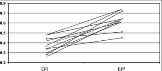
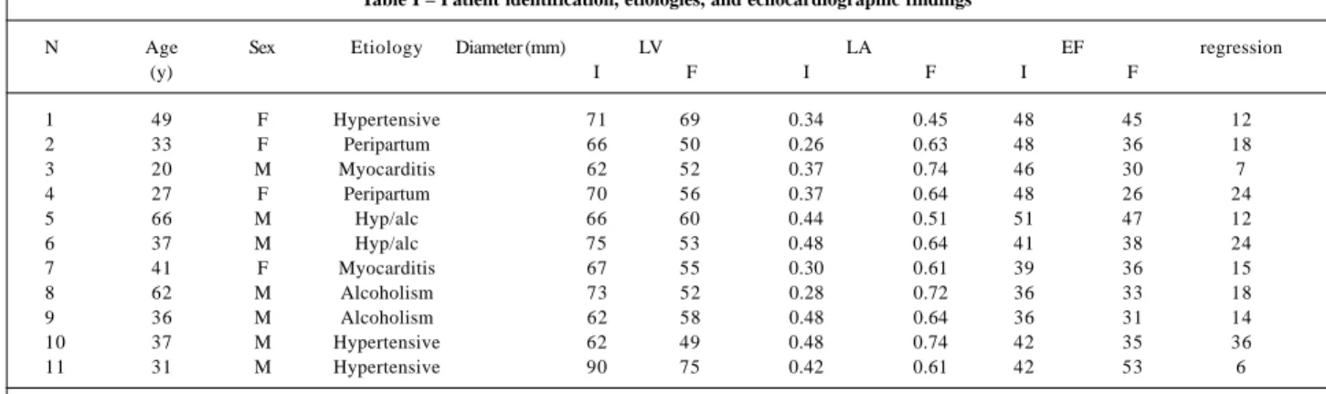
On the echocadiogram, left ventricular diastolic diame-ters ranged from 62 to 90mm (69.45-± 8.15mm average). Frac-tions ejected from the left ventricle ranged from 0.26 to 0.48 (0.38±0.08 average); left atria diameters ranged from 36 to 48mm (mean 43.36±5.1mm) (figures 1 and 2).
Heart failure was attributed to arterial hypertension in 5 cases, to peripartum cardiomyopathy in 2, to myocarditis of undetermined etiology (probably viral) in 2, and to alco-holic cardiomyopathy associated with arterial hypertension
in 2 (Table I). The cases of myocarditis and peripartum car-diomyopathy showed a positive gallium uptake response. It was possible to stop diuretic and digoxin adminis-tration to all patients with the exception of one who maintai-ned an ejection fraction of 0.45. The administration of angio-tensin-converting enzyme inhibitors was maintained even though it could have been suspended in most patients, who had ejection fractions above 0.45 after treatment, a va-lue considered the limit for inhibitor indication. The use of the inhibitors was nevertheless maintained because they had played an important role in the reversal of patients’ clini-cal picture and because many patients were reluctant to suspend this medication despite their significant clinical improvement. All patients tolerated the doses of inhibitors prescribed and did not have collateral effects, which could have indicated a need for suspension or dose reduction of the drugs.
Following an average period of 16.9±8.63 (6 to 36) months, reduction in left ventricular dilatation was ob-served, mean ventricular diameters changed from 69.45mm to 57.18mm; left atria diameters changed from 43.36mm to 37.27mm (Tables I and Figure 1). Mean ejec-tion fracejec-tion from the left ventricle increased from 0.38 to 0.63 (Figure 2).
Follow-up of the patients to the present day shows that none of them have a worsening in ventricular dysfunc-tion; no deaths have been recorded.
Discussion
Evidence has demonstrated that it is possible to mo-dify the natural history of cardiac failure with angiotensin-converting enzyme inhibitors 3-5, beta-blockers 6,7, and
spironolactone 8.
Experience has also shown that patients can experi-ence regression or a marked decrease in ventricular dys-function following such treatments. In the present work, we describe 11 cases showing this regression; the majority we-re highly symptomatic patients, and many had been sug-gested as candidates for possible heart transplantation. In these cases, significant improvement in ventricular function leading to practical normalization remains a rather rare out-come.
We should point out that the etiological diagnosis of our 11 cases falls into the category recognized as having reversible causes of ventricular dysfunction, namely arterial hypertension (5 cases), peripartum cardiomyopathy (2 ca-ses), myocarditis (2 caca-ses), and alcoholic cardiomyopathy (4 cases). Although this is a small cohort, it may be considered as expressive, because these patients were the only ones experiencing a regression in ventricular dysfunction of the many patients treated at this doctor’s office. It is our experi-ence that the large majority of cases showing symptom im-provement following treatment remain without great altera-tions in echocardiographic parameters. We did however observe that a significant percentage of patients stabilized by treatment remained clinically stable for years; however,
Fig. 1 - Left ventricle and left atrium diameters at the beginning and end of the follow-up period. Lvi- initial left ventricle diameter; LVf- final left ventricle diameter; Lai-initial left atrium diameter; Laf- final left atrium diameter.
Fig. 2 - Ejection fraction evaluated at the beginning and the end of the follow-up pe-riod. Efi- initial ejection fraction from the left ventricle; Eff- final ejection fraction from the left ventricle.
LVI LVf LAI LAf
EFi
Arq Bras Cardiol 2001; 77: 545-8.
Barretto, ACP Reversibility of ventricular dysfunction
5 4 7 regressions, as presently related, were only observed in
these 11 cases. Of them, arterial hypertension and alcoholic myocardiopathy were the most frequent. Poorly controlled hypertension by overloading the left ventricle may lead to ventricular dysfunction, a late complication observed in ca-ses of moderate and intense arterial hypertension. Accor-ding to the Hypertension Detection Follow-up study, mea-sures to lower blood pressure levels to less than 140/90m-mHg will control the near totality of these cases. Our 5 cases are an example of this 10.
Alcoholism is a cause of cardiomyopathy that, follo-wing interruption of alcohol consumption, may result in a re-gression in myocardial impairment 11. This regression
depends on the length and intensity of exposure to alcoho-lism as well as the type of the myocardial lesion. Prolonged, continuous aggression consequent to established fibrosis may lead to incomplete or even the lack of regression. In the majority of cases, avoidance of alcohol leads to regression in ventricular dysfunction, which however may reappear following a return to alcoholism. Although more frequent among drinkers of brazilian rum or sugar-cane rum, dysfunc-tion can also be due to the ingesdysfunc-tion of any kind of alcoholic beverage. In our cohort, all subjects were beer drinkers, but frequently also ingested distilled liquor.
The third type of diagnosis, myocarditis, probably in-cludes myocarditis proper as well as peripartum cardiomyo-pathy. Although the latter does not have a well-defined etiopathogeny, an inflammatory process could be detected in a significant percentage of patients, pointing towards the supposition that peripartum cardiomyopaths are a conse-quence of a myocarditic picture 12-14. Work from our
institu-tion has shown that the closer in time to parturiinstitu-tion that biopsies for peripartum cardiomyopaths are performed, the greater the incidence of inflammation becomes 12.
Cases of myocarditis are also recognized as reversible cardiomyopathies 13,14. Results indicate that the majority of
cases of myocarditis show spontaneous regression. Al-though a possible cause of dilated myocardiopathy, actual-ly a final phase of many cases of myocarditis that fail to rece-de, dilated cardiomyopathy is a rare complication within the universe of myocardities. Regression is so frequent
that studies using specific treatment with immunospressors, corticoid drugs, or antiimmunoglobulin antibodies, did not show results above those of placebo, which also had a prono-unced trend towards regression in ventricular dysfunction. For example, in the Myocarditis Trial ejection fraction rose from 0.36 to 0.46, regardless of treatment used, either placebo or corticosteroids in association with cyclosporine 15.
An interesting aspect of the experiment was the slow-ness of the regression ventricular dysfunction. On average, the period required for its normalization was 16.9 months, with a variation between 6 and 36 months. These figures are of importance because they provide guidance about the time intervals required for patient evaluation. Because re-gression does not occur very rapidly, monthly re-evalua-tions are not necessary. Our practice is to repeat the first evaluation after 3 months and every 6 months thereafter.
The prognosis was good in these cases, and no dea-ths occurred during the 3- to 5-year follow-up period. Such improved evolution of patients with ventricular dysfunction has already been referred to in the literature 4,16,17. In the
Ve-terans Heart Failure Study (VeHFT), the evolution of pati-ents who had increases in ejection fraction was much better than in those who remained stable or had a reduction 4.
In the results of the VeHFT Study 4, variations in
ejec-tion fracejec-tion were divided into 4 groups comprising, respec-tively, patients who got worse, who had no change, who had a 5% to 10% increase, and those having an increase over 10% increase. Survival times of these 4 groups were statistically different, the most expressive one being shown by the group having over 10% increases of their ejection fractions. At the end of the study, mortality in the 4 groups was, respectively, 21%, 11%, 6.7%, and 3%. Patients experi-encing the most significant improvement in ejection fraction also had the most significant clinical improvement.
Improved ventricular function in patients with re-cently acquired ventricular dysfunction has been described in over 37% of the cases treated at a transplantation clinic in California.
Steimle et al. 16 cited 13 patients in whom increases in
ejection fractions from 0.22 to 0.49 (mean of 0.27) were ob-served. This increase, similar to that shown in our study,
Table I – Patient identification, etiologies, and echocardiographic findings
N Age Sex Etiology Diameter (mm) LV LA EF regression
(y) I F I F I F
1 49 F Hypertensive 71 69 0.34 0.45 48 45 12
2 33 F Peripartum 66 50 0.26 0.63 48 36 18
3 20 M Myocarditis 62 52 0.37 0.74 46 30 7
4 27 F Peripartum 70 56 0.37 0.64 48 26 24
5 66 M Hyp/alc 66 60 0.44 0.51 51 47 12
6 37 M Hyp/alc 75 53 0.48 0.64 41 38 24
7 41 F Myocarditis 67 55 0.30 0.61 39 36 15
8 62 M Alcoholism 73 52 0.28 0.72 36 33 18
9 36 M Alcoholism 62 58 0.48 0.64 36 31 14
10 37 M Hypertensive 62 49 0.48 0.74 42 35 36
11 31 M Hypertensive 90 75 0.42 0.61 42 53 6
5 4 8
Barretto, ACP
Reversibility of ventricular dysfunction
Arq Bras Cardiol 2001; 77: 545-8.
was accompanied by a reduction in ventricular diameters from 67mm to 59mm. None of the patients died during the follow-up period that averaged 43 month (8 to 112 months). Kawai et al. 17 also observed a better evolution in
pati-ents, who evolved to improved ejection fraction 5. They
followed 88 patients who had idiopathic dilated cardiomyo-pathy with an ejection fraction below 25%, who had been discharged between 1984 and 1996 following hospitaliza-tion for compensahospitaliza-tion. Twenty (26%) of these patients had significant reverse remodeling, either a 5- mm reduction in ventricular diameter, or diameters remaining below 55mm and an ejection fraction above 25%, or with a reduction in left ventricular mass of over 10%. As a matter of fact, reverse re-modeling has been shown to be an important prognostic factor, characterizing groups with the best evolution 6-18. In
this work, patients were treated with angiotensin-converting
enzyme inhibitors (81%), and beta-blockers (59%). The im-proved cardiac function was an important prognostic factor, because during an evolutionary period of 50 months, none of the patients who had had reverse remodeling died or had to be hospitalized due to cardiac failure. In contrast, 16 (28%) patients not having such improvement died, and 22 (39%) had to be rehospitalized.
These data allow one to conclude that regression in ventricle dysfunction can occur even in the environment of a doctor’s office, potentially identifying patients having a better prognosis. Improvement in this dysfunction is not ra-pid and may require more than 1 year to reach completion. Patients having cardiomyopathies of a defined etiology like alcoholism, peripartum cardiomyopaths, myocarditis (in this case without defined etiology), or arterial hypertension were, in our experience, among those having this regression.
1. Bocchi EA. Situação atual das indicações e resultados do tratamento cirúrgico da insuficiência cardíaca. Arq Bras Cardiol 1994; 63: 523-30.
2. Oliveira Jr MT. Características clínicas e prognóstico de pacientes chagásicos e não chagásicos com insuficiência cardíaca congestiva avançada. Tese doutora-mento. Faculdade de Medicina da Universidade de São Paulo, São Paulo, 1999. 3. The SOLVD Investigators. Effect of enalapril on survival in patients with redu-ced left ventricular ejection fraction and congestive hear failure. N Engl J Med 1991; 325: 293-302.
4. Cintron G, Johnson G, Francis G, Cobb F, Cohn J for the V-HeFT VA. Cooperative Studies Group. Prognostic significance of serial changes in left ventricular ejec-tion fracejec-tion in patients with congestive heart failure. Circulaejec-tion 1993; 87(suppl VI):VI-17-V1-23.
5. The Acute Infarction Ramipril Efficacy (AIRE) Study Investigators. Effect of rami-pril on mortality and morbidity of survivors of acute myocardial infarction with clinical evidence of heart failure. Lancet 1993; 342: 821-8.
6. Packer M, Collucci WS, Sackner-Bernstein JD, et al. Double-blind, placebo-con-trolled study of the effects of carvedilol in patients with moderate to severe heart failure. The PRECISE trial. Circulation 1996; 94: 2793-9.
7. Cibis-II Investigators and committees. The cardiac insufficiency bisoprolol study (CIBIS-II): a randomized trial. Lancet 1999; 353: 9-13.
8. Pitt B, Segal R, Martinez FA, et al. Randomized trial of losartan versus captopril in patients over 65 with heart failure (Evaluation of Losartan in the Elderly Study, ELITE). Lancet 1997; 349: 747-52.
References
9. Massie B. 15 years of heart failure trials: what have we learned? Lancet 1998; 352(suppl I): 29-33.
10. Hypertension Detection and Follow-up Program Cooperative Group. The effect of treatment on mortality in mild hypertension N Engl J Med 1982; 307: 976-80. 11. Jacob AJ, McLaren KM, Boon NA. Effects of abstinence on alcoholic heart muscle
disease. Am J Cardiol 1991; 68: 805-7.
12. Mady C, Pereira Barretto AC, Bellotti G, et al. Biópsia endomiocárdica em pacien-tes portadores de miocardiopatia periparto. Arq Bras Cardiol 1986; 47: 403-5. 13. Glasier JJ, Constanzo-Nordin MR. Specific heart muscle disease. Cur Opin
Cardiol 1993; 8: 454-62.
14. Sliwa K, Skudicky D, Bergemann A, et al. Peripartum cardiomyopathy: Analysis of clinical outcome, left ventricular function, plasma levels of cytokines and Fas/ APO-1. J Am Coll Cardiol 2000; 35: 701-5.
15. Mason JW, O’Connell JB, Herskowitiz A, et al. A clinical trial of immunosuppres-sive therapy for myocarditis. N Engl J Med 1995; 333: 269-75.
16. Steimle AE, Stevenson LW, Fonarow GC, Hamilton MA, Moriguchi JD. Predic-tion of improvement in recent onset cardiomyopathy after referral heart transplan-tation. J Am Coll Cardiol 1994; 23: 553-9.
17. Kawai K, Takaoka H, Hata K, Yokota Y, Yokoyama M. Prevalence, predictors, and prognosis of reversal of maladaptive remodeling with intensive medical the-rapy in idiopathic dilated cardiomiopathy. Am J Cardiol 1999; 84: 671-6. 18. Patten RD, Udelson JE, Konstam MA. Ventricular remodeling and its prevention