5 8 7 5 8 7 5 8 7 5 8 7 5 8 7
Session editor: Alfredo José Mansur (ajmansur@incor.usp.br) Associate editors: Desiderio Favarato (dclfavarato@incor.usp.br) Vera Demarchi Aiello (anpvera@incor.usp.br)
Mailing address: Dante Marcelo Artigas Giorgi – InCor – Av. Dr. Enéas C. Aguiar, 44 05403-000 – São Paulo, SP, Brazil – E-mail: hipdante@incor.usp.br English version by Stela Maris C. e Gandour
Case 6/ 2001 – The pati ent i s an 11- year Case 6/ 2001 – The pati ent i s an 11- year Case 6/ 2001 – The pati ent i s an 11- year Case 6/ 2001 – The pati ent i s an 11- year
Case 6/ 2001 – The pati ent i s an 11- year --- ol d gi r l wi th Tol d gi r l wi th Tol d gi r l wi th Tol d gi r l wi th Tol d gi r l wi th Takakakak ayasu’ s ar ter i ti s and hear t fai l ur eakayasu’ s ar ter i ti s and hear t fai l ur eayasu’ s ar ter i ti s and hear t fai l ur eayasu’ s ar ter i ti s and hear t fai l ur eayasu’ s ar ter i ti s and hear t fai l ur e ( I nsti tuto do Cor ação do Hospi tal das Cl íni cas – FMUSP - São Paul o)
( I nsti tuto do Cor ação do Hospi tal das Cl íni cas – FMUSP - São Paul o) ( I nsti tuto do Cor ação do Hospi tal das Cl íni cas – FMUSP - São Paul o) ( I nsti tuto do Cor ação do Hospi tal das Cl íni cas – FMUSP - São Paul o) ( I nsti tuto do Cor ação do Hospi tal das Cl íni cas – FMUSP - São Paul o)
Clinicopathologic Session
The patient is an 11-year-old girl who sought medical assistance due to lumbar pain, nausea, and dyspnea.
One year earlier, this patient had been diagnosed with Takayasu’s arteritis, hypertension of renovascular origin, and marked left ventricular dysfunction. At that time, the patient underwent renal revascularization with a saphenous vein bypass graft between the aorta and both renal arteries; a postoperative occlusion of the graft occurred, requiring a new revascularization. Acute renal failure, elevation in urea levels up to 160mg/dL and creatinine levels up to 6.8mg/dL, which decreased later, complicated the interventions. On that occasion, the chest X-ray showed cardiomegaly and pulmonary congestion. The echocardiogram showed diffuse hypokinesia and marked left ventricular dilation, a diastolic diameter of 69mm, a systolic diameter of 60mm, and a shortening fraction of 13%.
The patient was discharged on the 21st postoperative
day, in good clinical condition, and with no complaints. Her heart rate was 104bpm, her blood pressure 90/60mmHg, and no other abnormalities were found on physical examination. Her creatinine level was 0.8mg/dL. The patient received the following prescription: 100mg of acetylsalicylic acid, 20mg of furosemide, 0.25mg of digoxin, and 18.5mg of captopril.
For 3 months the patient evolved with dyspnea on strenuous exertion and lumbar pain attributed to urolithia-sis, which was documented on ultrasonography. A few days later, complaining of nausea, dyspnea, lumbar pain, and ma-laise, the patient sought medical assistance.
On physical examination, the patient was in regular condition; her heart rate was 100bpm and blood pressure 120/80mmHg. Rhonchi and wheezings could be heard in both hemithoraces. Her cardiac rhythm was regular. The 2nd
cardiac sound was loud in the pulmonary area, and a systo-lic murmur (+/4+) could be heard in the mitral area. Her liver was palpated 2cm from the xiphoid process, and edema (++/ 4+) was present in her lower limbs.
The laboratory tests revealed anemia, neutrophilia with a high percentage of imature cells, and circulating antibodies of the IgG class for hepatitis A and cytomegalovirus. Serology was negative for hepatitis B and C, and mononucleosis (tab. I). After undergoing X-rays of the facial sinuses (frontal and sub-mental views), which showed no alterations, the patient was prescribed cefaclor and admitted to the hospital for diagnostic investigation and heart failure compensation.
The electrocardiogram revealed sinus rhythm, hyper-trophy of both atria and of the left ventricle, and secondary alterations in ventricular repolarization (fig. 1).
The echocardiogram showed marked left ventricular di-lation and hypokinesia (diastolic diameter of 68mm, systolic diameter of 59mm, and shortening fraction of 13%), distance between the mitral valve E point to the septum of 31mm, and low aortic flow (0.4m/s). The aortic valve showed signs of early closing. No anomaly was observed in the cardiac valves.
On hospital persisted heart failure and appeared pain in the anterior and posterior portions of the thorax. A few days later, severe dyspnea, hemoptysis, and worsening of the edema of the lower limbs occurred.
The electrocardiogram revealed sinus rhythm, a heart rate of 137bpm, reduction in voltage in the frontal plane (as compared with that in the previous electrocardiogram), left ventricular hypertrophy, and secondary alterations in ven-tricular repolarization (fig. 2). The chest X-ray showed pulmonary congestion.
A reduction in diuresis occurred, and the patient
Fabi o Gazel ato de Mel l o Fr anco, Dante Mar cel o Ar ti gas Gi or gi , Mar i a Cl em enti na Pi nto Gi or gi , Paul o Sam pai o Guti er r ez
São Paulo, SP - Brazil
5 8 8 5 8 8 5 8 8 5 8 8 5 8 8
required intravenous dobutamine (20µg/kg/min) and furo-semide (160mg). Because she did not improve, the patient was referred to the intensive care unit.
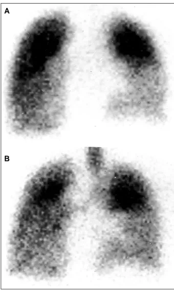
Pulmonary scintigraphy revealed a heterogeneous perfusion pattern, with hypoperfusion of the upper segment of the right lower lobe. During inhalation, a reduced uptake in the same area was also observed. Low probability of acute thromboembolism existed and the findings were compatible with a parenchymal process, which did not exclude the possi-bility of pulmonary infarction (fig. 3).
Table I – Laboratory tests
6/13/97 6/25/97 7/11/97 7/17/97 Red blood cells/mm³ 4,900,000 5,300,000 4,500,000 4,700,000 Hemoglobin (g/dL) 11.1 11.8 11 11.5
Hematocrit (%) 35 37.2 34 35
MCV (µm³) 71 70.2 76 74
MCH (pg) 23 31.6 24 24
Leukocytes/ mm³ 7,800 14,400 18,400 20,000 Band neutrophils (%) 24 63.8 4 4 Segmented neutrophils (%) 49 84 84
Eosinophils (%) 0 1.3 0 0
Lymphocytes (%) 23 26.9 8 10
Monocytes (%) 4 7.5 4 2
Platelets/mm³ 345,000 495,000 324,000 240,000 Creatinine (mg/dL) 0.8 0.8 1.2 1.8
Urea (mg/dL) 68 54 73 111
Sodium (mEq/L) 132 122 121
Potassium (mEq/L) 5.1 4.9 4.3
Chlorine (mEq/L) 79 101
Glucose (mg/dL) 94 71
Urine (EAS)
Density 1015 1011
pH 6.5 5
Proteins (g/L) 0.62 0.29
Leukocytes/mL 13,000 *
Red blood cells/mL 11,000 Covered field Electrophoresis of
blood proteins (g/dL)
Total 7.4
Albumin 3.5 2.8
Alpha-1 globulin 0.27 } 3.1
Alpha-2 globulin 0.7
Beta globulin 0.91
Gamma globulin 1.89
Prothrombin time (INR) 1.41 1.61 2.42 APTT (time ratio) 1.24 1.14 1.09 Factor V activity (%) 74
Fibrinogen (mg/dL) 383
Amylase 1.693
AST ((U/L) 44 105
Lactic dehydrogenase U/L 500 1680 Blood gas analysis Mask 5L O2 Venous
pH 7.48 7.18
pCO2 (mmHg) 31 54
pO2 (mmHg) 68 50
O2 saturation (%) 93 74.6
HCO3 (mEq/L) 22.5 19.3
Base excess (mEq/L) -0.2 -8.8
Fig. 1 – Electrocardiogram – hypertrophy of the right and left atria, and of the left ventricle.
Fig. 2 - Electrocardiogram – reduction in the voltage of the QRS complex in the fron-tal plane, and left ventricular hypertrophy.
A
B
5 8 9 5 8 9 5 8 9 5 8 9 5 8 9
Dynamic renal scintigraphy with 99 Tc-DTPA enhaced with furosemide showed low aortic flow with slightly decreased renal flow. The kidneys were enlarged, with bila-teral dilation of the pyelocaliceal system. After the use of fu-rosemide, an increase in excretion in the right kidney occur-red, but no adequate response was observed in the left kid-ney. The differential renal (semiquantitative) function was 52% in the right kidney and 48% in the left kidney.
The abdominal ultrasound revealed a markedly enlarged liver. The gallbladder was not identified. The spleen was normal. The kidneys measured 10cm, had a normal corticomedullary ratio and parenchymal echogenicity, and showed signs of left pyelocaliceal duplication. The urinary bladder was normal.
The thorax radiography revealed apical infiltration in the right lower lobe.
The patient evolved with shock, requiring the adminis-tration of high doses of catecholamines. On the following day, she developed acute pulmonary edema with reduction in her consciousness level. The patient required orotracheal in-tubation. Hemofiltration was considered to reduce excessive blood volume, due to oliguria, but it was not performed be-cause of hypotensive episodes. Leukocytosis persisted, being attributed to infection or to the inflammatory activity of arteritis. The patient received 500 mg of methylprednisolone. Clinical deterioration was refractory to treatment, and the patient underwent a cardiopulmonary arrest preceded by bradycardia, which was reversed with resuscitation ma-neuvers. Sodium bicarbonate was administered because of marked metabolic acidosis (Tab. I). Shock persisted, as did the metabolic acidosis, despite the frequent corrections with NaHCO3, and the patient died the following morning.
Discussion
Pulmonary perfusion and ventilation scintigraphy
-Pulmonary perfusion scintigraphy performed with ma-croaggregated albumin (MAA) labeled with technetium-99m showed hypoperfusion in the right inferior lobe (fig. 3A). The study of pulmonary ventilation (fig. 3B) was performed with an aerosol saline solution and diethylenetriamine pentaacetic acid (DTPA) labeled with technetium-99m and showed a radiopharmaceutical distribution pattern similar to that observed in the perfusion study (match pattern). This pattern is found in several pulmonary parenchymal pro-cesses, such as pneumonia, tumors, and pulmonary infarc-tions; however, it does not occur in acute embolism, except when the latter is accompanied by bronchospasm.
(Dr. Maria Clementina Pinto Giorgi)
Renogram – A relevant point in patients suspected of
having Takayasu’s arteritis is the time elapsed until the aorta is filled with contrast medium. In regard to our patient, this time was very long, suggesting marked obstruction to flow due to an aortic lesion.
(Dr. Dante M. A. Giorgi)
Clinical features – Takayasu’s arteritis is a chronic
in-flammatory disease, probably with an autoimmune cause, involving medium- and large-caliber arteries, mainly the aorta and its branches in their proximal portions. The most commonly involved vessels are the subclavian arteries, fol-lowed by the aortic arch, the ascending aorta, the carotid ar-teries, and the femoral arteries. The pulmonary and corona-ry arteries may also be affected.
Women are most frequently affected in a proportion of 8:1, and the mean age at diagnosis is around 29 years. In ¾ of the patients, the diagnosis is established during adolescence. An association of the disease and certain subtypes of HLA has been reported, even though its significance has not yet been clarified.
The lesions are purely stenotic in 85% of the cases, purely dilated in 2%, and mixed in 13%.
Morphological alterations are characterized by irregu-lar thickening of the aortic wall with intimal wrinkling. When the aortic arch is affected, the orifices of the major arteries to the upper portion of the body may be markedly narrowed or even occluded, which is why it is referred to as a “pulseless disease.”
In the acute phase, the symptoms may be unspecific, such as fever, anorexia, malaise, arthralgias, in addition to those related to ischemia in the area irrigated by the affected artery. Hypertension usually has a renovascular cause, complicating this disease in 50-60% of the cases. Congesti-ve heart failure occurs in 28% of the patients, and results from hypertension, or, more rarely, from aortic regurgitation. Usually, death is caused by cerebral strokes or heart failure. Our patient had already been diagnosed with Taka-yasu’s arteritis, with renal involvement (renovascular hy-pertension) and marked left ventricular dysfunction. She sought emergency medical assistance with clinical findings suggestive of cardiac decompensation. During hospitaliza-tion, she experienced chest pain, hemoptysis, and worse-ning of the dyspnea and of the edema of the lower limbs. She evolved with refractory cardiogenic shock and death.
The clinical and laboratory findings of our patient suggest active disease. In some patients, involvement of the pulmonary artery may occur, leading to its occlusion and consequent pulmonary infarction 1-3. The presence of
hemoptysis and dyspnea, in addition to hypoperfusion of the upper segment of the right lower lobe reinforce this idea. The previous ventricular dysfunction aggravated with the inflammatory activity and pulmonary infarction may have led to worsening of the ventricular function with refractory cardiogenic shock.
We cannot overlook the possibility of an associated infectious process, contributing to hemodynamic decom-pensation and death.
In some patients with Takayasu’s arteritis, left ventri-cular dysfunction may be secondary to myocarditis 4. The
5 9 0 5 9 0 5 9 0 5 9 0 5 9 0
shows that myocarditis is a very rare condition confirmed only in a few cases on autopsy.
We should still consider the possibility of myocardial ischemia due to coronary artery involvement, which may occur in as many as 10% of the patients with Takayasu’s ar-teritis.
(Dr. Fábio Gazelato de Mello Franco)
Diagnostic hypothesis – Active Takayasu’s arteritis,
pulmonary thromboembolism, ventricular dysfunction ag-gravated by pulmonary embolism, and, less probably, myo-cardial ischemia.
(Dr. Fábio Gazelato de Mello Franco)
Autopsy
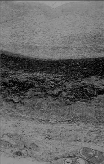
The major alterations on postmortem examination we-re found in the aorta, whose wall was very thickened from its beginning down to its abdominal portion, below the emer-gence of the renal arteries. Even though diffuse, the in-volvement was not regular; therefore, areas of important narrowing of the arterial lumen and other more preserved areas existed. The histologic sections showed chronic aor-titis (fig. 4), with destruction of the elastic fibers (fig. 5). The search for bacteria and fungi was negative. The diagnosis of Takayasu’s arteritis was established.
The branches of the aortic arch and the renal arteries were markedly compromised. The patient had undergone surgical renal revascularization because of the involve-ment of the renal arteries; the grafts were partially obs-tructed, and renal cortical necrosis existed. Signs of con-gestive heart failure were also present, with chronic pas-sive congestion of the lungs and liver, and thromboem-bolism with infarct in the middle and upper lobes of the right lung, with no inflammatory lesions in the pulmonary arterial tree. Finally, systemic signs of shock were present, such as centrolobular hepatic necrosis and pancreatic
steatonecrosis. Multisystem organ failure was considered the cause of death.
Even though occurring in an age bracket a little lower than the usual, this patient’s disease was well characterized as Takayasu’s arteritis, from both the clinical and morpho-logical points of view.
The final part of the patient’s evolution was not very well understood while she was still alive. Myocardial infarc-tion was suspected, but it was not confirmed on autopsy, which revealed the existence of another factor, pulmonary thromboembolism, which triggered the patient’s decom-pensation.
The factors that might account for heart failure as a complication of Takayasu’s arteritis 5-7 are the following:
im-pairment of the cardiac muscle, caused either by cardio-myopathy, reported in some patients, or by myocardial in-farction secondary to inflammation of the coronary arteries; valvar or supravalvar aortic stenosis; and decompensation of systemic arterial hypertension or hypervolemia, or both, when the renal arteries are involved. In the present case, because the coronary arteries and the myocardium were
Fig. 4 – Histological section of the middle layer of the aorta. Note the dense and diffuse inflammatory infiltrate with mononuclear cells. The elastic fibers present in the middle layer stain in black and are partially destructed and fragmented (HE, 16x magnification).
5 9 1 5 9 1 5 9 1 5 9 1 5 9 1
preserved, a combination of the remaining causes should have accounted for the functional alteration. It is worth no-ting that, even though all above-cited factors, isolated or in association, may have been involved in the pathogenesis of heart failure, this disorder appears most of the time in hy-pertensive patients, and with a greater incidence in younger patients. In a study carried out in Mexico with 12 children, hypertension was found in 11 patients and dyspnea on exertion in 9 5. Talwar et al 6 detected the disorder in 17 out of
31 individuals with Takayasu’s arteritis and heart failure below the age of 15 years.
(Dr. Paulo Sampaio Gutierrez)
Pathological diagnoses – Takayasu’s arteritis and
pulmonary infarct due to thromboembolism.
(Dr. Paulo Sampaio Gutierrez)
1. Bletry O . Severe pulmonary artery involvement of Takayasu arteritis. 3 cases and review of the literature. Arch Mal Coeur Vaiss 1991; 84: 817-22.
2. Nunes H. Pulmonary artery involvement in Takayasu arteritis. A case of right ventricular failure as presentation form. Rev Port Cardiol 1992; 11: 775-80. 3. Lupi E. Pulmonary artery involvement in Takayasu’s arteritis. Chest 1975; 67: 69-74. 4. Gay J. Myocardiopathy and Takayasu’s disease. Apropos of a case. Ann Cardiol
Angeiol (Paris) 1985; 34: 489-92.
References
5. Lupi Herrera E, Contreras R, Vela JE, Torres GS, Horwitz S. Arteritis inespecifica en la niñez. Observaciones clinicas y anatomopatologicas. Arch Inst Cardiol Mex 1972; 42: 477-93.
6. Talwar KK, Kumar K, Chopra P, et al. Cardiac involvement in nonspecific aortoarteritis (Takayasu’s arteritis). Am Heart J 1991; 122: 1666-70. 7. Panja M, Kar AK, Dutta AL, Chhetri M, Kumar S, Panja S. Cardiac involvement
in non-specific aorto-arteritis. Int J Cardiol 1992; 34: 289-95.
Editor da Seção de Fotografias Artísticas: Cícero Piva de Albuquerque
Correspondência: InCor - Av. Dr. Enéas C. Aguiar, 44 - 05403-000 - São Paulo, SP - E-mail: dclcicero@incor. usp.br

