Case Report
Right to Left Shunt Through a Patent Foramen Ovale without
Pulmonary Hypertension
Thiago Costa Lisboa, Clarice Daniele Oliveira Costa, Thiago Quedi Furian, Silvia Regina Rios Vieira
Hospital de Clínicas de Porto Alegre - Porto Alegre, RS, BrazilMailing Address: Thiago Lisboa • Rua Carazinho, 559/100
90460-190 – Porto Alegre, RS, Brazil E-mail: [email protected]
Manuscript received February 28, 2006; revised manuscript received May 27, 2006; accepted August 8, 2006.
Key words
Heart septal defects, atrial; anoxemia; dyspnea.
ECG without ischemic alterations; chest X-ray showing pleural calcifications; and chest angiotomography with no evidence of pulmonary thromboembolism.
He had progressive worsening of hypoxemia, and had to be admitted into the intensive care unit (ICU), at first with non-invasive mechanical ventilation and later with invasive mechanical ventilation, with two unsuccessful extubation attempts.
The investigation also included the following: pulmonary perfusion scintilography, with low probability of pulmonary thromboembolism; Doppler of lower limbs with no evidence of deep venous thrombosis; high resolution chest tomography showing no alterations that justified his degree of hypoxemia; transthoracic echocardiogram with air contrast, showing interatrial septal aneurysm, with intense and early passage of contrast to the left cavities, which is compatible with a patent foramen ovale, with no evidence of pulmonary arterial hypertension; enlarged left ventricle, with a relaxation deficit, and preserved global and segmental systolic function and with ejection fraction (EF) = 66%; transesophageal echocardiogram with Doppler, with evidences of a patent foramen ovale, with a predominant right to left flow, without intracavity thrombus; pulmonary arteriography with absence of intrapulmonary shunt.
With the presumptive diagnosis of hypoxemia secondary to the right to left shunt through the patent foramen ovale, we installed a pulmonary artery catheter and performed blood gas analysis. Later we performed cardiac catheterization and measured oxygen saturation in the heart chambers (Tab. 1).
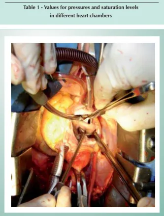
As the patient continued to present desaturation when in a seated position and therefore required mechanical ventilation despite the absence of a right left pressure gradient, he underwent surgical repair of the patent foramen ovale (atrioseptoplasty) (Fig. 1). In addition to the patent foramen ovale, no other alteration was identified in the interatrial septum. The patient progressed with substantial improvement in hypoxemia. He was extubated in the first postoperative period, and discharged from the ICU and from hospital without symptoms; his condition has been stable for six months.
Cases of hypoxemia secondary to right to left shunt through a patent foramen ovale in the absence of pulmonary hypertension are rarely described in the literature and may constitute a picture of platypnea-orthodeoxia syndrome1. In this paper we will
describe the case of a patient with this rare condition.
Case Report
A 71-year old white male patient, married, was admitted to the Emergency Room of HCPA due to major dyspnea at rest, with no associated symptoms. He reported having progressive dyspnea for ten months, which became more intense ten days before admission.
His previous medical history included: smoking from 31 to 35 years of age; thoracic bruise with no sequela 25 years ago; systemic arterial hypertension since 1985, treated with Amlodipine, Hydrochlorothiazide and Enalapril; dyslipidemia since 1996, treated with Sinvastatin and gastroesophageal reflux disease treated with Omeprazole.
On physical examination, the patient was conscious, oriented, had tachypnea (RR=28 breaths/minute), and O2SAT= 88% on room air, in a seated position. He presented
BP=140/80mmHg, HR=96bpm, with regular heart rhythm, with no extra sounds, normophonetic heart sounds, without murmurs. Peripheral pulse measurements of the four limbs were palpable and symmetrical; limb extremities were hot and cyanotic. He had no other alterations.
Tests showed: arterial blood gas analysis, on room air, with pH = 7.53; CO2P = 24.9; HCO3 = 20.3; O2P = 51.7; O2SAT= 90.5%; hematocrit = 50%, hemoglobin = 16.5 g/dl; normal white cell and platelet counts; normal heart enzymes; This paper describes a patient who presented with hypoxemia and platypnea and whose only finding on investigation was the presence of a patent foramen ovale with a right to left shunt, without pulmonary hypertension, the characteristic of the rare Platypnea-Orthodeoxia Syndrome which shows interesting physiopathological features. Therapeutic options have not yet been fully defined.
Case Report
Lisboa et alRIGHT TO LEFT SHUNT THROUGH A PATENT FORAMEN OVALE WITHOUT PULMONARY HYPERTENSION
Arq Bras Cardiol 2007; 88(1) : e9-e11
and documenting the presence of interatrial communication. After this, we performed right and left cardiac catheterization which confirmed the presence of normal intracardiac pressures. Arterial oxygen saturation showed to be appropriate in this assessment and we believe this finding was due to the mechanical ventilation and to the patient’s decubitus, since he presented desaturation when standing, which is compatible with the picture of platypnea-orthodeoxia.
The cause of the right to left shunt with normal intracardiac pressures through an interatrial communication has not yet been completely clarified and, although very complex diagnostic tools are available, a series of questions remain unanswered as to the physiopathology and the anatomical and physiological substrate that can explain this picture1-3.
The clinical syndrome described as platypnea-orthodeoxia is very infrequent but is also overlooked; patients present dyspnea and hypoxemia associated with supine and seated body position, with improvement of symptoms when the patient lies down. This picture was first described by Burchell in 1949 and less than a hundred cases have been described since then4.
Under normal conditions, an interatrial communication allows left to right flow, due to the pressure gradient and to the greater complacency of the right chambers as compared with the left chambers. When a right to left flow exists there are usually pressure alterations in the pulmonary circulation (for instance, massive pulmonary embolism, primary pulmonary hypertension), with pulmonary hypertension and reversal of the pressure gradient to right-left. However, some patients, such as the one described here, do not present either a pressure increase in the right chambers or pulmonary hypertension, which causes shunting to be done against a pressure gradient4.
Several attempts have been made to explain this phenomenon5-7. Although the mean atrial pressure is higher in the left atrium than in the right atrium, significant alterations in this gradient may take place in the beginning of diastole and during right ventricle isometric contraction. These gradient alterations can be intensified in situations such as deep inspiration, Valsalva, pulmonary embolism, hypoxemia, severe chronic obstructive pulmonary disease, right ventricle infarction, elevated positive end-expiratory pressure in patients in a seated position. In these cases there may be right to left shunt in the presence of interatrial communication with atrial septum aneurysm and patent foramen ovale6.
The anatomical substrate for shunting with normal pressures includes, in addition to an interatrial communication that allows it to happen, a phenomenon of the flow7. This
phenomenon would take place due to a preferential flow from the inferior vena cava towards the interatrial septum, as part of a remaining prenatal circulatory pattern.
In addition to this, the complacency of right cardiac cavities seems to decrease with age, which may generate a preferential flow through the interatrial communication. Other phenomena that seem to foster alterations in the flow include distortions in the anatomy of the relations between the inferior vena cava and the interatrial septum through the anticlockwise rotation of the heart and/or mediastinal deviation, which are
Discussion
This report describes a 71-year old male patient with a clinical picture of platypnea-orthodeoxia caused by right to left shunt through a patent foramen ovale with no increase in pulmonary circulation pressure.This is characteristic of a rare syndrome whose diagnosis depends on a high degree of suspicion, and that is frequently unrecognized. In this case, as in previous reports, the diagnosis was confirmed by a transesophageal echocardiography with contrast, with early passage of bubbles, demonstrating the presence of shunting
Parameter Pulmonary artery catheter
Cardiac catheterization
PASP/PADP 27/17 mmHg 26/8 mmHg PAOP 15 mmHg -RA Pressure 11 mmHg 8 mmHg LA Pressure - 11 mmHg RA O2S 73% 76% Pulmonary Artery
O2S 70% 69%
LA O2S - 93%
Arterial O2S 98% 97%
PASP - Pulmonary Artery Systolic Pressure; PADP - Pulmonary Artery Diastolic Pressure; PAOP - Pulmonary Artery Occlusion Pressure; RA - Right Atrium; LA - Left Atrium; O2S - Oxygen Saturation.
Table 1 - Values for pressures and saturation levels in different heart chambers
Fig. 1 -Foramen ovale seen transoperatively.
Case Report
Lisboa et al RIGHT TO LEFT SHUNT THROUGH A PATENT FORAMEN OVALE WITHOUT PULMONARY HYPERTENSIONArq Bras Cardiol 2007; 88(1) : e9-e11
more frequently caused by right pneumonectomy, pericardial effusion and alterations in the ascending aorta7,8.
Another approach that seeks to explain the causes of shunting relate to embryological aspects, and considers the atrial remodeling that takes place after the incorporation of the venous sinus to its structure and allows for independent flows from vena cava superior and vena cava inferior towards the interatrial septum. This could play a role in the origin of this phenomenon7.
It is essential to seek the origins of this phenomenon in these anatomical and physiological substrates and in embryological concepts. However, despite this knowledge, a full understanding of the mechanisms involved in the pathogenesis of right to left shunt with normal pressures has not yet been obtained.
Although the mechanisms involved in its pathogenesis have
not yet been clarified, there seems to be clear consensus as to the treatment of this clinical condition. Case reports1,2,8,9, as
well as the largest series available10 reporting on the treatment
of these patients include the percutaneous or surgical repair of the interatrial communication as a key point. We decided to perform the surgical repair of our patient’s defect, and obtained an excellent result.
The platypnea-orthodeoxia syndrome, with right to left shunt with normal pressure levels is a rare and complex condition whose physiopathological mechanism has not yet been fully clarified. However, this diagnosis should be considered when there is hypoxemia and dyspnea, especially in patients in supine/seated position, since their treatment with the repair of the atrial septal defect that allows shunting presents excellent results as reported in the literature and as exemplified in this description.
1. Khouzaie TA, Busser JR. A rare cause of dyspnea and arterial hypoxemia. Chest. 1997; 112: 1681-2.
2. Grutters JC, ten Berg JM, van der Zeijden J, Jaarsma W, Ernst JM, Westermann CJ. Patent foramen ovale causing position-dependent shunting in a patient, when laying down her corset. Eur Respir J. 2001; 18: 731-3.
3. Kubler P, Gibbs H, Garrahy P. Platypnoea-orthodeoxia syndrome. Heart. 2000; 83: 221-3.
4. Cheng TO. Mechanisms of platypnea-orthodeoxia: what causes water to flow uphill? Circulation. 2002; 105: 47
5. Medina A, de Lezo JS, Caballero E, Ortega JR. Platypnea-orthodeoxia due to aortic elongation. Circulation. 2001; 104:741.
6. Acharya SS, Kartan R. A case of Orthodeoxya caused by an atrial septal aneurysm. Chest. 2000; 118: 871-4.
7. Zanchetta M, Rigatelli G, Ho SY. A mistery featuring right-to-left shunting despite normal intracardiac pressure. Chest. 2005; 128: 998-1002.
8. Robin ED, McCauley RF. An analysis of platypnea-orthodeoxia syndrome including a “new” therapeutic approach. Chest. 1997; 112:1449–51
9. Hegland DD, Kunz GA, Harrison JK, Wang A. A hole in the argument. N Engl J Med. 2005; 353: 2385-90.
10. Godart F, Rey C, Prat A, Vincentelli A, Chmait A, Francart C, et al. Atrial right-to-left shunting causing severe hypoxaemia despite normal right-sided pressures. Eur Heart J. 2000; 21: 483-9.