Pulmonary artery aneurysms in Behçet disease
Aneurismas de artérias pulmonares na doença de Behçet
Shi-Min Yuan1
Abstract
Pulmonary artery aneurysms (PAAs) are the most common type of pulmonary involvement in Behçet’s disease. However, the relationships between clinical features and prognosis have not been suiciently evaluated. his article describes the results of a comprehensive review, revealing that PAAs have a predilection for hemoptysis manifestations, increased dimensions, right lower lobar location, multiplicity and concurrent intramural thrombus formation. Surgical intervention was needed in one third of patients. Patients with massive hemoptysis and PAA rupture warranted emergency operations. Conservatively treated patients were prone to PAA progression; interventional embolization was associated with higher risks of recurrence and reintervention for PAAs; and surgically treated patients exhibited the highest mortality rates. In conclusion, PAAs in Behçet’s disease are characterized by a predilection for hemoptysis manifestations, right lower lobar location, multiplicity, and concurrent intramural thrombus formation. Both the condition itself and the surgical operations it warrants are linked with high mortality due to PAA hemorrhage.
Keywords: hemoptysis; therapeutic embolization; vasculitis.
Resumo
Os aneurismas das artérias pulmonares (PAA) são as manifestações mais comuns dos pulmões na doença de Behçet. No entanto, as relações entre as características clínicas e o prognóstico ainda não foram devidamente explicadas. O objetivo do presente artigo foi fazer uma ampla revisão da literatura sobre esta questão. As fontes de dados contaram com uma ampla revisão bibliográica dos anos de 1990 a 2013, sobre os seguintes temas: doença de Behçet, síndrome de Hughes-Stovin, aneurisma de artéria pulmonar e pseudoaneurisma da artéria pulmonar. Os PAA evoluíram com predileção por hemoptise, aumento de dimensões, localização no lobo inferior direito, multiplicidade e formação de trombo intramural. A intervenção cirúrgica foi necessária em um terço dos pacientes. O tratamento cirúrgico emergencial foi indicado na vigência de ruptura do PAA e de hemoptise maciça. Os pacientes tratados conservadoramente evoluíram com propensão para a progressão do PAA. A embolização dos PAA foi associada a uma taxa maior de recidiva e de reintervenção. Houve diferença signiicativa entre os grupos quanto às taxas de mortalidade, tendo o grupo do tratamento cirúrgico apresentado a maior taxa. Dentre as variáveis citadas, a hemoptise, o envolvimento da artéria lobar e a ruptura do PAA foram fatores preditivos de maior risco de mortalidade. Houve diferenças signiicativas nas taxas de mortalidade entre os pacientes cirúrgicos e intervencionistas, e entre os três grupos de pacientes: cirúrgico, intervencionista e conservador. Os pacientes tiveram uma sobrevida global de 61,7% em um seguimento médio de 22,5 meses. Os PAA, na doença de Behçet, apresentaram as seguintes predileções: tendência a hemoptise, multiplicidade, localização no lobo inferior direito e presença de trombos intramurais. As rupturas e hemorragias dos PAA, aliadas ao necessário tratamento cirúrgico emergencial, resultaram no aumento de mortalidade destes pacientes.
Palavras-chave: hemoptise; embolização terapêutica; vasculite.
1Fujian Medical University, Teaching Hospital, he First Hospital of Putian, Department of Cardiothoracic Surgery, Putian, Fujian Province, People’s Republic of China.
Financial support: None.
Conlicts of interest: No conlicts of interest declared concerning the publication of this article. Submitted: 04.07.13. Accepted: 05.15.14.
INTRODUCTION
In Behçet’s disease, pulmonary involvement is
uncommon, with a prevalence of less than 5%.
1Pulmonary artery aneurysms (PAAs) are the most
common form of pulmonary involvement in Behçet’s
disease, followed by pulmonary artery thrombosis,
pulmonary infarction and pulmonary parenchymal
disorders.
2Thrombosis usually develops as a
consequence of the underlying extensive vasculitis.
3, 4Currently, pulmonary artery aneurysms are the
second most common type of arterial involvement
in Behçet’s disease, preceded by aortic aneurysms.
2Hemoptysis of varying degrees up to 500 ml was the
most common symptom of PAAs, observed in 79%.
2, 5Hemoptysis can sometimes be massive and lethal,
when PAAs rupture into the adjacent bronchus.
6Other manifestations of PAA include cough, dyspnea
and chest pain.
6Hughes-Stovin syndrome is a combination of
pulmonary artery thrombosis and aneurysms with
peripheral thrombophlebitis and is considered an
incomplete variant of Behçet’s disease. Characterized
by an association of multiple PAAs and peripheral
venous thrombosis, Hughes-Stovin syndrome
shares identical pulmonary manifestations with
Behçet’s disease.
7Thrombophlebitis, formation of
large pulmonary and/or bronchial aneurysms and
aneurysmal rupture leading to massive hemoptysis
and death are the three phases of the clinical paradigm
of Hughes-Stovin syndrome.
8Previous reports have described the clinical
features of PAAs in Behçet’s disease.
2, 9-11However,
the relationships between the clinical features and
prognosis have not been suficiently evaluated and
thus remain to be identiied. This article presents a
comprehensive literature review of the subject.
MATERIALS AND METHODS
Search strategies
A comprehensive literature search was conducted
on MEDLINE, Highwire Press and Google for the
year range 1990-2013. The search terms included
“Behçet’s disease”, “Hughes-Stovin syndrome” and
“pulmonary artery aneurysm” or “pulmonary artery
pseudoaneurysm”. Data were extracted from the
text, igures and/or tables, with details of the study
population, demographics, duration of Behçet’s
disease, characteristics of PAAs, management
strategies and pertinent indications, follow-up
duration and main outcomes (survival, recurrence,
complication, reintervention and mortality).
Definitions
Severity of hemoptysis was deined as: mild <5 ml
in 24 hours; moderate 5-600 ml/24 hours and massive
>600 ml/24 hours, or 100 ml/<24 hours to 1000 ml/
several days, or >50 ml per expectoration,
12and
sudden recurrent massive hemoptysis was deined
as life-threatening hemoptysis. Onset of action was
deined as the time for the immunosuppressive agents
to take effect (including symptom relief, decreased
inflammatory mediators, reduced pulmonary or
intracardiac thrombus and reduced PAA) after
administration; and cure time was the time interval
from drug administration to complete resolution of
the PAA and pulmonary or intracardiac thrombus.
Statistical analysis
Quantitative data were presented as mean ±
standard deviation with range and median values,
and intergroup differences were compared using the
unpaired
t
test. Frequencies were compared using
Fisher’s exact test. Univariate and multivariate
analyses were conducted to evaluate predictive
risk factors. Results with p<0.05 were considered
statistically signiicant.
RESULTS
The literature search yielded 107 reports
5, 10, 13-118covering 199 patients. Five case reports,
each describing the case of one patient, diagnosed
Hughes-Stovin syndrome.
37, 49, 51, 54, 99Among the
patients whose gender could be ascertained, there
were 166 (85.6%) males and 28 (14.4%) females,
giving a male-to-female ratio of 5.93:1. Patients
were aged 31.0±10.9 (range: 10-69; median: 30)
years (n=150). Male patients were aged 31.6±11.4
(range: 10-69; median: 30) (n=122) and females
were aged 28.6±8.5 (range: 14-48; median: 27)
(n=24). There was no signiicant difference in patient
age between male and female patients (p=0.2249).
Patients had presented symptoms of Behçet’s disease
for 5.0±4.8 (range: 0.25-26; median: 3) years (n=52)
and their diagnoses of Behçet’s disease had been
established for 4.7±2.9 (range: 0.83-10; median: 5)
years (n=34). No difference was detected between
the time since onset and time since Behçet’s disease
diagnosis (p=0.7522). On admission, 156 (78.4%)
patients presented with hemoptysis, while 43 (21.6%)
patients did not exhibit hemoptysis (
c
2=128.3,
p<0.0001). Hemoptysis was the only symptom at
addition to hemoptysis, fever, dyspnea, cough and
chest pain were also common symptoms in the PAA
patients. Comparison of the secondary symptoms of
hemoptysis patients with those of hemoptysis-free
patients revealed a signiicant intergroup difference
in prevalence of cervical or pedal edema (Table 1).
Hemoptysis volume was reported for 69 patients.
Hemoptysis was mild in 5 (7.2%) patients,
28, 48, 55, 65, 68moderate in 9 (13.0%),
37, 50, 57, 70, 71, 76, 95, 112, 118(1 patient
with increasing hemoptysis volumes
76), massive in
51 (73.9%)
5, 10, 14, 15, 21, 23, 24, 29, 34, 43, 45, 51, 53, 55, 56, 58, 59, 64, 74, 81, 84, 85, 87, 96, 98, 99, 109-111, 115(2 patients with increasing hemoptysis
volumes
84, 85) and life-threatening in 4 (5.8%)
patients
25, 34, 41, 42(
c
2=118.5, p<0.0001). Frequency of
hemoptysis was reported for 17 patients, as follows:
recurrent/repeated/iterative in 9 (52.9%),
14, 21, 54, 58, 59, 63, 65, 72, 79intermittent in 6 (35.3%)
21, 27, 57, 81, 113, 118and
persistent in 2 (11.8%) patients.
18, 63Additionally,
one patient was described as having hemoptysis of
unknown origin on admission.
47Overall, duration
of the PAA patients’ symptoms was 2.6±4.1
(range: 0.03-24; median: 1.5) months (n=35). One
exceptional patient exhibited clinical manifestations
14 days after admission.
26Erythrocyte sedimentation rate was tested for
43 patients. Results were elevated in 39 (90.7%)
patients
13, 14, 17, 18, 21, 26, 35, 38-40, 43, 45, 47, 50, 56, 57, 59-61, 65, 68-70, 73, 76, 81, 83, 88, 92, 93, 95, 98, 104, 106, 108, 111-113, 116, 118and normal in 4 (9.3%)
patients
21, 35, 37, 43(
c
2=57.0, p<0.0001). The quantitative
erythrocyte sedimentation rate for these patients
was 72.2±34.9 (range: 10-152; median: 71.5) mm/h
(n=38). Seventeen patients had C-reactive protein
tested and results showed normal C-reactive protein
in 1 (5.9%) patient
35and elevated C-reactive protein
levels in 16 (94.1%) patients, with a quantitative
value of 8.7±5.7 (range: 1.5-19.7; median:
7.6) mg/dl (n=15).
13, 21, 38, 39, 61, 62, 66, 76, 81, 92, 104, 106, 107, 116, 118Pulmonary artery aneurysms developed on the
right side in 111 patients (39.4%),
5, 10, 14, 16, 29, 32-34, 36-38, 56, 60, 61, 75, 76, 79, 85, 86, 107, 109, 112, 114-116, 118on the left side in 91
patients (32.3%)
5, 10, 13, 14, 16, 17, 30, 33, 35, 46, 48, 51, 55, 56, 64, 69, 72, 78, 87, 88, 94, 98, 100, 102, 109, 111, 118and on both sides in 72 patients
(25.5%)
14-16, 18, 20-28, 33, 34, 39-45, 47, 49, 50, 52-55, 57-59, 62, 63, 65, 68, 70, 73, 74, 77, 78, 80, 81, 83, 84, 89-91, 93, 95-97, 100, 101, 104, 105, 106, 108, 109, 113, 115(
c
2=12.5, p=0.00194). In 43 patients (30.1%) PAAs
were single, in 97 (67.8%) they were multiple, and
in 3 patients (2.1%), PAAs extended from the main
pulmonary branch to lobar (segmental) arteries
(
c
2=140.1, p<0.0001). With regard to singularity
versus multiplicity, the most common type of PAA
were multiple bilateral PAAs (Figure 1). Detailed
locations in the pulmonary zones were provided for
381 PAAs in 146 patients described in a total of 82
different reports,
5, 10, 13-18, 20-29, 32-35, 37-46, 48, 49, 51, 52-54, 56-62, 64, 65, 68-70, 72, 74-76, 78-81, 83, 87-89, 91, 93, 95, 96, 98, 102, 104-107, 109-116, 118showing that 293 (76.9%) PAAs originated from
the lobar arteries, 45 (11.8%) from the segmental
Table 1. Comparison of symptoms of 47 hemoptysis patients and 43 patients free from hemoptysis.
Symptom Hemoptysis patients (n=81) Hemoptysis-free patients (n = 67) c2
p value
Fever, n (%) 22 (27.2) (1 [4.5] was fever
of unknown origin)
26 (38.8) (1 [3.8] was fever of unknown origin)
2.3 0.13196
Dyspnea, n (%) 17 (21.0) 13 (19.4) 0.1 0.81134
Cough, n (%) 15 (18.5) 15 (22.4) 0.3 0.55998
Chest pain, n (%) 13 (16.0) 8 (11.9) 0.5 0.47579
Weight loss, n (%) 9 (11.1) 2 (3.0) 3.5 0.06066
Edema, n (%) 3 (3.7) (1 [33.3] was cervical
and 2 [66.7] were pedal edema)
0 (0) 3.9 0.04855
Loss of vision, n (%) 1 (1.2) 0 (0) 1.3 0.25688
Cardiac arrest, n (%) 1 (1.2) 0 (0) 1.3 0.25688
Palpitation, n (%) 0 (0) 1 (1.5) 1.8 0.18407
Epilepsy, n (%) 0 (0) 1 (1.5) 1.8 0.18407
Stroke, n (%) 0 (0) 1 (1.5) 1.8 0.18407
arteries, 40 (10.5%) from the main pulmonary
arteries and 3 (0.8%) extended from the main
pulmonary branches to the lobar (segmental) arteries
(
c
2=744.6, p<0.0001). Analysis of the distribution of
the PAAs across pulmonary zones revealed that the
right lower lobar arteries predominated, followed by
the left lower lobar arteries (Figure 2).
The mean dimension of PAAs was 4.0±2.4
(range: 0.5-13; median: 3.6) cm (n=60). The PAAs
were pseudoaneurysms in 5 (2.5%) patients,
72, 87, 88, 91, 118while all the remaining PAAs were true
aneurysms (
c
2=359.0, p<0.0001). A significant
difference in diameter was detected between false
and true aneurysms (8.0±3.5 [range: 4-13; median:
4] cm vs. 3.7±2.0 [range: 0.5-9; median: 3.1] cm,
p<0.0001). Ten patients (5.0%) suffered from PAA
rupture.
38, 55, 62, 64, 75, 84, 109, 115, 118In two large PAA patient
cohorts, concurrent pulmonary artery thrombus
accounted for 15/46 PAAs in 13 patients
110and 8/96
PAAs in 43 patients.
5, 109In another 35 reports,
5, 10, 13, 14, 16, 18, 21, 23, 27, 33, 34, 38, 46-49, 56, 58, 60, 63, 68-70, 74, 79, 81, 84, 94, 108-111, 114-116concurrent intramural thrombus was present in 53
PAAs in 41 (28.7%, 41/143) patients. Thrombi were
located in right, left and bilateral PAAs in 18 (43.9%),
12 (29.3%) and 10 (24.4%) patients, respectively, and
in a main pulmonary artery aneurysm in 1 (2.4%)
patient. Concurrent PAA and intramural thrombosis
was most often seen in the right lower lobar and left
lower lobar arteries (Figure 3).
Excluding pulmonary arterial thrombus,
intrapulmonary complications were present in 10
(5.0%) patients, including 5 (50%) pulmonary
infarcts, 4 (40%) pulmonary emboli and 1 (10%)
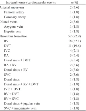
pulmonary emboli and infarct. Extrapulmonary
cardiovascular events occurred in 56 (28.1%)
patients, including thrombus formation in 52
(92.9%), and arterial aneurysm and vein dilation in
2 (3.6%) patients, each (Table 2).
Management strategies for the PAAs were not
described for 70 patients. Of the remaining 129 patients,
82 (63.6%) were treated conservatively,
10, 13-15, 17-19, 22, 23, 25, 27-29, 31, 37, 38, 40, 41, 46, 49-56, 61-63, 65, 68-70, 73, 75, 78, 81, 83, 84, 86, 91, 94, 95,97, 99, 100, 102-104, 105, 108, 112-116, 118
22 (17.1%) were treated
interventionally
10, 24, 32, 34, 42, 47, 60, 64, 71, 72, 74-76, 79, 87, 89, 92, 96and
Figure 2. Distribution of pulmonary artery aneurysms. LPA: left pulmonary artery; MPA: main pulmonary artery; RPA: right pulmonary artery.
Figure 3. Distribution of concurrent intramural thromboses of pulmonary artery aneurysms. a: artery; LPA: left pulmonary artery; MPA: main pulmonary artery; RPA: right pulmonary artery.
Table 2. Extrapulmonary cardiovascular events in 56 patients.
Extrapulmonary cardiovascular events n (%)
Arterial aneurysm 2 (3.6)
Femoral artery 1 (1.8)
Coronary artery 1 (1.8)
Dilated veins 2 (3.6)
Azygous vein 1 (1.8)
Hepatic vein 1 (1.8)
Thrombus formation 52 (92.9)
RV 18 (32.1)
DVT 11 (19.6)
IVC 4 (7.1)
RA 3 (5.4)
Dural sinus + DVT 3 (5.4)
RA + RV 2 (3.6)
Dural sinus + RV 2 (3.6)
SVC 2 (3.6)
Dural sinus 1 (1.8)
Dural sinus + RV + DVT 1 (1.8)
IVC + DVT 1 (1.8)
RV + DVT 1 (1.8)
RV + SVC 1 (1.8)
Dural sinus + jugular vein 1 (1.8)
SVC + innominate vein 1 (1.8)
25 (19.4%) were treated surgically with concurrent
immunosuppressive agents
21, 26, 30, 35, 36, 43, 55, 58, 59, 78, 85, 88, 98, 100, 106, 107, 110, 111, 115, 118(
c
2=79.7, p<0.0001). Two patients
given conservative treatment were eventually
treated for PAA interventionally because of PAA
progression
40, 41and one patient who originally
underwent interventional embolization of the PAA
was later subjected to emergency pulmonary artery
ligation because of PAA recurrence with severe
hemoptysis and respiratory distress.
24There were a
total of 24 interventional and 26 surgical procedures
for PAAs. Additionally, one patient underwent
resection of endomyocardial ibrosis plus tricuspid
valve repair
101and one patient underwent surgical
excision of intracardiac mass.
56Of the whole patient sample, immunosuppressive
treatment strategies were described in 114 patients.
Steroids were administered to 101 (88.6%),
cyclophosphamide to 62 (54.4%), azathioprine
(150 mg/day or 2 mg/kg/day) to 25 (21.9%), colchicine
(0.5-1.5 mg/day) to 36 (31.6%), anticoagulants to 22
(19.3%), methotrexate to 3 (2.6%) and cyclosporine
A was administered to 2 (1.8%) patients. Alternative
conservative treatments included infliximab, a
tumor necrosis factor neutralizing agent, 5 mg/
kg at 0, 2, 6, 14 and 22 weeks in 3 (2.6%) cases,
adalimumab, a human monoclonal antibody
against tumor necrosis factor-α, in 2 (1.8%),
hematopoietic stem cell transplantation in 2 (1.8%)
and mycophenolate mofetil in 1 case (0.9%). Onset
of action was soon after drug administration in 5
(4.4%) patients.
22, 52, 58, 104, 118In another 31 patients,
13, 15, 17, 18, 20, 25, 37, 46-48, 50, 51, 61, 65, 69, 81, 94, 97, 103, 109, 116onset of
action took 13.6±23.5 (range: 0.25-120; median: 6)
months (n=44). Cure time was 6.4±5.5 (range: 2-24;
median: 5) months (n=18).
14, 25, 34, 38, 46, 47, 54, 56, 59, 61, 63, 65, 73, 81, 91, 95, 113, 116In 11 cases PAA embolization materials
were described, as follows: N-butyl cyanoacrylate
(n=3, 27.3%),
34, 41Nester® Embolization Coils (n=3,
27.3%),
74, 76Amplatzer devices (n=3, 27.3%),
64, 66, 72Ethylene Vinyl Alcohol Copolymer (n=1, 9.1%)
71and
Guglielmi detachable coils (n=1, 9.1%).
60In patients
with multiple PAAs, the number of embolization
coils deployed in a single patient varied from 2,
74through 6,
74to 10.
76Details of surgical operations
performed on PAA patients are listed in Table 3,
showing that lobectomy was the most common
surgical operation, accounting for 42.3% of surgical
procedures.
The indications for conservative treatment in
severe patients were extensive PAA locations,
55, 97failed interventional embolization
95and patient
reluctance to undergo interventional embolization
or surgical operations.
29, 68, 114Hematopoietic stem cell
transplantation
86and tumor necrosis factor-α plus
inliximab
25were indicated for patients refractory to
conventional immunosuppressive therapy. Indications
for interventional embolization included bilateral
pulmonary artery involvement,
74giant pulmonary
artery pseudoaneurysm,
87ruptured PAAs,
64failed
conservative treatment,
34to avoid potential surgical
complications
60and patients’ reluctance to undergo
surgical operations.
72Patients with rapidly expanding
pulmonary artery (pseudo)aneurysms
24, 26, 107or with
recurrent PAA following successful percutaneous
embolization
24usually warranted surgical operations;
while patients with uncontrolled massive hemoptysis
with impending PAA rupture often required an
emergency operation.
55, 59, 106, 110A total of 9 patients
received intervention/surgery for the treatment of
PAA on an urgent basis including 1 interventional
embolization
41and 8 open thoracic operations.
24, 35, 55, 85, 110, 115, 118In all 9 of these patients an urgent procedure
was indicated for massive hemoptysis, and 4 (44.4%)
patients of whom suffered from a PAA rupture.
55, 110, 115, 118Patients were followed-up for 22.5±33.5 (range:
Table 3. Surgical operations for pulmonary artery aneurysms in 26 patients.
Surgical operation n (%)
Lobectomy 11 (42.3)
Pneumonectomy 3 (11.5)
PAA excision 2 (7.7)
Pulmonary artery ligation 2 (7.7)
Pulmonary artery aneurysmorrhaphy 1 (3.8)
Pulmonary artery aneurysmorrhaphy + pneumonectomy 1 (3.8)
Pulmonary artery aneurysmectomy + pneumonectomy 1 (3.8)
Thoracotomy (patient died during thoracotomy with no time for further interventions) 1 (3.8)
Lobectomy + pulmonary arterioplasty 1 (3.8)
Left pulmonary artery replacement under cardiopulmonary bypass 1 (3.8)
Left and right pulmonary artery operations (details not stated) 1 (3.8)
0.75-204; median: 15) months (n=94). Outcomes
were not described for 24 patients. Of the remaining
175 patients, 108 (61.7%) were event-free survivals
and 44 (25.1%) patients died. Time to death was
reported for 30 patients, 11 (36.7%) of whom were
early deaths and 19 (63.3%) of whom were late
deaths (
c
2=4.3, p=0.03887). Mean time to death
was 6.1±7.2 (range: 0-24; median: 3) months after
intervention or after discharge (a “0” indicates that
one patient died of uncontrolled massive bleeding
during the operation
83). Cause of death was described
for 27 patients: massive hemoptysis in 20 (74.1%),
PAA rupture in 4 (14.8%), pulmonary hemorrhage
as evidenced by bronchoscopy in 2 (7.4%),
postoperative bleeding in 1 (3.7%) patient and
septicemia in 1 patient (3.7%) (
c
2=47.2, p<0.0001).
Outcomes were reported for 175 patients and
comparison of outcomes across different treatments
revealed that conservatively treated patients
were prone to suffer from PAA progression, that
interventional embolization was associated with
higher risks of recurrence and reintervention for
PAAs, and that surgically treated patients had the
highest overall and early mortality rates. However,
there were no signiicant differences in late mortality
between these three groups (Table 4). The time to
death of surgically treated patients was much shorter
than among conservatively treated patients (2.3±5.9
months vs. 7.8±6.4 months, p=0.04650). There were
24 interventional procedures, 23 (95.8%) of which
were elective including 1 (4.3%) postoperative
death, while 1 (4.2%) was an emergency intervention
and the patient survived. There were 26 surgical
procedures, 21 (80.8%) of which were elective,
including 9 (42.9%) deaths; whereas 5 (19.2%)
were emergency surgeries, including 3 (60%)
deaths (
c
2=0.5, p=0.48953). Ten patients suffered
PAA rupture. Four of them had been managed
conservatively, 4 had been managed surgically and
2 had been managed interventionally, with only
2 surgically-treated patients surviving, giving an
overall survival rate of 20% and an emergency
surgery survival rate of 50% (
c
2=5.0, p=0.08416).
Univariate analysis revealed that presence of
hemoptysis (p<0.0001), lobar artery involvement
(p=0.00200) and PAA rupture (p=0.00900) were
statistically signiicant adverse prognostic factors
indicative of poor prognosis in PAA patients, whereas
intramural pulmonary artery thrombus (p=0.16000),
surgical intervention (p=0.20900), emergency
operation (p=0.23100) and PAA multiplicity
(p=0.97400) were not predictive. Attempts to identify
independent risk factors for mortality using multiple
logistic regression employed hemoptysis, lobar
arterial aneurysm, multiplicity, concurrent intramural
thrombus and surgical/interventional therapy as
input variables, overall model it parameters were:
c
2=14.1580, df=5, p=0.0146. Mortality correlated
signiicantly with lobar artery involvement, and there
was a quasi-correlation with hemoptysis (Table 5).
DISCUSSION
The pulmonary vasculitis seen in PAA associated
with Behçet’s disease is primarily located in the vasa
vasorum. Histopathological observations revealed
pulmonary vasculitis involving all layers of pulmonary
arteries and veins,
3resulting in thrombosis, stenosis,
aneurysm formation and rupture. Mononuclear
inlammatory cells, predominantly lymphocytes,
are responsible for inlammatory iniltration in and
around the vessel wall. Additionally, impaired natural
killer cell activity and immune system dysregulation
have also been observed in patients with Behçet’s
disease and pulmonary manifestations.
119Marked
intimal thickening with degenerative changes in the
Table 4. A comparison of the outcomes of 173 patients with pulmonary artery aneurysm: patients treated conservatively versus surgically versus interventionally.
Outcome Conservative (n=83)
Surgical (n=25) Interventional (n=24)
Treatment not indicated (n=66)
c2
p value
Event-free survival, n (%) 47 (56.6) 10 (40) 12 (50) 39 (59.1) 9.3 0.00970
Progression, n (%) 7 (8.4)** 0 (0) 0 (0) 1 (1.5) 6.7 0.03473
Recurrence, n (%) 4 (4.8) 1 (4) 5 (20.8)† 4 (6.1) 8.4 0.01464
Complication, n (%) 2 (2.4) ‡ 1 (4) 2 (8.3) † 0 (0) 2.1 0.34975
Reintervention, n (%) 0 (0) 0 (0) 7 (29.2) 0 (0) 42.1 <0.00001
Outcome not stated, n (%) 6 (7.2) 1 (4) 2 (8.3) 14 (21.2) --
--Death, n (%) 20 (24.1) 12 (48) 3 (12.5) 8 (12.1) 8.0 0.01872
Early death 2 (10) 5 (41.7) 1 (33.3) 1 (12.5) 10.4 0.00548
Late death 18 (90) 7 (58.3) 2 (66.7) 7 (87.5) 2.7 0.26446
medial layer from the lobar branches to the arterioles
can explain hemoptyisis and PAA rupture.
120Bronchopulmonary istulas found during autopsies
of patients who died of sudden hemoptysis
43and
in surgical specimens resected from patients with
massive hemoptysis
10, 110were apparently the
causative etiology of life-threatening hemoptysis.
Aneurysms may be single or multiple, unilateral
or bilateral, saccular or fusiform, and may be
located in the main pulmonary artery or lobar
or segmental arteries.
2Pulmonary vasculitis in
Behçet’s disease may also result in thrombosis,
stenosis or occlusion of lung vessels, but thrombosis
rather than embolism was usually associated with
PAAs.
121Other pulmonary problems seen in Behçet’s
disease patients, including pleural effusion and
chylous pleural effusions, are the result of vascular
complications.
11Focal hemorrhages or infarct areas
can present in the lung parenchyma adjacent to the
aneurysms.
120Thrombosed aneurysms cause ischemia
and infarction in the pulmonary parenchyma.
6Pulmonary artery aneurysms may manifest as hilar
enlargement or round, lobulated opacities on chest
radiographs.
16Computed tomography has largely
replaced angiography as the diagnostic tool and
magnetic resonance imaging can also be useful for
diagnosis of PAAs, but appears to be less sensitive
than computed tomography for diagnosis of small
PAAs.
2Erkan et al.
2reported that PAAs are most often
located in the right lower lobar arteries, followed by
bilateral main pulmonary branches. Tunaci et al.
109proposed that the pulmonary arteries of the lower
lobe were the most common site of involvement,
and a mural thrombus was observed in 85% of PAAs.
Aneurysms were most frequently located in the lobar
artery in the right lower lobe (35%), followed by the
lobar artery in the left lower lobe (19%) and right
main pulmonary artery (17%), and 33% of aneurysms
had partial or total thrombus within the aneurysm
itself.
109The present study showed similar results to
those published by Tunaci et al.
109Another important
inding of Tunaci et al.
109was complete or partial
PAA resolution in response to immunosuppressive
treatments: 76% of PAAs completely disappeared in
3-42 (mean: 21) months and 24% of PAAs regressed
in 4-28 (mean: 17) months after treatment.
109It was
reported that the largest aneurysm was 7 cm,
109and
the mean diameter of aneurysms was 2.3±1.1 cm.
122The present study, however, revealed much larger
dimensions of PAAs with a mean diameter of 4.0 cm.
In Behçet’s disease, immunosuppressants should
be the irst line treatment of choice and provoke
regression of the PAAs and associated thrombus in a
majority of the patients.
21Pulse methylprednisolone
followed by oral prednisolone is one routine
regimen for Behçet’s disease.
48, 50, 59Monthly
intravenous cyclophosphamide 1 g, and azathioprine
2.5 mg/kg/day may also be employed.
88Tumor
necrosis factor-neutralizing monoclonal antibody
inliximab was an effective treatment for a case
of potentially lethal PAA in Behçet’s disease.
25Two patients with resistant PAA were treated with
hematopoietic stem cell transplantation.
86Other
tumor necrosis factor-
a
blockers, which include
etanercept and adalimumab, would also be effective
for the treatment of PAA in Behçet’s disease
patients.
77Pulmonary artery aneurysm patients with
thrombophlebitis and/or pulmonary emboli who are
on anticoagulants are at risk of massive bleeding and
so anticoagulants should be used with great caution,
especially in patients with hemoptysis.
76Alternative management strategies to conservative
treatment include surgical and transcatheter
interventions. Diseased lung tissue resection is
preferred to pulmonary artery reconstruction in
surgical treatment of PAAs. Surgical treatment
mostly consists of major anatomical resection rather
than preserving lung tissue. Immediate operation
for PAAs should be performed when systemic
inflammation is under control after short-term
immunosuppressive therapy.
21Patients with recurrent
or massive hemoptysis, in particular patients with
ruptured PAAs, warrant emergency surgery, which
is often associated with very high mortality.
21Endovascular embolization has been shown to be
Table 5. Odds ratios and 95% conidence intervals for multivariate regression analysis of risk factors predictive of mortality.
Variable Odds ratio 95% conidence interval p value
Hemoptysis 2.6030 0.9573-7.0779 0.0609
Lobar arterial involvement 0.3025 0.1062-0.8614 0.0251
Multiplicity of the pulmonary artery aneurysm 1.4231 0.6332-3.1986 0.3931
Concurrent pulmonary artery aneurysm and intramural thrombus
0.7596 0.2986-1.9323 0.5638
effective at controlling PAA hemorrhage.
34The
Amplatzer duct occluder is currently the most
commonly used device for management of large
aneurysms.
88It can be employed in most cases and
recurrent PAAs may be curable with reintervention.
32Pulmonary artery aneurysms were the major
contributing factor to overall mortality from Behçet’s
disease.
121In the early years, short-term survival of
patients with PAAs associated with Behçet’s disease
was only 50%.
3A decade later, 5-year survival had
increased to 62%.
4The present study displayed an
overall survival of 76.9% at a mean follow-up of
21.6 months.
The present study revealed that PAAs have
predilections for hemoptysis manifestations, right
lower lobar locations, multiplicity and concurrent
intramural thrombus formation. Surgical intervention
therapy is warranted in most of the patients. All
these variables were shown to be predictive risk
factors for higher mortality in PAAs. For refractory
cases that respond poorly to conservative treatment,
endovascular embolization and Amplatzer occluder
device are good choices for PAA management.
Hemorrhaging PAAs can also be controlled by
pneumonectomy, lobectomy, pulmonary artery
aneurysmectomy, or pulmonary arterial ligation, but
these options may be associated with higher mortality
compared with interventionally and conservatively
treated patients.
The most important limitations of this review are
related to the non-availability of numbers, locations
and dimensions of the arteries involved by PAAs in
a large proportion of the references cited. Further
prospective studies with more detailed information
of PAAs are essential if more precise conclusions
are to be drawn.
In conclusion, PAAs in Behçet’s disease are
characterized by a predilection for hemoptysis
manifestations, right lower lobar location, multiplicity
and concurrent intramural thrombus formation. Both
the condition itself and the surgical operations it
warrants are linked with high mortality due to PAA
hemorrhage.
REFERENCES
1. Seyahi E, Yurdakul S. Behçet’s syndrome and thrombosis. Mediterr J Hematol Infect Dis. 2011;3(1):e2011026. http://dx.doi. org/10.4084/MJHID.2011.026. PMid:21869912
2. Erkan F, Gül A, Tasali E. Pulmonary manifestations of Behçet’s disease. horax. 2001;56(7):572-8. http://dx.doi.org/10.1136/ thorax.56.7.572. PMid:11413359
3. Hamuryudan V, Yurdakul S, Moral F, et al. Pulmonary arterial aneurysms in Behçet’s syndrome: a report of 24 cases. Br
J Rheumatol. 1994;33(1):48-51. http://dx.doi.org/10.1093/ rheumatology/33.1.48. PMid:8162457
4. Hamuryudan V, Er T, Seyahi E, et al. Pulmonary artery aneurysms in Behçet syndrome. Am J Med. 2004;117(11):867-70. http:// dx.doi.org/10.1016/j.amjmed.2004.05.027. PMid:15589493 5. Seyahi E, Melikoglu M, Akman C, et al. Pulmonary artery
involvement and associated lung disease in Behçet disease: a series of 47 patients. Medicine (Baltimore). 2012;91(1):35-48. http://dx.doi.org/10.1097/MD.0b013e318242f37. PMid:22210555 6. Erkan F, Kiyan E, Tunaci A. Pulmonary complications of Behçet’s disease. Clin Chest Med. 2002;23(2):493-503. http://dx.doi. org/10.1016/S0272-5231(01)00014-4. PMid:12092042
7. Saadoun D, Wechsler B. Hughes-Stovin syndrome. Orphanet: he Portal of Rare Diseases and Orphan Drugs. 2011 Dec [cited 2013 July 04]. http://www.orpha.net/consor/cgi-bin/OC_Exp. php?lng=en&Expert=228116.
8. Khalid U, Saleem T. Hughes-Stovin syndrome. Orphanet J Rare Dis. 2011;6(1):15. http://dx.doi.org/10.1186/1750-1172-6-15. PMid:21489283
9. Uzun O. Pulmonary involvement in Behçet’s disease and Takayasu’s arteritis. Eur Respir Mon. 2011;54:32-45.
10. Uzun O, Erkan L, Akpolat I, Findik S, Atici AG, Akpolat T. Pulmonary involvement in Behçet’s disease. Respiration. 2008;75(3):310-21. http://dx.doi.org/10.1159/000101954. PMid:17446699
11. Uzun O, Akpolat T, Erkan L. Pulmonary vasculitis in behcet disease: a cumulative analysis. Chest. 2005;127(6):2243-53. http:// dx.doi.org/10.1378/chest.127.6.2243. PMid:15947344
12. Hemoptysis. [cited 2013 July 04]. http://thelungcenter.co.in/ yahoo_site_admin/assets/docs/hemoptysis.107184533.pdf. 13. Aamar S, Peleg H, Leibowitz D, Chajek-Shaul T, Hiller N, Heyman
SN. Efficacy of adalimumab therapy for life-threatening pulmonary vasculitis in Behçet’s disease. Rheumatol Int. 2014;34(6):857-60. PMid:23412691
14. Acican T, Gürkan OU. Azathiopine-steroid combination therapy for pulmonary arterial aneurysms in Behçet’s disease. Rheumatol Int. 2001;20(4):171-4. http://dx.doi.org/10.1007/s002960100102. PMid:11411965
15. Agha A, Bella AM, Assiri AH, Al-Hakami M. Can Behcet’s disease related pulmonary arterial aneurysms be completely resolved? Open Rheumatol J. 2011;5(1):88-90. http://dx.doi.org/10.2174/1 874312901105010088. PMid:22216070
16. Ahn JM, Im JG, Ryoo JW, et al. horacic manifestations of Behçet syndrome: radiographic and CT findings in nine patients. Radiology. 1995;194(1):199-203. PMid:7997553.
17. Aksu K, Koçanaoğullari H, Keser G, et al. A case of Behçet’s disease with pulmonary arterial aneurysm and secondary amyloidosis. Rheumatology (Oxford). 2002;41(7):831-2. http:// dx.doi.org/10.1093/rheumatology/41.7.831-a. PMid:12096241 18. Aktoğu S, Erer OF, Urpek G, Soy O, Tibet G. Multiple
pulmonary arterial aneurysms in Behçet’s disease: clinical and radiologic remission after cyclophosphamide and corticosteroid therapy. Respiration. 2002;69(2):178-81. http:// dx.doi.org/10.1159/000056324. PMid:11961435
19. Alkaabi JK, Pathare A. Pattern and outcome of vascular involvement of Omani patients with Behcet’s disease. Rheumatol Int. 2011;31(6):731-5. http://dx.doi.org/10.1007/s00296-010-1363-z. PMid:20130881
org/10.1164/ajrccm-conference.2013.187.1_MeetingAbstracts. A4604.
21. Aroussi AA, Redai M, El Ouardi F, Mehadji BE. Bilateral pulmonary artery aneurysm in Behçet syndrome: report of two operative cases. J horac Cardiovasc Surg. 2005;129(5):1170-1. http://dx.doi. org/10.1016/j.jtcvs.2004.08.038. PMid:15867796
22. Aşker S, Aşker M, Gürsu O, Mercan R, Timuçin OB. A Behcet’s disease patient with right ventricular thrombus, pulmonary artery aneurysms, and deep vein thrombosis complicating recurrent pulmonary thromboembolism. Case Rep Pulmonol. 2013;2013. http://dx.doi.org/10.1155/2013/492321
23. Atalay F, Ernam D, Okten F, Akar N. Elevated FVIII and FIX level in a Behçet’s disease patient with intracardiac thrombosis and pulmonary arterial aneurysms. hromb Res. 2005;115(1-2):159-61. http://dx.doi.org/10.1016/j.thromres.2004.08.010. PMid:15567468
24. Attia R, Reidy J, D’Cruz D, Lang-Lazdunski L. Pulmonary artery ligation with lung preservation in Behcet disease: report of a case with prolonged survival. J Thorac Cardiovasc Surg. 2010;139(4):e93-5. http://dx.doi.org/10.1016/j.jtcvs.2009.07.033. PMid:19744671
25. Baki K, Villiger PM, Jenni D, Meyer T, Beer JH. Behcet’s disease with life-threatening haemoptoe and pulmonary aneurysms: complete remission after inliximab treatment. Ann Rheum Dis. 2006;65(11):1531-2. http://dx.doi.org/10.1136/ard.2005.045195. PMid:17038456
26. Başak M, Gül S, Yazgan Y, et al. A case of rapidly progressive pulmonary aneurysm as a rare complication of Behçet’s syndrome—a case report. Angiology. 1998;49(5):403-8. http:// dx.doi.org/10.1177/000331979804900510. PMid:9591533 27. Basoglu T, Canbaz F, Bernay I, Danaci M. Bilateral pulmonary
artery aneurysms in a patient with Behcet syndrome: evaluation with radionuclide angiography and V/Q lung scanning. Clin Nucl Med. 1998;23(11):735-8. http://dx.doi.org/10.1097/00003072-199811000-00002. PMid:9814558
28. Bastos AL, de Brito ILA. Pulmonary artery aneurysms in Behçet’s disease: case report. Radiol Bras. 2011;44(6):396-8. http://dx.doi. org/10.1590/S0100-39842011000600012.
29. Berkan O, Oztürkcan S, Doğan K, Onen A, Başel H, Hatipoğu A. Pulmonary arterial aneurysm in Behçet’s disease. J Eur Acad Dermatol Venereol. 1999;13(2):140-1. http://dx.doi. org/10.1111/j.1468-3083.1999.tb00869.x. PMid:10568496 30. Berkmen T. MR angiography of aneurysms in Behçet disease: a
report of four cases. J Comput Assist Tomogr. 1998;22(2):202-6. http://dx.doi.org/10.1097/00004728-199803000-00007. PMid:9530379
31. Bilginer Y, Besbas N, Aktay Ayvaz N, Bakkaloglu A, Ozen S. Behcet disease: treatment of vascular involvement in children. Pediatr Rheumatol. 2008;6(Suppl 1):P262. http://dx.doi. org/10.1186/1546-0096-6-S1-P262.
32. Bozkurt AK. Embolisation in Behçet’s disease. Thorax. 2002;57(5):469-70. http://dx.doi.org/10.1136/thorax.57.5.469-a. PMid:11978931
33. Caglar M, Ergun E, Emri S. 99Tcm-MAA lung scintigraphy in patients with Behçet’s disease: its value and correlation with clinical course and other diagnostic modalities. Nucl Med Commun. 2000;21(2):171-9. http://dx.doi.org/10.1097/00006231-200002000-00009. PMid:10758613
34. Cantasdemir M, Kantarci F, Mihmanli I, et al. Emergency endovascular management of pulmonary artery aneurysms in Behçet’s disease: report of two cases and a review of the
literature. Cardiovasc Intervent Radiol. 2002;25(6):533-7. http:// dx.doi.org/10.1007/s00270-002-1967-0. PMid:12042999 35. Cebi N, Johannes F, Botsios S, Walterbusch G. Intraparenchymal
replacement of the left pulmonary artery with implantation of segmental arteries in a 26-year-old patient. J horac Cardiovasc Surg. 2003;126(6):2074-7. http://dx.doi.org/10.1016/S0022-5223(03)00932-2. PMid:14688729
36. Ceyran H, Akçali Y, Kahraman C. Surgical treatment of vasculo-Behçet’s disease. A review of patients with concomitant multiple aneurysms and venous lesions. Vasa. 2003;32(3):149-53. http:// dx.doi.org/10.1024/0301-1526.32.3.149. PMid:14524035 37. Chalazonitis AN, Lachanis SB, Mitseas P, et al. Hughes-Stovin
syndrome: a case report and review of the literature. Cases J. 2009;2(1):98. http://dx.doi.org/10.1186/1757-1626-2-98. PMid:19178695
38. Chang JE, Lee YH, Lee J. Multiple cardiovascular complications in a patient with Behcet’s disease. Korean J Intern Med. 2008;23(2):100-2. http://dx.doi.org/10.3904/kjim.2008.23.2.100. PMid:18646513
39. Cho SB, Yun M, Lee JH, Kim J, Shim WH, Bang D. Detection of cardiovascular system involvement in Behçet’s disease using fluorodeoxyglucose positron emission tomography. Semin Arthritis Rheum. 2011;40(5):461-6. http://dx.doi.org/10.1016/j. semarthrit.2010.05.006. PMid:20822800.
40. Cil BE, Geyik S, Akmangit I, Cekirge S, Besbas N, Balkanci F. Embolization of a giant pulmonary artery aneurysm from Behcet disease with use of cyanoacrylate and the “bubble technique”. J Vasc Interv Radiol. 2005;16(11):1545-9. http://dx.doi. org/10.1097/01.RVI.0000171692.61294.91. PMid:16319165 41. Cil BE, Turkbey B, Canyiğit M, Kumbasar OO, Celik G, Demirkazik
FB. Transformation of a ruptured giant pulmonary artery aneurysm into an air cavity after transcatheter embolization in a Behçet’s patient. Cardiovasc Intervent Radiol. 2006;29(1):151-4. http://dx.doi.org/10.1007/s00270-005-0225-7. PMid:16328688 42. Cohle SD, Colby T. Fatal hemoptysis from Behcet’s disease
in a child. Cardiovasc Pathol. 2002;11(5):296-9. http://dx.doi. org/10.1016/S1054-8807(02)00117-5. PMid:12361841
43. de Montpréville VT, Macchiarini P, Dartevelle PG, Dulmet EM . L arge bilateral pulmonar y ar ter y aneur ysms in Behçet’s disease: rupture of the contralateral lesion after aneurysmorrhaphy. Respiration. 1996;63(1):49-51. http://dx.doi. org/10.1159/000196515. PMid:8833993
44. Denecke T, Staeck O, Amthauer H, Hänninen EL. PET/ CT visualises inflammatory activity of pulmonary artery aneurysms in Behçet disease. Eur J Nucl Med Mol Imaging. 2007;34(6):970. http://dx.doi.org/10.1007/s00259-007-0429-y. PMid:17447060.
45. Dikensoy O, Bayram NG, Filiz A. Massive haemoptysis in a young woman. Postgrad Med J. 2002;78(917):183, 187-8. http://dx.doi. org/10.1136/pmj.78.917.183-a. PMid:11884709
46. Düzgün N, Anil C, Ozer F, Acican T. The disappearance of pulmonary artery aneurysms and intracardiac thrombus with immunosupressive treatment in a patient with Behçet’s disease. Clin Exp Rheumatol. 2002;20(4, Suppl 26):S56-7. PMid:12371638. 47. El Houari T, Oukerraj L, Ghzaiel L, et al. Management of
Behçet disease with multiple complications. Hellenic J Cardiol. 2009;50(5):420-2. PMid:19767285.
49. Emad Y, Ragab Y, Shawki A-H, Gheita T, El-Marakbi A, Salama MH. Hughes-Stovin syndrome: is it incomplete Behçet’s? Report of two cases and review of the literature. Clin Rheumatol. 2007;26(11):1993-6. http://dx.doi.org/10.1007/s10067-007-0609-y. PMid:17457658
50. Endo LM, Rowe SM, Romp RL, Buckmaster MA, Atkinson TP. Pulmonary aneurysms and intracardiac thrombi due to Behçet’s disease in an African-American adolescent with oculocutaneous albinism. Clin Rheumatol. 2007;26(9):1537-9. http://dx.doi. org/10.1007/s10067-006-0426-8. PMid:17047893
51. Erkan D, Yazici Y, Sanders A, Trost D, Yazici H. Is Hughes-Stovin syndrome Behçet’s disease? Clin Exp Rheumatol. 2004;22(4, Suppl 34):S64-8. PMid:15515789.
52. Ernam D, Atalay F, Alp A, Hasanoğlu HC. A Behçet’s disease patient with intracardiac thrombus, pulmonary artery aneurysms complicating recurrent pulmonary thromboembolism. Tuberk Toraks. 2006;54(2):168-71. PMid:16924574.
53. Filiz A, Dikensoy O. Lethal aneurysm formation of pulmonary arteries in a woman with Behçet’s disease. Rheumatology (Oxford). 2000;39(2):222-4. http://dx.doi.org/10.1093/ rheumatology/39.2.222A. PMid:10725085
54. Fischer A, Korman DS, West SG. Radiologic vignette: Hughes-Stovin syndrome. Arthritis Rheum. 2005;53(1):114-6. http:// dx.doi.org/10.1002/art.20907. PMid:15696563
55. Gebitekin C, Yilmaz M, Senkaya I, Saba D, Sağdiç K, Ozer G. Fatal haemoptysis due to pulmonary artery aneurysm in Behçet’s disease. Eur J Vasc Endovasc Surg. 1997;13(2):233-6. http://dx.doi. org/10.1016/S1078-5884(97)80027-5. PMid:9091163
56. Gönlügür U, Atalar MH, Kaptanoğlu M, et al. Intracardiac thrombus and co-existing pulmonary artery aneurysm in Behçet’s disease: two case reports. Turk Respir J. 2003;4(3):153-5. 57. Gopathi S, Hurt RT, Guardiola J. Intracardiac thrombus in Behcet’s disease: a rare case in the United States. Respir Med CME. 2011;4(4):154-6. http://dx.doi.org/10.1016/j.rmedc.2011.07.001. 58. Greene RM, Saleh A, Taylor AK, et al. Non-invasive assessment
of bleeding pulmonary artery aneurysms due to Behçet disease. Eur Radiol. 1998;8(3):359-63. http://dx.doi.org/10.1007/ s003300050394. PMid:9510565
59. Gül A, Yilmazbayhan D, Büyükbabani N, et al. Organizing pneumonia associated with pulmonary artery aneurysms in Behçet’s disease. Rheumatology (Oxford). 1999;38(12):1285-9. http://dx.doi.org/10.1093/rheumatology/38.12.1285. PMid:10587562
60. Hammad AM, Al-Qahtani SM, Al-Zahrani MA. Huge pulmonary artery aneurysm. Can Respir J. 2009;16(3):93-5. PMid:19557216. 61. Hammami S, Mahjoub S, Ben-Hamda K, Brahem R, Gamra H, Ben
Farhat M. Intracardiac thrombus in Behçet’s disease: two case reports. hromb J. 2005;3(1):9. http://dx.doi.org/10.1186/1477-9560-3-9. PMid:16042810
62. Hirohata S, Kikuchi H. Histopathology of the ruptured pulmonary artery aneurysm in a patient with Behçet’s disease. Clin Exp Rheumatol. 2009;27(2, Suppl 53):S91-5. PMid:19796542. 63. Houman M, Ksontini I, Ben Ghorbel I, et al. Association of right heart thrombosis, endomyocardial fibrosis, and pulmonary artery aneurysm in Behçet’s disease. Eur J Intern Med. 2002;13(7):455-7. http://dx.doi.org/10.1016/S0953-6205(02)00134-6. PMid:12384136
64. Ianniello A, Carraiello G, Nicotera P, Vaghi A, Cazzulani A. Endovascular treatment of a ruptured pulmonary artery aneurysm in a patient with Behçet’s disease using the Amplatzer Vascular Plug 4. Korean J Radiol. 2013;14(2):283-6. http://dx.doi. org/10.3348/kjr.2013.14.2.283. PMid:23482415
65. Ilvan A, Okutan O, Kartaloglu Z, et al. A case of Behcet’s disease with pulmonary artery aneurysm and thrombosis. Int J Angiol. 2002;11(2):92-4. http://dx.doi.org/10.1007/BF01616373. 66. Jayachandran NV, Rajasekhar L, Chandrasekhara PK ,
Kanchinadham S, Narsimulu G. Multiple peripheral arterial and aortic aneurysms in Behcet’s syndrome: a case report. Clin Rheumatol. 2008;27(2):265-7. http://dx.doi.org/10.1007/s10067-007-0713-z. PMid:17929077
67. Kaçar Güveli T, Tamam M, Ergür S, Özülker T, Mülazimoğlu M, Özpaçaci T. Behcet’s disease with complete obstruction of right pulmonary artery by a thrombus. Turk J Nucl Med. 2009;18(2):53-4.
68. Kanchinadham S, Potikuri D. Multiple pulmonary arterial aneurysms in a young male patient with incomplete Behçet’s syndrome. Lung India. 2013;30(1):76-7. http://dx.doi. org/10.4103/0970-2113.106121. PMid:23661925
69. Kasikcioglu E, Akhan H, Cuhadaroglu C, Erkan F. Pulmonary artery aneurysm in Behcet’s disease: a case report. Heart Vessels. 2004;19(3):157-9. http://dx.doi.org/10.1007/s00380-003-0742-8. PMid:15168067
70. Kaya A, Ertan C, Gürkan OU, et al. Behçet’s disease with right ventricle thrombus and bilateral pulmonary artery aneurysms—a case report. Angiology. 2004;55(5):573-5. http:// dx.doi.org/10.1177/000331970405500516. PMid:15378123 71. Khalil A, Parrot A, Fartoukh M, Djibre M, Tassart M, Carette
MF. Pulmonary artery occlusion with ethylene vinyl alcohol copolymer in patients with hemoptysis: initial experience in 12 cases. AJR Am J Roentgenol. 2012;198(1):207-12. http://dx.doi. org/10.2214/AJR.10.5370. PMid:22194499
72. Kojuri J, Aslani A, Shahrzad S. A large pulmonary artery pseudoaneurysm in a patient with Behcet’s disease. J Cardiovasc Med (Hagerstown). 2007;8(12):1073-5. http://dx.doi.org/10.2459/ JCM.0b013e328028fe5e. PMid:18163026
73. Kömürcüoğlu B, Gayaf M, Büyüksirin M, Celikten E. Case of Behçet’s disease presenting with bilateral multiple pulmonary arterial aneurysms. Monaldi Arch Chest Dis. 2003;59(3):216-9. PMid:15065318.
74. Lacombe P, Qanadli SD, Jondeau G, et al. Treatment of hemoptysis in Behçet syndrome with pulmonary and bronchial embolization. J Vasc Interv Radiol. 1997;8(6):1043-7. http://dx.doi. org/10.1016/S1051-0443(97)70708-5. PMid:9399476
75. Lê Thi Huong D, Wechsler B, Papo T, et al. Arterial lesions in Behçet’s disease. A study in 25 patients. J Rheumatol. 1995;22(11):2103-13. PMid:8596152.
76. Lee JM, Ahn J, Hwang YJ, et al. A case of Behcet’s disease complicated with a pulmonary artery aneurysm and deep vein thrombosis, separately. J Rheum Dis. 2013;20(1):52-5. http:// dx.doi.org/10.4078/jrd.2013.20.1.52.
77. Lee SW, Lee SY, Kim KN, Jung JK, Chung WT. Adalimumab treatment for life threatening pulmonary artery aneurysm in Behçet disease: a case report. Clin Rheumatol. 2010;29(1):91-3. http://dx.doi.org/10.1007/s10067-009-1272-2. PMid:19816754 78. Leibowitz D, Planer D, Chajek-Shaul T. Echocardiographic
manifestations of Adamantiades-Behcet’s disease. Eur J Echocardiogr. 2007;8(6):457-62. http://dx.doi.org/10.1016/j. euje.2006.07.013. PMid:17011238
79. Loh H, Yung G, Bui C, Mansberg R, Comsa M. Pulmonary artery aneurysm with false-positive FDG PET in a patient with Behcet disease. Clin Nucl Med. 2010;35(4):286-8. http://dx.doi. org/10.1097/RLU.0b013e3181d18f21. PMid:20305427
2005;353(4):400. http://dx.doi.org/10.1056/NEJMicm041070. PMid:16049212
81. Louali FE, Tamdy A, Soufiani A, et al. Cardiac thrombosis as a manifestation of Behçet syndrome. Tex Heart Inst J. 2010;37(5):568-71. PMid:20978571.
82. Mahendran C, Singh P, Mani NB, Jindal SK. Successful treatment of pulmonary artery aneurysms secondary to possible Behçet’s disease. Respiration. 2002;69(4):355-8. http://dx.doi. org/10.1159/000063262. PMid:12169752
83. Malik KJ, Weber SL, Sohail S, Balaan MR. Hilar mass and papilledema on presentation. Chest. 1998;113(1):227-9. http:// dx.doi.org/10.1378/chest.113.1.227. PMid:9440594
84. Marchiori E, Zanetti G, Mano CM. Fulminant evolution of Behçet disease. AJR Am J Roentgenol. 2010;195(4):W311-2. http://dx.doi. org/10.2214/AJR.10.4602. PMid:20858797
85. Marzban M, Mandegar MH, Karimi A, et al. Cardiac and great vessel involvement in “Behcet’s disease”. J Card Surg. 2008;23(6):765-8. http://dx.doi.org/10.1111/j.1540-8191.2008.00607.x. PMid:19017008
86. Maurer B, Hensel M, Max R, Fiehn C, Ho AD, Lorenz HM. Autologous haematopoietic stem cell transplantation for Behcet’s disease with pulmonary involvement: analysis after 5 years of follow up. Ann Rheum Dis. 2006;65(1):127-9. http:// dx.doi.org/10.1136/ard.2005.035410. PMid:15919675
87. Meshksar A, Farahangiz S, Assadsangabi R, Nabavizadeh SA. Bilateral pulmonary artery involvement in Behcet’s yndrome (a case report). Iran J Radiol. 2008;5(S1):7-8.
88. Modaghegh MH, Kazemzadeh GH, Jokar MH. A case of Behçet disease with pulmonary artery pseudoaneurysm: long term follow-up. East Mediterr Health J. 2010;16(3):346-9. PMid:20795454.
89. Mouas H, Lortholary O, Lacombe P, et al. Embolization of multiple pulmonary arterial aneurysms in Behçet’s disease. Scand J Rheumatol. 1996;25(1):58-60. http://dx.doi. org/10.3109/03009749609082670. PMid:8774558
90. Nemattallah W. Pulmonary artery aneurysms in patient with Behcet’s. MyPACS.net: Radiology Teaching Files: Case 5175797. 2009 Dec [cited 2013 July 04]. http://www.mypacs.net/cases/ PULMONARY-ARTERY-ANEURYSMS-IN-PATIENT-WITH-BEHCETS-5175797.html.
91. Ozcan H, Aytac SK, Yagmurlu B, Kaya A. Color Doppler examination of a regressing pulmonary artery pseudoaneurysm due to Behçet disease. J Ultrasound Med. 2002;21(6):697-700. PMid:12054310.
92. Ozen S, Bilginer Y, Besbas N, Ayaz NA, Bakkaloglu A. Behçet disease: treatment of vascular involvement in children. Eur J Pediatr. 2010;169(4):427-30. http://dx.doi.org/10.1007/s00431-009-1040-y. PMid:19756733
93. Ozge C, Calikoğlu M, Yildiz A, Türsen U, Tamer L. Bilateral pulmonary artery aneurysms with protein C and protein S deiciency in a patient with Behçet’s disease. Scand J Rheumatol. 2004;33(1):52-4. http://dx.doi.org/10.1080/03009740310004694. PMid:15124944
94. Ozkaya S, Sahin U, Gumus A, Taşçı F, Cınarka H, Yavuz A. In situ thrombosis in pulmonary arterial aneurysms due to Behçet’s disease and eicacy of ımmunosuppressive therapy. Multidiscip Respir Med. 2012;7(1):33. http://dx.doi.org/10.1186/2049-6958-7-33. PMid:23078955
95. Park JY, Park JG, Won JH, Lee JM, Kim NS, Jung TH. Efects of corticosteroid and chlorambucil on multiple pulmonary artery aneurysms in Behcet’s syndrome. A case report. J Korean Med Sci. 1995;10(6):470-3. PMid:8924235.
96. Peall AF, Jones SM. Hemoptysis and Behcet’s Syndrome. J Rheumatol. 2009;36(4):848-9. http://dx.doi.org/10.3899/ jrheum.080900. PMid:19342723
97. Pereira de Godoy JM, Batigália F. Bilateral pulmonary artery aneurysm associated with bilateral pulmonary thromboembolism, superior vena caval thrombosis, and Chagas’ disease—a case report. Angiology. 2000;51(7):609-14. http:// dx.doi.org/10.1177/000331970005100711. PMid:10917587 98. Rutherford RM, O’Keefe D, Gilmartin JJ. An unusual case of
non-speciic interstitial pneumonitis. Respiration. 2004;71(2):202-5. http://dx.doi.org/10.1159/000076687. PMid:15031581 99. Saadoun D, Wechsler B, Desseaux K, et al. Mortality in Behçet’s
disease. Arthritis Rheum. 2010;62(9):2806-12. http://dx.doi. org/10.1002/art.27568. PMid:20496419
100. Saba D, Saricaoğlu H, Bayram AS, et al. Arterial lesions in Behçet’s disease. Vasa. 2003;32(2):75-81. http://dx.doi.org/10.1024/0301-1526.32.2.75. PMid:12945099
101. Sacré K, Ducrocq G, Hernigou A, Laissy JP, Papo T. Unusual cardiovascular events in Behçet’s disease. Clin Exp Rheumatol. 2010;28(4, Suppl 60):S82-5. PMid:20868577.
102. Sahin AA, Kalyoncu AF, Selçuk ZT, Cöplü L, Celebi C, Baris YI. Behcet’s disease with half and half nail and pulmonary artery aneurysm. Chest. 1990;97(5):1277. http://dx.doi.org/10.1378/ chest.97.5.1277a. PMid:2331944
103. Santana AN, Antunes T, Barros JM, Kairalla RA, Carvalho CR, Barbas CS. [Pulmonary involvement in Behcet’s disease: a positive single-center experience with the use of immunosuppressive therapy]. J Bras Pneumol. 2008;34(6):362-6. PMid:18622502. 104. Schreiber BE, Noor N, Juli CF, Haskard DO. Resolution of
Behçet’s syndrome associated pulmonary arterial aneurysms with inliximab. Semin Arthritis Rheum. 2011;41(3):482-7. http:// dx.doi.org/10.1016/j.semarthrit.2011.02.006. PMid:21546064 105. Sirmali M, Aloğlu HV, Ozçakar L, Kaya S. Bilateral giant
pulmonary artery aneurysms early in Behçet’s disease. Eur J Cardiothorac Surg. 2003;24(6):1033. http://dx.doi.org/10.1016/ S1010-7940(03)00614-6. PMid:14643828
106. Steward MJ, D’Cruz DP, Lang-Lazdunski L, Chambers J. Fever, haemoptysis and a mass in the heart. J R Soc Med. 2007;100(2):105-6. http://dx.doi.org/10.1258/jrsm.100.2.105. PMid:17277285
107. Takahama M, Yamamoto R, Nakajima R, Tada H. Successful surgical treatment of pulmonary artery aneurysm in Behçet’s syndrome. Interact Cardiovasc horac Surg. 2009;8(3):390-2. http://dx.doi.org/10.1510/icvts.2008.194647. PMid:19064583 108. Tolosa-Vilella C, Capela CA, Monteagudo-Jiménez M,
Marí-Alfonso B. Inliximab for life-threatening pulmonary artery aneurysms in Behçet’s disease. A case report. Clin Exp Rheumatol. 2011;29(4, Suppl 67):S94-5. PMid:21385550. 109. Tunaci M, Ozkorkmaz B, Tunaci A, Gül A, Engin G, Acunaş B.
CT indings of pulmonary artery aneurysms during treatment for Behçet’s disease. AJR Am J Roentgenol. 1999;172(3):729-33. http://dx.doi.org/10.2214/ajr.172.3.10063870. PMid:10063870 110. Tüzün H, Hamuryudan V, Yildirim S, et al. Surgical therapy
of pulmonary arterial aneurysms in Behçet’s syndrome. Ann horac Surg. 1996;61(2):733-5. http://dx.doi.org/10.1016/0003-4975(95)00913-2. PMid:8572805
112. Vansteenkiste JF, Peene P, Verschakelen JA, van de Woestijne KP. Cyclosporin treatment in rapidly progressive pulmonary thromboembolic Behçet’s disease. horax. 1990;45(4):295-6. http://dx.doi.org/10.1136/thx.45.4.295. PMid:2356556 113. Vivante A, Bujanover Y, Jacobson J, Padeh S, Berkun Y.
Intracardiac thrombus and pulmonary aneurysms in an adolescent with Behçet disease. Rheumatol Int. 2009;29(5):575-7. http://dx.doi.org/10.1007/s00296-008-0730-5. PMid:18850100 114. Wu CT, Lim KE. An unusual cause of large thrombosed
pulmonary artery aneurysms associated with chronic thromboembolism. Tzu Chi Med J. 2006;18(3):205-9. 115. Yakut ZI, Odev K. Pulmonary and cardiac involvement in
Behçet disease: 3 case reports. Clin Appl hromb Hemost. 2007;13(3):318-22. http://dx.doi.org/10.1177/1076029607302437. PMid:17636195
116. Yeung PHJ, Lau KKP, Lac A. Behçet’s disease with resolving pulmonary artery aneurysm and intracardiac thrombus. Hong Kong J Radiol. 2011;14:167-9.
117. Yilmaz S, Cimen KA. Pulmonary artery aneurysms in Behçet’s disease. Rheumatol Int. 2010;30(10):1401-3. http://dx.doi. org/10.1007/s00296-009-1092-3. PMid:19693504
118. Yoon YH, Kim KH, Baek WK, et al. Pulmonary artery pseudoaneurysm in a patient with Behçet disease. J horac Cardiovasc Surg. 2004;127(2):590-2. http://dx.doi.org/10.1016/j. jtcvs.2003.09.043. PMid:14762379
119. Hamzaoui K, Berraies A, Kaabachi W, Ammar J, Hamzaoui A. Pulmonary manifestations in Behçet disease: impaired natural killer cells activity. Multidiscip Respir Med. 2013;8(1):29. http:// dx.doi.org/10.1186/2049-6958-8-29. PMid:23556512
120. Hamuryudan V, Oz B, Tüzün H, Yazici H. The menacing pulmonary artery aneurysms of Behçet’s syndrome. Clin Exp Rheumatol. 2004;22(4, Suppl 34):S1-3. PMid:15515774.
121. Calamia KT, Schirmer M, Melikoglu M . Major vessel involvement in Behçet’s disease: an update. Curr Opin Rheumatol. 2011;23(1):24-31. http://dx.doi.org/10.1097/ BOR.0b013e3283410088. PMid:21124084
122. Sayin AG, Vural FS, Bozkurt AK, Oz B, Uygun N. Right atrial thrombus mimicking myxoma and bilateral pulmonary artery aneurysms in a patient with Behçet’s disease—a case report. Angiology. 1993;44(11):915-8. http://dx.doi. org/10.1177/000331979304401111. PMid:8239064
Correspondence Shi-Min Yuan Department of Cardiothoracic Surgery, he First Hospital of Putian, Teaching Hospital, Fujian Medical University 389 Longdejing Street - Chengxiang District Putian 351100, Fujian Province, People’s Republic of China Tel.: 86 594 6923117 E-mail: [email protected]
Author information S-MY is a cardiac surgeon at he First Hospital of Putian, Teaching Hospital, Fujian Medical University.
Author contribution Conception and design: S-MY Analysis and interpretation: S-MY Data collection: S-MY Writing the article: S-MY Critical revision of the article: S-MY Final approval of the article*: S-MY Statistical analysis: S-MY Overall responsibility: S-MY Obtained funding: None