Dysfunction in the fellow eyes of strabismic and anisometropic amblyopic
children assessed by visually evoked potentials
Alterações dos potenciais visuais evocados nos olhos contralaterais de crianças
com ambliopia estrabísmica e anisometrópica
Eric PinhEiro AndrAdE1, AdriAnA BErEzovsky1, PAulA yuri sAcAi1, JosEnilson MArtins PErEirA1, dAniEl MArtins rochA1, solAngE rios sAloMão1
Submitted for publication: January 12, 2016 Accepted for publication: May 30, 2016
1 Department of Opththalmology and Visual Sciences, Escola Paulista de Medicina (EPM),
Univer-sidade Federal de São Paulo (UNIFESP), São Paulo, SP, Brazil.
Funding: No specific financial support was received for this study.
Disclosure of potential conflicts of interest: None of the authors have any potential conflict of interest to disclose.
Corresponding author: Eric Pinheiro de Andrade. Rua Catão, 804/61 - São Paulo, SP - 05049-000 - Brazil - E-mail: [email protected]
Approved by the following research ethics committee: Federal University of São Paulo (approval no 0503/08).
ABSTRACT
Purpose: To evaluate visual acuity and transient pattern reversal (PR) visual evoked potentials (VEPs) in the fellow eyes of children with strabismic and/or anisometropic amblyopia.
Methods: Children diagnosed with strabismic and/or anisometropic amblyopia were recruited for electrophysiological assessment by VEPs.Monocular grating and optotype acuity were measured using sweep-VEPs and an Early Treatment Diabetic Retinopathy Study chart, respectively. During the same visit, transient PR-VEPs of each eye were recorded using stimuli subtending with a visual angle of 60’, 15’, and 7.5’. Parameters of amplitude (in μV) and latency (in ms) were deter-mined from VEP recordings.
Results: A group of40 strabismic and/or anisometropic amblyopic children (22 females: 55%, mean age= 8.7 ± 2.2 years, median= 8 years) was examined. A control group of 19 healthy children (13 females: 68.4%, mean age= 8.2 ± 2.6 years, median= 8 years) was also included. The fellow eyes of all amblyopes had significantly worse optotype acuity (p=0.021) than the control group, regardless of whether they were strabismic (p=0.040) or anisometropic (p=0.048). Overall, grating acuity was significantly worse in the fellow eyes of amblyopes (p=0.016) than in healthy controls. Statistically prolonged latency for visual angles of 15’ and 7.5’ (p=0.018 and 0.002, respectively) was found in the strabismic group when compared with the control group. For the smaller visual stimulus (7.5’), statistically prolonged latency was found among all fellow eyes of amblyopic children (p<0.001). Conclusions: The felloweyes of amblyopic children showed worse optotype and grating acuity, with subtle abnormalities in the PR-VEP detected as prolonged latencies for smaller size stimuli when compared with eyes of healthy children. These findings show the deleterious effects of amblyopia in several distinct visual functions, mainly those related to spatial vision.
Keywords: Visual acuity; Evoked potentials, visual; Electrophysiology; Amblyopia; Refractive errors; Child
RESUMO
Objetivo: Avaliar a acuidade visual e os potenciais visuais evocados transientes por reversão de padrões no olho contralateral de crianças com ambliopia estrabísmica e/ou anisometrópica.
Métodos: Foram avaliados os potenciais visuais evocados de crianças com amblio-pia estrabísmica e/ou anisometrópica. As acuidades visuais monoculares de grades e de optotipos foram mensuradas utilizando o PVE de varredura e a tabela EDTRS, respectivamente. Na mesma visita, foram registrados os PVERP transients de cada olho usando estímulos de ângulo visual de 60’; 15’ e 7,5’. Parâmetros de amplitude (em microvolts) e latência (em milissegundos) foram determinados para os registros dos potenciais visuais evocados.
Resultados: Um grupo de 40 crianças amblíopes estrábicas e/ou anisometrópicas (22 meninas - 55%, media idade= 8,7 ± 2,2, mediana= 8) foi examinado. Um grupo de 19 crianças saudáveis (13 meninas 68,4%, media idade= 8,2 ± 2,6, mediana= 8) de controle também foi incluído. A acuidade visual por optotipos foi significativamente pior (p=0,021) nos olhos contralaterais de todos os amblíopes, quando comparado com o grupo controle, independentemente se estrábico (p=0,040) ou anisometrópico (p=0,048). No geral, a acuidade visual por grades foi significativamente pior nos olhos contralaterais dos amblíopes (p=0,016), quando comparados com o grupo controle. Foi encontrada latência estatisticamente prolongada para ângulos visuais de 15’ (p=0,018) e 7,5’ (p=0,002) no grupo estrábico, quando comparado com o grupo controle. Para o menor estímulo visual (7,5’) foi encontrada latência estatisticamente prolongada nos olhos contralaterais de todas crianças amblíopes (p<0,001).
Conclusões: Os olhos contralaterais de crianças amblíopes mostraram pior acuidade visual de optotipo e de resolução de grades, com alterações sutis nos PVERP, detectadas pelas latências prolongadas para estímulos de menor tamanho, quando comparados com os olhos de crianças saudáveis. Estes resultados mostram os efeitos deletérios da ambliopia em várias funções visuais distintas, principalmente relacionadas à visão espacial.
Descritores: Acuidade visual; Potenciais visuais evocados; Eletrofisiologia; Ambliopia; Erros de refração; Criança
INTRODUCTION
Amblyopia is a developmental disorder that occurs when the visual input from the two eyes is poorly correlated during early de-velopment. Such poor correlation may be due to a chronically blurred image in one eye (anisometropia), a turned eye (strabismus), or
de-privation of one or both eyes(1).
In anisometropic amblyopia, the connections between the retina and cortex do not form an accurate cortical topographic map, as in a normal subject, which results in widespread visual loss in the afected
eye(2-4). In strabismus, a new ixation point is formed mainly for
esotro-pia and creates new connections with the visual cortex. As the den-sity of retinal ganglion cells decreases and the center of receptive ields becomes larger as they move away from the fovea, visual acuity in this new setting is limited and the “good” eye image suppresses visualization by the eye with poorer acuity to avoid diplopia and
ge-neration of amblyopia(5).
cortical areas, possibly even as early as the lateral geniculate nucleus. Accordingly, sensory deicits include loss of visual acuity, stereopsis, position acuity, and contrast sensitivity, particularly at high spatial frequencies(6).
Several diferent treatments have been proposed for amblyopia therapy over the last century. Of these, patching therapy has been used to treat amblyopia for several years, even though this therapeutic option has many shortcomings and compliance is poor because of the diiculty of forcing a child to wear a patch combined with the impaired vision experienced by the child when the patch is in place. Moreover, the use of full-time occlusion can cause psychophysical dysfunction in the
fellow eye; therefore, partial occlusion is more beneicial(7).
Physiological and electrophysiological parameters are markedly attenuated in the amblyopic eye, mainly the amplitude of small pattern elements of the transient pattern reversal visual evoked po-tentials (PR-VEPs). A small, but statistically signiicant, increase in the
latency of the transient PR-VEP also occurs in the amblyopic eye(8).
While the amblyopic eye has been overly studied(2-11) in children with
strabismic and anisometropic amblyopia, there are few studies about the fellow eye(11-15).
In the present study, the hypothesis that fellow eyes of children with amblyopia are not fully normal was tested by visual acuity mea-sured behaviorally with an optotype chart, grating acuity meamea-sured electrophysiologically by sweep-VEP, and by assessment of the inte-grity of the maculo-occipital pathway tested by electrophysiological parameters of transient PR-VEP.
METHODS
The main outcome measures of this prospective cross-sectional observational study were optotype acuity, grating acuity, and PR-VEP parameters of amplitude and latency. The study protocol was appro-ved by the Committee of Ethics in Research of the Federal University of São Paulo (approval number 0502/08) and conducted in accor-dance with the tenets of the Declaration of Helsinki, and informed consent was obtained from the parents of each child before testing.
Children with anisometropic and/or strabismic amblyopia who participated in the study were recruited from pediatric private practi-ce and the Strabismus Section of the Department of Ophthalmology and Visual Sciences, Federal University of São Paulo. Inclusion criteria were a previous diagnosis of amblyopia by ophthalmic examination, inter-ocular acuity diference of at least two lines using the conven-tional printed Snellen chart, best optical correction, and normal fun-dus. Anisometropia was determined by one or more of the following: a diference of at least 1.00 diopter (D) in the spherical component; a diference of 0.75 D in the cylindrical component; or a spherical equivalent diference of more than 1.50 D. The exclusion criteria were the presence of any other eye condition that could decrease vi sual acuity; any neurological disease; or a history of seizures or use of anti-seizure medication.
The type of occlusion therapy was noted for each participant as full-time (patching during all awake hours) or part-time (patching during part of awake hours), the eye to be patched (if alternate or not), and the compliance of the prescribed patching.
A comprehensive ophthalmic examination, which included an external eye examination, ocular motility assessment, biomicroscopy, cycloplegic refraction, and fundus assessment with indirect binocular ophthalmoscopy, was performed before electrophysiological testing by a pediatric ophthalmologist.
O
PTOTYPEACUITYThe optotype acuity of each eye was measured, with best optical correction, using an ETDRS “tumble E” retro-illuminated chart pre-sented at a distance of 4 m. Visual acuity scores are prepre-sented as the logarithmic minimum angle of resolution (logMAR).
G
RATINGACUITYMEASUREDBYSWEEPVEP
SGrating acuity of each eye was measured using the PowerDiva (digital infant vision assessment) sweep-VEP system developed by
Norcia and Tyler in 1999. The stimuli were phase-reversal sine-wave gratings presented on a 29 x 38-cm, high-resolution, monochromatic, video monitor. The mean luminance was maintained at 140 cd/m² throughout the session. Responses were obtained from electroen-cephalogram (EEG) electrodes attached to the scalp with electrode cream and cotton pads.
The EEG was recorded from two bipolar placements positioned at O1
(left occiput) and O2 (right occiput), 2-3 cm to the left and right of a
ground electrode placed 1 cm above the inion on the midline. A
re-ference electrode was placed at the vertex (Cz). Diferences in electric
potentials were ampliied (gain= 10,000; -3 dB cutof at 1 and 100 Hz). The stimuli were presented at a constant average luminance of
120.97 to 142.35 cd/m2, contrast of 80%, and temporal modulation
rate of 6Hz (12 reversals/s). The subject was positioned at a distance of 30 to 150 cm from the monitor (depending on the quality of ixation and age) and visual attention was drawn to small toys and objects shown in the center of the monitor.
Fixation was maintained during each trial with small toys presen-ted hanging in front of the TV monitor and small music boxes to focus the infant’s attention. When the subject was alert and ixated on the monitor (judged by the position of the image of the monitor cente-red in the subject’s pupil), the observer began recording the EEG from two channels that were adaptively iltered (bandpass) in real time (sampling rate = 397 Hz) to isolate the VEPs. The amplitude and phase of the irst and second harmonics of the stimulus frequency were calculated for each channel. The test was performed in a dark room so that the primary cues for accommodation were provided by the display. Grating acuity was measured by sweeping spatial frequency at a ixed contrast of 80%. Ten linearly-spaced spatial frequencies at 1/s were presented starting at a low spatial frequency. Patterns were temporally alternated in counterphase with a temporal modulation of 6.6 Hz for all tests.
Three to 12 repetitions of the sweep were obtained and the vectors were averaged. Grating acuity was estimated with an automated al-gorithm, which performs a linear it and extrapolation to zero ampli-tude for the inal descending limb of the function related to the VEPs in the second harmonic amplitude to the linear spatial frequency. A signal-to-noise ratio (SNR) at peak mean amplitude of 3:1 was requi-red. In all cases, two thresholds (one for each channel) were obtained. The inal acuity score was calculated in logMAR using the results of the better threshold channel with the highest SNR.
PR-VEPS
PR-VEPs of each eye were recorded with natural pupils and the par ticipant using the best optical correction. During the examination, each patient remained comfortably seated and the scalp was cleaned
with Nuprep® abrasive gel (DO Weaver & Co., Aurora, CO, USA). Each
channel was processed using gold disk electrodes (Grass Model F-E5GH; Astro-Med, Inc., West Warwick, RI, USA) that were placed accor-ding to the 10-20 system of electro-encephalography. One active
electrode was placed 2 cm above the inion (Oz), a reference electrode
was placed on the forehead (FPz), and a ground electrode was placed
at the vertex (Cz). Each electrode was ixed in position with EC2
con-ductive paste (Astro-Med, Inc.) and an elastic band.
The screen of the stimulator had a visual ield of 17° × 17° and consisted of a black and white checkerboard pattern with subten-ding visual angles of 7.5’, 15’, and 60’. The temporal frequency was set at 1.9 Hz. Maximum contrast (100%) and constant luminance were used. The occipital responses were recorded using the UTAS E-3000 system (LKC Technologies Inc., Gaithesburg, MD, USA). An average of 100 individual responses to the PR-VEPs were recorded from each eye. Latencies of the largest positive peak (P100) were determined in ms. Peak-to-peak amplitude was deined as the diference between the peaks of N75 and P100 in µV. A representative PR-VEP waveform is shown in igure 1.
S
TATISTICALANALYSISFigure 1. A representative transient PR-VEP waveform of a 9-year-old healthy girl.
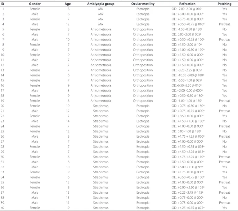
Table 1. Demographic and clinical features of amblyopic children
ID Gender Age Amblyopia group Ocular motility Refraction Patching
01 Female 08 Mix Esotropia OD: -2.00 -2.00 @ 010º Yes
02 Male 06 Mix Esotropia OD: +3.00 -0.00 @ 000º Yes
03 Female 07 Mix Esotropia OD: +3.75 -0.00 @ 000º Yes
04 Male 12 Mix Esotropia OD: +0.50 +0.75 @ 010º Pretreat
05 Female 08 Anisometropia Orthoposition OD: -1.50 -0.50 @ 180º No
06 Male 07 Anisometropia Orthoposition OD: 0.00 -2.00 @ 005º Yes
07 Male 10 Anisometropia Orthoposition OD: +0.50 +0.25 @ 100º Pretreat
08 Female 07 Anisometropia Orthoposition OD: +1.50 -2.00 @ 10º No
09 Male 07 Anisometropia Orthoposition OD: +1.00 +0.50 @ 170º No
10 Female 09 Anisometropia Orthoposition OD: +1.50 -0.00 @ 000º No
11 Male 07 Anisometropia Orthoposition OD: +1.50 -0.00 @ 000º No
12 Male 07 Anisometropia Orthoposition OD: +1.50 -0.00 @ 000º No
13 Female 07 Anisometropia Orthoposition OD: -0.25 -2.25 @ 005º No
14 Female 06 Anisometropia Orthoposition OD: -10.50 -3.00 @ 180º Yes
15 Female 07 Anisometropia Orthoposition OD: -6.50 -1.00 @ 035º Yes
16 Female 09 Anisometropia Orthoposition OD:-6.50 -5.50 @ 010º Yes
17 Male 08 Anisometropia Orthoposition OD:+2.00 -0.00 @ 000º Yes
18 Female 09 Anisometropia Orthoposition OD: +0.50 -0.50 @ 180º Yes
19 Female 08 Anisometropia Orthoposition OD: -1.00 -1.00 @ 180º Pretreat
20 Female 10 Strabismus Esotropia OD: +0.75 +0.50 @ 180º No
21 Male 12 Strabismus Esotropia OD: +0.75 +0.75 @ 090º Pretreat
22 Female 07 Strabismus Esotropia OD: +8.50 -0.00 @ 000º Yes
23 Male 14 Strabismus Esotropia OD: +1.50 +1.00 @ 180º No
24 Female 07 Strabismus Exotropia OD: +1.00 -0.00 @ 000º Pretreat
25 Female 12 Strabismus Esotropia OD: 0.00 -1.00 @ 180º No
26 Male 08 Strabismus Esotropia OD: +1.75 +1.25 @ 060º Pretreat
27 Male 07 Strabismus Esotropia OD: +1.00 -0.00 @ 000º No
28 Female 07 Strabismus Esotropia OD: +1.50 +0.75 @ 095º No
29 Male 07 Strabismus Esotropia OD: +4.50 +2.25 @ 015º No
30 Female 08 Strabismus Esotropia OD: +4.75 +2.25 @ 110º Pretreat
31 Male 08 Strabismus Esotropia OD: +1.50 -0.00 @ 000º Pretreat
32 Male 10 Strabismus Esotropia OD: +6.00 +1.00 @ 95º Yes
33 Female 09 Strabismus Exotropia OD: +1.75 -0.00 @ 000º Yes
34 Female 06 Strabismus Esotropia OD: +3.50 +0.75 @ 100º Yes
35 Female 13 Strabismus Esotropia OD: +1.00 -0.00 @ 000º Pretreat
36 Female 08 Strabismus Esotropia OD: +2.00 +2.50 @ 105º Yes
37 Male 13 Strabismus Esotropia OD: +2.25 -3.75 @ 175º Pretreat
38 Male 13 Strabismus Esotropia OD: +0.75 -0.00 @ 000º No
39 Male 11 Strabismus Esotropia OD: +0.75 -0.00 @ 000º Pretreat
40 Female 09 Strabismus Esotropia OD: +4.25 +0.75 @ 075º Yes
ID= identiication; OD= right eye; OS= left eye.
USA). The following statistical models were used: the Student’s t-test,
one-way analysis of variance (ANOVA), the paired t-test, the Pearson
correlation coeicient, and multiple linear regression analysis. When there was no normal distribution of variables, the nonparametric Mann-Whitney and Kruskal-Wallis ANOVA tests were used. A
proba-bility (p) value of ≤0.05 with a two-tailed rejection region, according
to the common general pattern in medical and biological areas, was considered statistically signiicant.
RESULTS
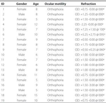
Table 2. Demographic and clinical features of the control group
ID Gender Age Ocular motility Refraction
01 Female 08 Orthophoria OD: +0.75 -0.00 @ 000º
02 Male 08 Orthophoria OD: +1.25 -0.00 @ 000º
03 Female 05 Orthophoria OD: +1.50 -0.00 @ 000º
04 Female 12 Orthophoria OD: -2.25 -0.00 @ 000º
05 Female 07 Orthophoria OD: +7.25 +1.50 @ 100º
06 Male 10 Orthophoria OD: +5.25 +2.75 @ 095º
07 Female 09 Orthophoria OD: +0.50 -0.00 @ 000º
08 Female 08 Orthophoria OD: +1.75 -0.00 @ 000º
09 Female 07 Orthophoria OD: +0.50 +0.25 @ 060º
10 Male 06 Orthophoria OD: +1.00 -0.00 @ 000º
11 Female 07 Orthophoria OD: +1.50 -0.00 @ 000º 12 Female 06 Orthophoria OD: +1.00 -0.00 @ 000º
13 Male 08 Orthophoria OD: +1.00 -0.00 @ 000º
14 Female 11 Orthophoria OD: +0.75 -0.00 @ 000º
15 Female 05 Orthophoria OD: +1.50 -0.00 @ 000º
16 Male 09 Orthophoria OD: +1.50 -3.00 @ 180º
17 Male 05 Orthophoria OD: +1.50 -0.00 @ 000º
18 Female 15 Orthophoria OD: +0.50 -0.00 @ 000º
19 Female 10 Orthophoria OD: +0.75 -0.00 @ 000º
ID= identiication; OD= right eye; OS= left eye.
ongoing in 15 (37.5%) children at the time of examination, whereas 14 (35.0%) children had never had previous patching therapy and 11 (27.5%) have had it previously, but it had been discontinued before the examinations.
The control group consisted of 19 healthy children with normal ophthalmic exam results, consisting of six males (31.6%) and 13 fe-males (68.4%) with a mean age of 8.2 ± 2.6 (range, 5-15) years. The demographic and clinical features of this group are shown in table 2.
V
ISUALACUITYOptotype acuity
Optotype acuity ranged from 0.00 (20/20) to 0.24 (20/34) logMAR in the fellow eye of amblyopic children and from 0.00 (20/20) to 0.00 (20/20) logMAR in those of the control group. Overall, optotype acuity
was signiicantly worse (p=0.021) in the fellow eyes of all amblyopic
patients than in the control group (0.04 ± 0.07 logMAR, median 0.0 logMAR vs. 0.0 ± 0.0 logMAR, median 0.0 logMAR, respectively). The
same trend was found in the strabismic group (p=0.040, 0.04 ± 0.08
logMAR, median 0.0 logMAR) and the anisometropic group (p=0.048,
0.04 ± 0.07 logMAR, median 0.0 logMAR). Optotype acuity was signi-icantly better in control eyes than in fellow eyes in the groups with and without occlusion therapy (p<0.05, both).
Grating acuity
Grating acuity ranged from -0.01 (20/19) to 0.21 (20/32) logMAR in the fellow eyes of amblyopic children and from 0.01 (20/20) to 0.20 (20/31) logMAR in those of the control group. Grating acuity was also
signiicantly worse (p=0.016) in fellow eyes of all amblyopic subjects
than in the control group (0.07 ± 0.05 logMAR, median 0.07 logMAR vs. 0.05 ± 0.04 logMAR, median 0.05 logMAR, respectively). Regarding occlusion, the control group had signiicantly better optotype acuity
than the fellow eyes in the group without occlusion (p<0.05).
Figure 2 shows the individual scores of optotype (A) and grating acuity (B) for the fellow eyes of amblyopic children (illed symbols)
and a randomly selected eye of the control group for optotype acuity A
B
Figure 2. Individual scores of optotype (A) and grating acuity (B) of the fellow eyes of amblyopic children and the better-vision eye of control children.
and the eye with better vision of the control children (open symbols) for grating acuity.
P
ATTERNREVERSALTRANSIENTVISUALLYEVOKEDPOTENTIALSP100 latency
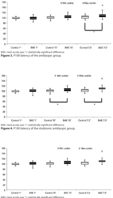
There was a statistically prolonged latency of visual stimuli of 15’ (p=0.018, 106.81 ± 7.99 ms, median 106.00 ms) and 7.5’ (p=0.002, 112.93 ± 11.38 ms, median 110.00 ms) of the fellow eyes in the strabismic group, as compared to the control group (101.42 ± 5.46 ms, median 102.50 ms for visual stimulus of 15’, and 103.21 ± 6.82 ms, median 104.00 ms for visual stimulus of 7.5’). For the smaller visual stimulus, a statistically
prolonged latency was also found in all amblyopic children (p<0.001,
110.89 ± 11.42 ms, median 109.50 ms) (Figures 3, 4, and 5).
Regarding occlusion, there was no statistically signiicant dife-rence between control group and fellow eyes at 60’ and 15’ compared to those with occlusion, without occlusion, and previously treated. The fellow eyes showed prolonged latencies for stimuli at 7.5’ in the
group previously treated (p<0.05), as compared to controls.
Comparing the N75-P100 amplitude of the fellow eyes with that of the control group, there were no diferences between any stimuli and groups, including occlusion therapy.
DISCUSSION
Deicits in visual function of fellow eyes have been studied for
more than three decades. Kandel et al.(13) unjustiiably classiied the
sound eye of amblyopic patients as normal based on the inding that these eyes have reduced contrast sensitivity, low visual acuity, and horizontal eccentric ixation when compared to normal eyes with normal binocular ixation.
A retrospective study evaluating visual acuity and the maturation of the fellow eye was conducted by reviewing the medical records of 112 children with unilateral amblyopia secondary to anisometropia,
strabismus, or both conditions with previous patching therapy(11).
Cor-roborating the indings of this past study, the current study found sta-tistically worse optotype acuity of the fellow eyes than of eyes of the control group , especially among patients with strabismic amblyopia.
Diferent results were observed when analyzing grating acuity, where the strabismic and anisometropic amblyopia groups showed similar results to the control group. However, when we analyzed the results of all amblyopes, signiicantly worse values were observed
than those of the control group. The small patient cohort was a limi-tation to the present study, thus a larger sample of cases may provide diferent results.
Grating acuity may be overestimated by sweep VEP, especially with
lower visual acuity(12). By comparing the values of the contralateral
eye of all amblyopes (N=40), we found statistically worse values than those of control group. This fact conirms the hypothesis that the fellow amblyopic eye is not completely normal. As indicated by the results of latency and amplitude of PR-VEPs of the fellow eyes in this study, only patients with strabismic amblyopia showed increased P100 latency for the smaller visual stimuli, as compared to the control group. Although few studies have evaluated the parameters of PR-VEPs
in the fellow eyes of patients with amblyopia, Mendonça et al.(16)
reported a case of mixed amblyopia in an 11-year-old child with delayed P100 latency in the amblyopic and fellow eyes to stimuli at 60’, 30’, and 15’. This inding was attributed to the loss of contrast sensitivity at high spatial frequencies, which can be intensiied in anisometropic amblyopia.
However, patching therapy for amblyopia, particularly occluding the fellow eye, can have a negative efect, as shown in animal expe-riments, in which monocular deprivation has the greatest efect on the primary visual cortex rather than the retina and geniculate lateral
body(13). It is worthwhile to note that about one-third of the children
in the current study were receiving patching therapy and another third had discontinued patching therapy. Occlusion, somehow, may have contributed to the results of the sound eye, especially in the parvocellular pathway.
When compared to healthy children, the fellow eyes of amblyopic children showed worse optotype and grating acuity, with subtle abnormalities in the PR-VEP detected as prolonged latencies for smaller size stimuli, especially in children with a history of therapy at the time of the exam. These indings conirm those of previous studies showing that the fellow eyes of amblyopic patients were not fully normal and patching therapy can cause physiological defects in sound eyes.
REFERENCES
1. Allen B, Spiegel DP, Thompson B, Pestilli F, Rokers B. Altered white matter in early visual pathways of humans with amblyopia. Vision Res. 2015;114:48-55.
2. Kiorpes L, Kiper DC, O’Keefe LP, Cavanaugh JR, Movshon JA. Neuronal correlates of amblyopia in the visual cortex of macaque monkeys with experimental strabismus and anisometropia. J Neurosci. 1998;18(16):6411-24.
3. Kiorpes L, McKee SP. Neural mechanisms underlying amblyopia. Curr Opin Neurobiol. 1999;9(4):480-6.
4. Birch EE. Amblyopia and binocular vision. Progr Retin Eye Res. 2013;33:67-84. 5. Daw NW. Visual development. New York: Spring Science; 2006.
6. Levi DM, Knill DC, Bavelier D. Stereopsis and amblyopia: a mini-review. Vision Res. 2015;114:17-30.
7. Mendonça RH, Abbruzzese S, Bagolini B, Nofroni I, Ferreira EL, Odom JV. Visual evoked potential importance in the complex mechanism of amblyopia. Int Ophthalmol. 2013; 33(5):515-9.
8. Oner A, Coskun M, Evereklioglu C, Dogan H. Pattern VEP is a useful technique in mo-nitoring the efectiveness of occlusion therapy in amblyopic eyes under occlusion therapy. Doc Ophthalmol. 2004;109(3):223-7.
9. Hess RF, Thompson B. Amblyopia and the binocular approach toits therapy. Vision Res. 2015;114:4-16.
10. Sokol S. Abnormal evoked potential latencies in amblyopia. Br J Ophthalmol. 1983;67(5): 310-4.
11. Brémond-Gignac D, Copin H, Lapillonne A, Milazzo S, European Network of Study and Research in Eye Development. Visual development in infants: physiological and pathological mechanisms. Curr Opin Ophthalmol. 2011;22(1):S1-S8.
12. Ridder III WH, Rouse MW. Predicting potencials acuities in amblyopes. Doc Ophthalmol. 2007;114(4):135-45.
13. Kandel GL, Grattan PE, Bedell HE. Are the dominant eyes of amblyopes normal? Am J Optom Physiol Opt. 1980;57(1):1-6.
14. Varadharajan S, Hussaindeen JR. Visual acuity deicits in the fellow eyes of children with unilateral amblyopia. J AAPOS. 2012;16(1):41-5.
15. Leguire LE, Rogers GL, Bremer DL. Amblyopia: The normal eye is not normal. J Pediatr Ophthalmol Strabismus. 1990;27(1): 32-8.
16. Mendonça RH, Ferreira EL. Visual evoked potentials (VEP) and visual acuity improve-ment after cytidine 52-diphosphocholine (CDP-Choline) therapy in amblyopic pa tient. Rev Bras Oftalmol. 2012;71(5):328-30.
BAE= best acuity eye; *= statistically signiicant diference. Figure 3. P100 latency of the amblyopic group.
BAE= best acuity eye; *= statistically signiicant diference. Figure 4. P100 latency of the strabismic amblyopic group.
BAE= best acuity eye; *= statistically signiicant diference.