1
Original Article
Effects of Atorvastatin, Fluvastatin, Pravastatin,
and Simvastatin on Endothelial Function, Lipid
Peroxidation, and Aortic Atherosclerosis in
Hypercholesterolemic Rabbits
Paulo Afonso Ribeiro Jorge, Eros Antonio de Almeida, Michiko R. Ozaki, Mariana Jorge,
Adriano Carneiro
Campinas, SP - Brazil
Faculdade de Ciências Médicas da Unicamp, Campinas, SP Mailing address: Paulo Afonso Ribeiro Jorge - Rua Guilherme da Silva, 397 - Cep 13025-070 - Campinas, SP, Brazil
E-mail: [email protected] Sent for publication: 04/04/2004 Accepted for publication: 10/29/2004 English version by Stela Maris Costalonga
Objective
To compare the effects of atorvastatin, fluvastatin, pravasta-tin, and simvastatin on endothelial function, aortic atheroscle-rosis, and the content of malondialdehyde (MDA) in native and oxidized LDL and in the arterial wall of hypercholesterolemic rabbits after adjusting the dosages of those statins to reduce total serum cholesterol levels to similar values.
Methods
Male rabbits were divided into the following 6 groups of 10 animals (n=10): 1) GH (control) - hypercholesterolemic animals; 2) GA - atorvastatin; 3) GF - fluvastatin; 4) GP - pravastatin; 5) GS - simvastatin; and 6) GN - normal. The animals were fed a standard food preparation enriched with 0.5% cholesterol and 2% coconut oil for 45 days. Fifteen days after beginning the experiment, atorvastatin, fluvastatin, pravastatin and simvastatin were administered for 15 days through gavage and the dosages were adjusted to obtain similar cholesterol values in each group. At the end of the experiment, a blood sample was withdrawn for determining total cholesterol and separating the lipoproteins, and a segment of the thoracic aorta was removed to be used for studying endothelial function and lipid peroxidation, and for measuring aortic atherosclerosis in histological sections.
Results
The statins significantly reduced total serum cholesterol levels, LDL-cholesterol levels, and aortic atherosclerosis. The MDA con-tent was also significantly reduced in native and oxidized LDL, as well as in the arterial wall. Endothelium-dependent relaxation was significantly greater in the treated group compared with that in the hypercholesterolemic group.
Conclusion
The statins, at dosages adjusted, had a significant and simi-lar effect in reducing lipid peroxidation in native and oxidized LDL-C and in arterial walls, in decreasing aortic atherosclerosis, and in reverting endothelial dysfunction.
Key words
HMG-CoA reductase inhibitors, aortic atherosclerosis, lipid peroxidation
The HMG-CoA reductase inhibitors effectively reduce mortality and coronary events 1-6, representing a powerful instrument in
preventing and controlling atherosclerosis. One of the major actions of these drugs is reducing serum LDL-cholesterol, interfering with the biosynthesis of cholesterol and increasing the number of hepatic receptors for ApoB100 7.
More recently, additional effects of these drugs have been demonstrated and considered important for stabilizing the athe-rosclerotic plaque. Such effects, called pleiotropic, refer to endo-thelial protection, lipid peroxidation reduction, and control of in-flammatory reactions and hemostasia 8-11.
Some adverse effects of the statins have been reported and include cephalea, myalgia, pharyngitis, and interaction with other drugs metabolized in cytochrome P-450 12-14.
Although statins have a similar mechanism of action, they differ in their potency for reducing serum cholesterol, their solubility and metabolism 15,16.
Previous studies 17,18have assessed the effect of simvastatin
and pravastatin on endothelial function, tissue cholesterol, and the number of foam cells in aortic rings of hypercholesterolemic rabbits. Those studies showed that pravastatin was more effective in reversing endothelial dysfunction caused by hypercholesterole-mia, in addition to more intensely reducing the number of foam cells in the histological sections obtained from the thoracic aorta. Those findings were carefully interpreted because they derived from an experimental study on animals, and the differences between the effects of those statins encompassed the controversial discussion about the pharmacological difference of the HMG-CoA reductase inhibitors.
The present study aimed at comparing the effects of 4 statins (atorvastatin, fluvastatin, pravastatin, and simvastatin) on endo-thelial function, lipid peroxidation, and aortic atherosclerosis in hypercholesterolemic rabbits. The dosages of statins were adjusted to reduce total serum cholesterol levels to similar values.
Methods
2
Sixty male rabbits of the New Zealand breed, aged approxi-mately 12 weeks and weighing from 1.6 to 2.4 kg, were divided into the following 6 groups (n=10): 1) normal group (GN); 2) hypercholesterolemic (GH); 3) atorvastatin (GA); 4) fluvastatin (GF); 5) pravastatin (GP); and 6) simvastatin (GS). The animals were separated in individual cages and fed a standard preparation of Purina brand food, enriched with 0.5% cholesterol and 2% coconut oil for 45 days. They received 100 g of food preparation per day and unlimited water. After 15 days, a blood sample was obtained through cardiac puncture for determining serum choles-terol. Then atorvastatin (10 mg/day), fluvastatin (20 mg/day), pravastatin (20 mg/day), and simvastatin (15 mg/day) were admi-nistered to the respective groups. The dosages were based on previous studies 17,18 and the drugs administered once a day through
gavage. After 15 days, another blood sample was obtained for determining serum cholesterol levels, and the dosages of the statins were adjusted to obtain similar values of serum cholesterol in each group. The final dosages were 10 mg/day, 15 mg/day, 15 mg/day, and 10 mg/day for atorvastatin, fluvastatin, pravastatin, and simvastatin, respectively.
At the end of the experiment, another blood sample was ob-tained for determining serum cholesterol levels and for separating the lipoproteins. The animals were euthanized by having their necks severed. Median thoracotomy was then performed, and the aorta was removed to obtain the rings for assessing endothelial function and MDA content, and for measuring aortic atherosclerosis on histological sections. Liver and muscle fragments were also obtained for histological examination.
The thoracic aorta, free from connective tissue, was sectioned into rings of approximately 5 mm, with special care not to damage the endothelium. In some rings, however, the endothelium was mechanically damaged by a small forceps. The aortic rings were suspended in 10 mL of the Krebs-Henseleit solution at pH 7.4 (composition in mmol/L: NaCl, 113; CaCl2, 2.19; NaHCO3, 25.0; MgSO4, 0.44; KH2PO4,1.18; EDTA, 0.03; glucose, 11.0). The solution was maintained at 37°C and continuously aerated with a gas mixture containing 95% O2 and 5% CO2. The rings were mounted on 2 metallic supports coupled to a force transducer (Narco Byosystem) and distended at a baseline tension of 1g. The aortic rings were allowed to stabilize for 60 minutes, with Krebs Henseleit solution exchange every 20 minutes. To prevent the synthesis of prostaglandins, the experiments were performed in the presence of 10 µM of indomethacin.
The rings of the thoracic aorta with and without endothelium were contracted with noradrenaline (NA, 10-7 M). When the
con-traction was kept stable, acetylcholine (ACH) was added to the solution in a cumulative form (10-8-10-5.5M) to obtain the
concentration-effect curves. Then, the solution was replaced for the Krebs-Henseleit solution, and the tension was stabilized at baseline values with frequent renewal of the solution. After 30 minutes, the aortic rings were contracted with NA (10-7 M), and
other concentration-effect curves were obtained with sodium ni-troprusside (SNP, 10- 8 - 10- 5.5 M).
The 5-mm-thick aortic rings were obtained in a standardized form from defined areas of the superior part of the thoracic aorta. Fragments from the liver and thigh muscle were also obtained. The tissues were fixed in 10% formalin for 24 hours, processed,
and embedded in paraffin. Serial sections were mounted on slides and stained with hematoxylin-eosin.
Total serum cholesterol level was measured with commercial enzymatic kits in a spectrophotometer (Genesys 10, Spectronics) and the results were expressed in mg/dL. The GOT and GPT levels were also measured with the commercial enzymatic kit, and the results were expressed in U/L.
Lipid peroxidation was assessed by use of MDA measurement, one of the end-products of peroxidation. The aortic samples were homogenized in cold trichloroacetic acid (TCA) (1 mg of tissue per mL of 10% TCA). After centrifugation, a portion of the super-natant was added to an equal volume of thiobarbituric acid (0.6% v/v), and the mixture was heated at 100°C for 20 minutes. The MDA concentration was calculated by use of a spectrophotometer, with absorption of 532 nm and a molar extinction coefficient of 1.49x10⁻⁵, and the results were expressed in nmol/mg of dry tissue
x 10-7 19. The lipoproteins were separated by plasma sequential
ultracentrifugation according to the method by Havel et al 20. The
plasma was centrifuged at 40000 rpm for 18 hours at 4°C in a Ti50 rotor (Beckman), and the VLDL layer (d < 1,006 g/mL) was removed. The supernatant was adjusted to a density of 1063 g/ mL by using potassium bromide, being then centrifuged at 40,000 rpm for 20 hours. The isolated LDL-C was dialyzed in 0.01 M of phosphate-buffered saline (PBS), pH 7.4 at 8°C for 24 hours, with frequent buffer washing. The LDL protein concentration was determined by use of the method of Lowry et al 21 with bovine
albumin as the standard. The LDL oxidation was performed by use of incubation with 5 µM copper sulfate (100 µg of protein/ mL, 1 M PBS) for 24 hours at 37°C 22. The concentration of
peroxide lipids of the native and oxidized LDL was measured by the reaction of the thiobarbituric acid (TBARS), as reported by Buege and Aust 23. The TBARS values were expressed as MDA
equivalents (nmol/protein LDL) using the standard solution of 1,1,3,3 tetramethoxypropane.
Images of the histological sections of the aortic rings were obtained with a Leica DMLD microscope and imported to a com-puter. By use of the Scion program 24, areas of the arterial wall
and of the extracellular liposomes were manually selected. The number of pixels was determined and used as a measurement. The results were expressed as % of extracellular liposomes in relation to the arterial wall 25.
The acetylcholine chloride, noradrenaline bitartrate, sodium nitroprusside, trichloroacetic acid, thiobarbituric acid, and indo-methacin were obtained from Sigma Chemical Co (St. Louis, MO, USA). The reagents of the Krebs-Henseleit solution were obtained from Merck Chemicals.
The descriptive analyses were performed by use of the position and dispersion measurements for the continuous variables. For comparing the groups of treatment through the variables collected, the simple analysis of variance (one-way ANOVA) was used, with transformation of the variables according to ranks. For identifying the differences, the Tukey test, a multiple comparisons procedure, was used.
3
Results
At the beginning of the study, the mean weights of the rabbits in the different groups were as follows: 1) GN: 1.80±0.2 kg; 2) GH: 2.0±0.18 kg; 3) GA: 1.75± 0.12 kg; 4) GF: 1.85±0.14 kg; 5) GP: 2.09±0.23 kg; and 6) GS: 1.79±0.10 kg. All animals gained weight during the study, but no significant difference was observed between the groups.
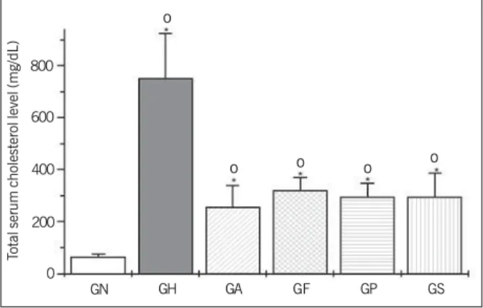
Figure 1 shows the occurrence of marked elevation in total serum cholesterol levels in the hypercholesterolemic group (GH) when compared with the total serum cholesterol levels in the normal group (GN). The treatment with statins significantly redu-ced serum cholesterol levels. No difference was observed between the groups, and the same occurred in regard to LDL-cholesterol levels (fig. 2).
The content of malondialdehyde (MDA) (nmol/mg x 10-7M) in
the oxidized LDL particles and aortic wall is shown in figures 3 and 4. The MDA content of the aortic wall and oxidized LDL particles was significantly reduced in the treated group when com-pared with that in the hypercholesterolemic group (P < 0.05). No significant difference was observed between the treated groups. A similar response was obtained for the MDA content of native LDL particles.
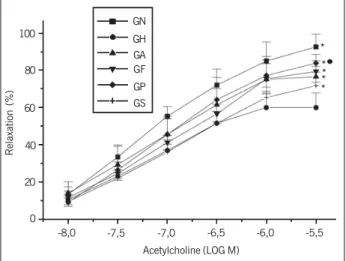
Figure 5 expresses the percentage of relaxation of the aortic rings of hypercholesterolemic rabbits in response to acetylcholine.
It was significantly smaller than that of the animals in the normal group. All statins reversed the relaxation until close to normal in varied degrees, pravastatin being the most effective. Maximum relaxation with nitroprusside was 100% in all groups. No relaxation was observed with ACH in the aortic segments whose endothelium had been mechanically removed. The intensity of contraction with NE was similar between the groups.
The histological examination showed the presence of extra-cellular liposomes in the intimal layer of the aorta of hypercholes-terolemic rabbits and no inflammatory cells. The percentages of extracellular liposomes in the vessel wall in the hypercholestero-lemic, atorvastatin, fluvastatin, simvastatin, and pravastatin groups were as follows, respectively: 21.0±0.15, 2.58±1.35, 3.83± 0.89, 2.95±0.91, and 3.84±1.08. When comparing the hyper-cholesterolemic group with the treated groups, statins significantly (P<0.05) reduced aortic atherosclerosis, and no difference was observed between the treated groups.
Figure 6, in its left lower portion, illustrates the evolution of atherosclerosis in animals treated with a 2% diet. A greater con-centration of extracellular liposomes is observed in the intimal layer. The histological study of liver fragments revealed moderate steatosis in all groups studied, while the muscle tissue appeared normal on microscopic examination.
The GOT and GPT levels were within the normal range in all treated groups.
Total serum cholesterol level (mg/dL)
800
600
400
200
0
GN GH GA GF GP GS
o
o o o o
Fig. 1 - Total serum cholesterol levels. Values expressed as mean and SD. The groups were as follows: GN = normal, GH = hypercholesterolemic, GA = atorvastatin, GF = fluvastatin, GP = pravastatin, GS = simvastatin. * P < 0.05 for GH; P < 0.05 for GN.
LDL-cholesterol (mg/dL)
700
600
500
400
300
200
100
0
o o o o
Fig. 2 - Serum LDL-cholesterol levels (mg/dL). Values expressed as mean and SD. The groups were as follows: GN = normal, GH = hypercholesterolemic, GA = atorvastatin, GF = fluvastatin, GP = pravastatin, GS = simvastatin. * P < 0.05 for GH; P < 0.05 for GN.
o
MDA-Aorta (nmol/mg tissue x 10
-7) 10
9
8
7 6
5 4 3
o o o
Fig. 3 - Concentration of malondialdehyde (MDA) in the aortic wall (x 10-7 nmol/
mg/tissue). Values expressed as mean and SD. The groups were as follows: GN = normal, GH = hypercholesterolemic, GA = atorvastatin, GF = fluvastatin, GP = pravastatin, GS = simvastatin. * P < 0.05 for GH; P < 0.05 for GN.
2
1 0
GN GH GA GF GP GS
o
MDA OXI-LDL (nmol/mg proteín)
65 60 55 50 45 40 35 30
Fig. 4 - Malondialdehyde in the oxidized LDL particles (nmol/mg protein). Values expressed as mean and SD. The groups were as follows: GN = normal, GH = hypercholesterolemic, GA = atorvastatin, GF = fluvastatin, GP = pravastatin, GS = simvastatin. * P < 0.05 for GH.
25 20 15 10 5 0
4
Discussion
Statins vary considerably in their pharmacological and lipophilic characteristics, their mean life, metabolism, and other proper-ties28. Pravastatin is hydrophilic, while the other statins are
lipo-philic. Atorvastatin, simvastatin, and fluvastatin circulate in the blood bound to protein in 95 to 98%, while pravastatin circulates bound to protein in 50% 29. Except for pravastatin, the statins are
metabolized in cytochrome P-450-3A4 (fluvastatin is metabolized in cytochrome P-450-2C9). Atorvastatin, fluvastatin, and simvas-tatin, but not pravassimvas-tatin, decrease the migration and proliferation of smooth muscle cells in the arterial wall 30, probably by blocking
RhoA- and Rac1-, which are cell signalers 31. Evident alterations
in inflammatory response have been reported with the use of statins, including the inhibition of cytokines, C-reactive protein, expression of the metalloproteinases of the matrix, and decrease in the adhesion of monocytes to the endothelial cell 32. Recent
studies about the pleiotropic properties of statins have revealed their capacity to inhibit the synthesis of important intermediates
of the isoprenoids, which serve as bindings to a variety of proteins implicated in intracellular signaling 33,34. On the other hand,
well-conducted studies have revealed a difference in the potential of statins to reduce cholesterol and serum LDL particles 15,16;
diffe-rences in their pleiotropic effects and supposed tissular selectivity have been discussed.
The major objective of this study was to assess the pleiotropic effects of different statins, considering the controversial results reported in the literature. Pentikainem et al 35 reported that
pra-vastatin is an HMG-CoA reductase inhibitor with greater affinity to peripheral tissues, which was confirmed by Germershausen et al36. The latter authors attributed that finding to the active species
derived from pravastatin, which bind to proteins in approximately 50%, while the other statins are intensely bound to proteins (>95%); thus, drugs that bind to proteins in smaller amounts have greater penetration in tissues. The hydrophilic HMG-CoA reductase inhibitors, on the other hand, may reach a greater concentration in endothelial cells, therefore increasing the NO synthase activity. However, the tissular selectivity of HMG-CoA reductase inhibitors is controversial 14,37. Thiery et al 38, investigating
the extension of atherogenesis in hypercholesterolemic rabbits treated with lovastatin, simvastatin, and pravastatin (15 mg/day), reported a smaller concentration of tissular cholesterol in animals treated with lovastatin and simvastatin than that in animals treated with pravastatin. De Vries et al 39 stated that the synthesis of
cholesterol in the lens is 100 times more effectively inhibited by pravastatin than by simvastatin. Tesfamarian et al 40 reported
dif-ferent effects of pravastatin, simvastatin, and atorvastatin in the release of cytoplasmic Ca2+and in vascular reactivity, while
Jouk-hadar et al 41 reported a similar effect for atorvastatin, simvastatin,
and pravastatin in some parameters relating to hemostasia and inflammation in the plasma of hypercholesterolemic patients.
However, the supposed differences between statins apparently do not have a major clinical significance, because the great rando-mized studies have shown a significant reduction in mortality and coronary events with the use of the different statins studied 1,3,4,6.
Our results showed that all statins were effective in reducing lipid peroxidation in native or oxidized LDL and also in the arterial wall. No significant difference was observed in the intensity of the effect between the different drugs. The antioxidizing effect of sim-vastatin42, fluvastatin 43, pravastatin 44, and atorvastatin 45 has
been reported in the literature and represents an important step in preserving endothelial function 34.
In the group of hypercholesterolemic animals, all statins were effective in partially reversing endothelial dysfunction. The effect was similar in the different drugs, except for pravastatin, whose effect on endothelium-dependent relaxation was more significant. The mild difference in the intensity of endothelium-dependent relaxation observed with pravastatin was interpreted as due to its hydrophilic characteristic, but this finding should be reassessed in further studies.
The increase in endothelium-dependent relaxation observed in the aortic rings of rabbits treated with statins should be related to the reduction in tissular cholesterol and oxidative stress in the arterial wall 46, in addition to the action on the mediators of
cellular transcription, which was not studied in this experimental model. In a previous study 18, we observed that the reversion of
endothelial dysfunction in hypercholesterolemic rabbits treated
Relaxation (%)
100
80
60
40
20
0
GN
GH GA GF
GP GS
-8,0 -7,5 -7,0 -6,5 -6,0 -5,5
Acetylcholine (LOG M)
Fig. 5 - Endothelium-dependent relaxation of the aortic rings precontracted with norepinephrine (NE) in response to acetylcholine (ACH). Relaxation expressed as percentage of NE-induced contraction (10-7). The points represent the mean and
SD of 10 rabbits per group. * P < 0.05 comparing the hypercholesterolemic and normal groups. P < 0.05 between the groups.
Fig. 6 - Histological section of the aorta of hypercholesterolemic animals and those treated with atorvastatin, fluvastatin, pravastatin, and simvastatin. Note the significant regression of extracellular liposomes in the animals treated.
0.5% cholesterol/45 days
HYPERCHOLESTEROLEMIA
2% cholesterol/45 days ATORVASTATIN 10mg/days SIMVASTATIN 10mg/days
5
with pravastatin and simvastatin occurred rapidly, which is bene-ficial to the treatment of acute coronary syndrome.
Another point that deserves to be stressed in the present study due to clinical implications is the regression of intimal thickening, represented by the build up of extracellular liposomes. The LDL particles enter the endothelial cell due to the mechanism of pino-cytosis, forming vesicles of pinopino-cytosis, which are incorporated as endosomes and liposomes in the cell cytoplasm. In hypercholeste-rolemia, the lipid material carried by the endosomes accumulates in extracellular liposomes in the intimal layer of the artery. Consi-dering that the presence of extracellular liposomes are the earliest morphological expression of atherosclerosis, their regression may represent a potential mechanism of control of the atherosclerotic disease. The regression of the atherosclerotic plaque caused by statins has been demonstrated in several experimental studies 47-49.
Studies with magnetic resonance imaging and spectroscopy have reported mobilization of the lipid content in the atherosclerotic plaques of animals treated with different drugs 50-52.
More recently, the REVERSE study 53 has confirmed the
inter-ruption of progression, and even a small regression, of the atheros-clerotic plaque (0.4% of median reduction) evidenced on coronary ultrasound in patients with stable coronary disease. Those results open an exceptional perspective in the management of atherosclerosis. The present study showed no significant difference in atheros-clerosis regression between the different statins studied. However, it is worth emphasizing that the dosages of statins were adjusted for obtaining similar values of serum cholesterol in the different groups considering the different potencies of the drugs. Atorvastatin was the most potent, because a lower dose of atorvastatin was required for a similar reduction in serum cholesterol in the groups; an eventual difference in the effect of regression could not be assessed considering the intensity of the reduction in serum cho-lesterol and LDL particles. More recently, a more intense reduction was observed in serum LDL in patients with acute coronary syn-dromes leading to a greater benefit regarding a reduction in mor-tality rate and other coronary events 54. This initiated the discussion
about the best LDL serum level for controlling atherosclerosis, stressing the significance of the lipid-lowering potency of statins. The most intense reduction in serum lipids is significant when
one considers that the major causal factor in atherosclerosis is the afflux of LDL particles and their epitopes to the intimal region of the vessel through the endothelial cell. This causes an immune response mediated by several factors related to inflammation, with mobilization of monocytes, neutrophils, and lymphocytes. This restricts the lipid core through a predominantly proliferative reaction or makes the plaque unstable due to a greater cellular and enzy-matic response 55. Probably the magnitude of the reduction in
cholesterol and LDL serum levels, in addition to the other factors already mentioned, has a beneficial effect on reversing endothelial dysfunction and mobilizing the lipid core of the atherosclerotic plaque, mainly in the earliest phase. The safe limit to which serum LDL can be lowered has yet to be established.
The authors of the REVERSE 53 and PROVE-IT 54 study have
also emphasized the existence of a pharmacological difference between the statins studied, atorvastatin and pravastatin. A well-conducted experimental study 55 using fluvastatin has also
consi-dered the existence of an indirect effect of the drug, independently of the reduction in serum cholesterol. This reinitiates the discussion about a possible pharmacological difference and tissular specificity between these drugs, independently of the reduction in lipids.
In this study, the statins did not alter the enzymatic activity of the liver. The histological examinations showed moderate hepatic steatosis and a normal appearance of the muscle tissue, probably consequent to the elevated serum concentration of cholesterol. Areas neither of necrosis nor of inflammation were observed. These findings suggest that the drugs had no toxic effect at the dosages administered.
The lack of inflammatory reaction in the intimal layer of the vascular wall, despite the presence of extracellular liposomes in hypercholesterolemic rabbits, reinforces the idea that the inflam-matory response occurs later to repair the inadequate presence of LDL and its epitopes in the intimal layer of the artery 56. This,
once more, reinforces the impression that a more intense reduction in tissue LDL will have a more favorable effect on disease control. The results of this study showed that all statins were effective in reducing serum total cholesterol, the MDA content in the arte-rial wall and in native and oxidized LDL, and in reversing the endothelial dysfunction in hypercholesterolemic rabbits.
References
1. Shepherd J, Cobbe SM et al. Prevention of coronary heart disease with pravastatin in men with hypercholesterolemia. West Scotland Coronary Prevention Study (WOSCOPS). N Engl J Med 1995; 333:1301-7.
2. AFCAPS/TEXCAPS, Domws JR, Clearfield M et al. Primary prevention of acute co-ronary events with lovastatin in men and women with average cholesterol levels. N Engl J Med 1998;339:1349-57.
3. LIPID (Long -Term Intervention with Pravastatin in Ischemic Disease - LIPID Study Group. N Engl J Med 1998;339:1349-57.
4. 4S (Scadinavian Simvastatin Survival Study Group). Randomized trial of cholesterol lowering in 4,444 patients with coronary heart disease. Lancet 1994;344: 1383-9. 5. Sacks FM, Pfeffer MA et al. The effect of pravastatin on coronary events after myocardial infaction in patients with average cholesterol levels. Care Study N Engl J Med 1966;14:1001-9.
6. The Heart Protection Collaborative Group. MRC/BHF Heart Protection Study of antioxidant vitamin supplementation in 20,536 high risk individuals: a randomized placebo-controlled trial. Lancet 2002; 360:23-33.
7. Vaughan CJ, Gotto AM, Basson CT. The envolving role of statins in the manage-ment of atherosclerosis. J Am Coll Cardiol 2000;35:1-10.
8. Ricker PM, Rifail N, Lowenthal SP. Rapid reduction in C-reactive protein with
ce-rivastatin among 785 patients with primary hyperchoelsterolemia. Circulation 2001; 103:1191-3.
9. Bellosta S, Bernini F, Ferri N, Quarato P, Canavesi M, Arnaboldi L. Direct vascular effects of HMGCoA reductase inhibitors. Atherosclerosis 137:S101-S109,1998. 10. Scalia R, Appel JZ, Lefer AM. Leukocyte-endothelium interaction during the early stages of hypercholesterolemia in the rabbit. Arterioscl Thromb Vasc Biol 1998;18:1093-100.
11. Dangas GJ, Badimon L, Smith DA, Levine A, Ambrose JA. Pravastatin therapy in hyperlipidemia. Effects on thrombus formation and the systemic hemostatic pro-file. Am Coll Cardiol 1999; 33:1294-304.
12. Lefer AM, Scalia R, Lefer DJ. Vascular effects of HMG-COA reductase inhibitors unrelated to cholesterol lowering: new concepts for cardiovascular disease. Car-diovasc Res 2001;49:281-7.
13. Williams D, Feely J. Pharmacokinetic-pharmacodynamic drug interactions with HMG-CoA reductase inhibitors. Clin Pharmacokinet 2002;41:343-370. 14. Corsini A, Maggi F, Catapano AL. Pharmacology of competitive inhibitors of
HMGCoA reductase. Pharmacological Res 1995; 31:9-27.
6
16. Jones P, Kafonek S, Laurora I, Hunninghake, Curves investigators. Comparative dose efficacy study of atorvastatin versus simvastatin, pravastatin, lovastatin and fluvastatin in patients with hypercholesterolemia.(The Curves Study). Am J Cardiol 1998; 81:582-7.
17. Ribeiro JPA, Ozaki MK. Effects of simvastatin and pravastatin on endothelium-depen-dent relaxation in hypercholesterolemic rabbits. Exp Toxic Pathol 1994; 46: 465-9. 18. Ribeiro JPA, Eros AA, Ozaki MR et al. Rapid reversal of endothelial dysfunction in
hypercholeterolemic rabbits treated with sinvastatin and pravastatin. Clin Exp Pharmacol Physiol 1997; 24:948-95.
19. Cassini AF, Ferrali M, Pompella A, Maellaro E, Comporti M. Lipid peroxidation and cellular damage in extrahepatic tissue of bromobenzene-intoxicated mice. Am J Pathol 1986; 123:520-531.
20. Havel RJ, Eder HA, Bragdon JH. The distribution and chemical composition of ultra-centrifugally separated lipoproteins in human serum. J Clin Invest 1955;34: 1345-53. 21. Lowry OH, Rosebrough NJ, Farr AL, Randall RL. Protein measurement with the
Folin phenol reagent. J Biol Chem 1951:193:265-75.
22. Parthasarathy S, Khoo JC, Miller E, Barnett J, Witztum J, Steinberg DE. Low den-sity lipoprotein rich in oleic acid is preotected against oxidative modifications: im-plications for dietary prevention of atherosclerosis. Porc Natl. Acad Sci USA 1990:87:3894-8.
23. Buege, JA, Aust, SD. Microsomal lipid peroxidation. Methods Enzymol 1978;52: 302-310.
24. Lehr HA, Mankoff DA, Corwin D. Application of Photoshop-based analysis to quantification of hormone receptor expression in breast cancer. J Histochem Cy-tochem 1997; 45: 1559-65.
25. Lehr HA, Van der Loos CM, Teeling P. Complete chromogen separation and analysis in double immunohistochemical stains using Photoshop-based image analysis. J Histochem Cytochem 1999; 47: 119-126.
26. Fleiss JL, Statistical Methods for Rates and Proportions. New York: John Wiley, 2nd
ed (1981).
27. Milliken GA & Johnson DE, Analysis of Messy Data. Vol I: Designed Experiments. New York: Van Nostrand Reinhold (1984).
28. Motti C, Gnasso A, Cortese C. Statins: similarities and differences in the pharma-cological, clinical and laboratory aspects. Ann Ital Med Int 2000, 15: 96-102. 29. Corsini A, Maggi FM, Catapano AL. Pharmacology of competitive inhibitors of
HMG-CoA reductase. Pharmacol Res 1995; 31: 9-27.
30. Negre-Aminou P, van Erck M, van Leeuwen RE, Collard JG, Cohen LH. Differencial effect of simvastatin on various signal transduction intermediates in cultured hu-man smooth muscle cells. Biochm Pharmacol 2002; 61: 991-8.
31. Davignon J, Mabile L. Mechanisms of action of statins and their pleiotropic ef-fects. Ann Endocrinol(Paris) 2001; 62: 101-2.
32. Koh KK. Effects of statins on vascular wall: vasomotor function, inflammation and plaque stability. Cardiovasc Res 2000; 47: 648-57.
33. Takemoto M, Lia JK. Pleiotropic effects of 3-hydroxy-3-methylglutaryl coenzyme a reductase inhibitors. Arterioscler Thrombo Vasc Biol 2001;21: 1712-9.
34. Hughes AD. The role of isoprenoids in vascular smooth muscle: potential benefits of statins unrelated to cholesterol lowering. J Hum Hypertens 1996;10:387-90. 35. Carneado J, Alvarez de Sotomayor M, Perez-Guerrero C et al. Simvastatin improves
endothelial function in spontaneiusly hypertensive rats through a superoxide dis-mutase mediated antioxidant effect. J Hypertens 2002, 20: 429-37.
36. Suzumura K, Tanaka K, Yasuhara M, Narita H. Inhibitory effects of fluvastastin and its metabolites on hydrogen peroxide induced oxidative destruction of hemin and low desnsity lipoprotein. Biol Pharmacol Bull 2000:23:837-78.
37. Salonen R, Nyyssonen K, Porkkala-Sarataho. The Kuopio Atherosclerosis Preven-tion Study: effects of pravastatin teatment on lipids, oxidaPreven-tion resistance of lipo-proteins and atherosclerosis progression. Am J Cardiol 1995; 76: 3.
38. Wassmann S, Laufs U, Muller K et al. Cellular antioxidant effects of atorvastatin in vitro and in vivo. Arterioscler Thromb Vasc Biol 2002; 22:300-5.
39. Laufs V, Fata Vita BA, Plutzky J, Liao K. Up regulation of endothelial nitric oxide after lipid lowering therapy in hypercholesterolemic patients. Circulation 1998; 98:211-6. 40. Pentikainen PJ, Saraheimo M, Swartz JI. Comparative pharmacokinetics of lovasta-tin, simvastatin and pravastatin in humans. L Clin Pharmacol 1992;32: 136-40. 41. Germershausern JI, Hunt VM, Bostedor RG. Tissue selectivity of the
cholesterol-lowering agents lovastatin, simvastatin and pravastatin in rats in vivo. Biochem Biophys Res Comm 1989; 158:667-75.
42. Endo A, Tsujita Y, Kuroda M, Tanzaka K. Effects of ML-236B on cholesterol meta-bolism in mice and rats: lack of hypocholesterolemic activity in normal animals. Biochem Biophys Acta 1979; 576: 266-76.
43. Thiery J, Nebendhal K, Walli AK. Effects of three different HMGCoA reductase inhi-bitors on hypercholesterolemia and atherosclerosis in cholesterol fed-rabbits. Ar-terioscler Tromb 1991; 11: 148.
44. De Vries AC, Vermeer MA, Bloemendal H, Coehn LH. Pravastatin and simvastatin differently inhibit cholesterol biossynthesis humanas lens. Invest Ophthalmol Vis Sci 1993; 34:377-84.
45. Tesfamariam B, Frohlich BH, Gregg RE. Differencial effects of pravastatin, simvas-tatin, and atorvastatin on Ca++ release and vascular reactivity. J Cardiovasc Pharmacol 1999; 34:95-101.
46. Joukhadar C, Klein N, Schrolnberger C, Vukovich T, Wolzt M, Dorner GT. Similar effects of atorvastatin, simvastatin, simvastatin and pravastatin on thrombogenic and inflammatory parameters in patients with hypercholesterolemia. Thromb Haemost 2001;85:47-51.
47. Armstrong ML. Regression of coronary atheromatosis in rhesus monkeys. Circ Res 1975; 36: 256.
48. Vesselinovith D. Reversal of advanced atherosclerosis in reshus monkeys. Light mi-croscopic studies. Atherosclerosis 1976; 23:155.
49. David AS. Regression of advanced atherosclerosis in swine. Arch Pathol Lab Med 1976; 100: 372.
50. Helf G, Worthley SG, Fuster V, Fayad ZA, Zaman AG, Corti R, Fallon JT, Badimon JJ. Progression and regression of atherosclerotic lesions: monitoring with serial no-ninvasive magnetic resonance imaging. Circulation 2002; 105:993-8. 51. Moreno PR, Lodder RA, Purusshothaman KR, Charash WE, Oconnor WN, Muller
JE. Detection of lipid pool,thin fibrous cap,and inflamatory cells in human aortic atherosclerotic plaque by near-infrared spectroscpiy. Circulation 2002; 105:923-7. 52. Van de Pooll SW, Delsin DJ, Jukema JW et al. Raman spectroscopic investigation of atorvastatin, amlodipine, on atherosclerotic plaque development in ApoE3 Leiden transgenic mice. Atherosclerosis 2000; 164:65-71.
53. Nissen SE, Tuzcu EM, Soenhagen P et al. Effects of intensive compared with mod-erate lipid-lowering therapy on progression of coronary atherosclerosis: a rando-mized controlled trial. JAMA 2004; 291:1071-80.
54. Cannon PC, Braunwald E, McCabe CH et al. Comparison of intensive and moderate lipid lowering with statins after acute coronary syndroms. N Engl J Med 2004; 350:15. 55. Mitoni H, Egashira K, Kimura M. Fluvastatin has cholesterol-lowering independ-ent “direct effects” on atherosclerotic vessels in high cholesterol diet-fed rabbits. Pharmacol Res 2003; 48: 417-27.