Arquivos Brasileiros de Cardiologia - Volume 85, Nº 4, October 2005
Case Report
Case Report
Case Report
Case Report
Case Report
Case Report
Percutaneous Management of Penetrating
Aortic Ulcer
M a i l i n g A d d r e s s : J o s é R a m o s F i l h o • R u a B e n j a m i n A r r u d a , 1 2 6 / 1 - 1 2 9 1 4 - 5 6 0 - B r a g a n ç a P a u l i s t a , S P - B r a z i l E-mail: [email protected] Received on 02/11/05 • Accepted on 04/13/05
Penetrating aortic ulcer is a variant of classic aortic dissection having distinct histopathological characteristics. If not appropriately treated, it evolves to combined morbidity and mortality rates as high as those of classic dissection. This condition, therefore, warrants special attention with accurate diagnosis and treatment. Percutaneous management using endoprosthesis is the method of choice, since the patients are usually elderly and have comorbidities that would increase the complication rates of traditional surgery. A 78-year-old woman complaining of thoracic pain was admitted to the hospital; her pain had been radiating to the interscapular and left lumbar regions for four months. Upon diagnostic investigation, a penetrating aortic ulcer was found, and after being successfully treated percutaneously with stent implantation, the patient became asymptomatic and is under outpatient follow-up.
With the expanding technological development in the field of imaging techniques, the variants of aortic dissection can be characterized as: penetrating ulcer and intramural hematoma1-3.The first refers to a serious
condition that, if not appropriately treated, may cause massive bleeding as well as a persistent, refractory, and extremely limiting pain.
Dissection of the aorta is defined as the delamination of its walls by blood infiltration in the space formed between the adventitia and intima. Aortic diseases are associated with high cardiovascular mortality and morbidity rates and, despite all technological advances achieved in diagnostic and therapeutic methods, they remain a challenge for clinical cardiologists, interventionists and cardiovascular surgeons. If left untreated, early mortality rate is estimated at 1% per hour within the first 48 hours and up to 75% by the end of the second week1-6.
Penetrating aortic ulcer is defined as an ulcerating atherosclerotic lesion that invades the internal elastic
Gustavo Carvalho, Maurício de Nassau Machado, Robson B. de Carvalho, Antônio Carvalho Leme Neto
Faculdade de Medicina de São José do Rio Preto e Santa Casa de Misericórdia de Goiânia São José do Rio Preto, SP – Goiânia, GO - Brazil
lamina of the aortic wall. This condition predisposes to hematoma formation within the aortic media layer7. This
lesion is similar to the peptic ulcer observed in the esophageal-gastroduodenal seriography.
Penetrating atherosclerotic ulcers occur most commonly in the thoracic aorta, particularly in its mid and distal thirds. Radiographic findings of focal ulcer with adjacent subintimal hematoma on computed tomography corroborate this diagnosis8.
Currently, considering the profile of patients with penetrating aortic ulcer, regarding age group and associated comorbidities, endoprosthesis implantation via the femoral artery is the preferred treatment. The classical surgical procedure should be reserved only for the more severe cases in which the percutaneous approach is difficult to perform3,6.
C
ASE
R
EPORT
A seventy-eight-year-old white female, born and residing in the city of São José do Rio Preto (SP) with a long-standing history of systemic arterial hypertension and taking medications regularly for 25 years. The patient was admitted with a complaint of continuous pain in the interscapular region of insidious onset which commenced about two years earlier and had gradually worsened over the past four months, radiating to the left lumbar region of the ipsilateral flank. Her pain was not related to physical exertion. She had sought medical assistance several times, and on all these occasion was medicated unsuccessfully with non-hormonal anti-inflammatory drugs.
Arquivos Brasileiros de Cardiologia - Volume 85, Nº 4, October 2005 PERCUTANEOUS MANAGEMENT OF PENETRATING AORTIC ULCER
Based on the clinical condition described, the following diagnostic hypotheses were considered: atypical acute coronary syndrome, compressive radiculopathy from intervertebral disc herniation, and urolithiasis (despite the continuous nature of the pain).
Blood cell count and blood chemistry were found to be normal, as well as the myocardial injury markers. Chest and abdomen radiographs were non-specific, with the presence of some osteophytes and parietal calcifications of the aorta. Serial electrocardiograms showed sinus rhythm with diffuse, non-specific changes of ventricular repolarization without dynamic changes in the ST segment. The echocardiogram showed diastolic dysfunction, such as abnormal relaxation. The abdominal ultrasonography was normal. A thoracoabdominal CT scan revealed the absence of compressive radiculopathy and the presence of aneurysmal dilatation at the distal third of the descending thoracic aorta, the image being suggestive of penetrating aortic ulcer or saccular aneurysm with intraluminal thrombus at T-8 level (fig. 1).
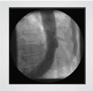
Therefore, aortography was performed and confirmed the presence of penetrating aortic ulcer in the left posterior lateral wall of the descending thoracic aorta with intraluminal thrombus, determining saccular aneurysm of the aorta as the differential diagnosis (fig 2).
In view of the persistent symptomatic clinical condition, and also considering the patient comorbidities, it was decided for a percutaneous approach using a self-expandable aortic stent-graft, which was successfully performed (figs. 3, 4, and 5).
On clinical follow-up, a complete resolution of symptoms with reduced risk of hemorrhagic complications was noted.
D
ISCUSSION
Aortic dissection is one of the most feared cardiovascular emergencies, as it results in degeneration
of the media layer and thus the delamination of its walls, creating a false lumen between the intima and adventitia. Its incidence has been estimated at about 15 cases per million inhabitants per year. Mortality rate from untreated dissection of the ascending aorta is 1% per hour in the first 48 hours, 74% within 2 weeks, and 90% within three months2,6.
At the end of the 20th century, with the remarkable
technological advances in the field of imaging techniques, the variants of aorta dissection were rediscovered. First described by Shennan in 1934, penetrating aortic ulcers were characterized later by Stanson in 19863,4.
These lesions result from the ulceration of atheromatous plaques that disrupt the aortic wall layers, penetrating deeply into the adventitia. According to Stanson et al, the majority of aortic ulcers are single lesions4.
Most patients with this variant of aortic dissection are between the sixth and eighth decades of life, different from the age group of patients with classic type-A aortic dissection and similar to those with type-B dissection. These patients have systemic hypertension, pulmonary emphysema, dislipidemias, chronic renal failure, and diabetes mellitus. Aortic ulcers usually occur in a larger descending aorta, irrespective of gender. Thoracic pain radiating to the caudal regions is a common clinical finding, following the involvement of the descending thoracic aorta3,6.
Imaging techniques are essential for the diagnosis of penetrating aortic ulcer. A common issue refers to the examination that should be performed first, and this actually depends on the clinical condition and on the availability at the institution in which the patient is being treated. Angiography, despite being an invasive method, is considered the golden standard for diagnosis of vascular diseases. Some institutions offer less invasive techniques, just as effective as angiography, such as magnetic
Fig. 1 - Chest tomography showing penetrating aortic ulcer, a variant of classic aortic dissection
Arquivos Brasileiros de Cardiologia - Volume 85, Nº 4, October 2005
Fig. 3 - Aortography showing penetrating ulcer of the descending thoracic aorta, treated with self-expandable aortic endoprosthesis
PERCUTANEOUS MANAGEMENT OF PENETRATING AORTIC ULCER
resonance imaging and ultra-fast helical computed tomography (multi-slice), which are extremely useful in the diagnosis of these conditions3,6,9.
Aortic ulcers are now recognized as a potential source of massive hemorrhage or even chronic, refractory pain. Therefore, treatment should be instituted whenever possible, using either traditional surgery or percutaneous endovascular stenting via the femoral artery. The latter has been shown to be a safer procedure, as it results in
Fig. 5 - Control TC of the chest after stent implantation in the aorta
Fig. 4 - Control TC of the chest after stent implantation in the aorta
lower complication rates for these patients who are at a higher surgical risk due to their more advanced age and associated comorbidities3,5.
In this specific case the patient is an elderly and hypertensive woman with chronic thoracic pain refractory to non-hormonal anti-inflammatory drugs and who, incidentally, was diagnosed with a penetrating aortic ulcer in its descending thoracic segment. The presence of unstable myocardial ischemic syndrome, disc herniation and urolithiasis was properly investigated and ruled out. The differential diagnosis between penetrating atherosclerotic aortic ulcer and saccular aneurysm, which are often mistaken, is confirmed by the pathological study. These conditions can be associated. The management and complications of this condition, however, follow the same line of thought, thus requiring expeditious and appropriate diagnosis and treatment.
Due to the age group and comorbidities, a percuta-neous approach using self-expandable aortic stenting was performed, with angiographic and clinical success. Having no complaints and free of impending risk of the hemorrhagic complications associated with the penetrating aortic ulcer, the patient is now under outpatient follow-up.
While uncommon, penetrating aortic ulcer should be always suspected in elderly patients with heart disease and systemic hypertension presenting with subacute and chronic thoracic pain difficult to be clinically controlled and no evidence of myocardial ischemia. Under these circumstances, percutaneous treatment by means of endovascular stenting via the femoral artery is preferred because of the excellent angiographic and clinical results achieved, as in the case just discussed.
R
EFERENCES
1. Carvalho AC, Almeida DR, Lima GP. Quadro clínico e classificação das dissecções aórticas. Rev Soc Cardiol Estado de São Paulo. 2001; 11(6): 1044-50.
2. Car valho ACC, Souza AJM. Doenças da Aor ta. Manual de
Cardiologia: SOCESP. São Paulo: Editora Ateneu, 2000: 222-8. 3. Buffolo E, Pessa CJN. Variantes da dissecção aórtica: úlcera penetrante
Arquivos Brasileiros de Cardiologia - Volume 85, Nº 4, October 2005
4. Stanson AW, Kasmier FJ, Hollier LH. Penetrating atherosclerotic ulcers of the thoracic aorta: natural history and clinicopathologic correlations. Ann Vasc Surg 1986; 1: 15-23.
5. Brittenden J, MacBride K, McInnes G, Gillespie I, Bradbury AW. The use of endovascular stents in the treatment of penetrating ulcers of the thoracic aorta. J Vasc Surgery. 1999; 30(5): 946-9. 6. Albuquerque LC, Palma JH, Braile D. Diretrizes para a cirurgia das
doenças da aorta. Arq Bras Cardiol 2004; 82 (Supl.V): 35-50.
PERCUTANEOUS MANAGEMENT OF PENETRATING AORTIC ULCER
7. Stanson AW, Kazmier FJ, Hollier LH et al. Penetrating atherosclerotic ulcers of the thoracic aorta: natural history and clinicopathologic correlations. Ann Vasc Surg 1986; 1: 15-23.
8. Kazerooni EA, Bree RL, Williams DM. Penetrating atherosclerotic ulcers of the descending aorta: evaluation with CT and distinction from aortic dissection. Radiology 1992; 183: 759-65.