480
Pediatric Cardiovascular Surgery Service in São José do Rio Preto – Hospital de Base – Medical School in São José do Rio Preto. Correspondence: Ulisses Alexandre Croti
Hospital de Base – FAMERP – Avenida Brigadeiro Faria Lima, 5544 CEP 15090-000 – São José do Rio Preto – São Paulo
Phone (Fax): 17 - 3201 5025 / 9772 6560 E-mail: [email protected]
Ulisses Alexandre CROTI, Domingo Marcolino BRAILE, Gustavo Eduardo DIAZ SUAREZ, Valdester Cavalcante PINTO JÚNIOR
Braz J Cardiovasc Surg 2006; 21(4): 480-481
CLINICAL-SURGICAL CORRELATION
RBCCV 44205-860
Operação de Ross no tratamento da valva aórtica bivalvulada calcificada
The Ross operation in the treatment of calcified
bicuspid aortic valves
CLINICAL DATA
The case of a 45-year-old female Caucasian patient weighing 97 kg and measuring 1.67 m in height is reported. The symptoms started with dyspnea at moderate effort one year previously with a significant deterioration over the last 3 months with episodes of fainting and precordial pain. The patient was referred to a cardiologist who diagnosed aortic valve disease and introduced adequate medication. The patient was in a good general state, conscious, eupneic, hydrated, ruddy but obese. The lungs presented with bilateral vesicular murmurs but without adventitious sounds. Auscultation showed a regular rhythm and systolic murmur at an aortic focus ++++/6+ were recorded. The abdomen was globous with normal tension, without pain on palpation or signs of hepatomegaly and with normal fluid-air sounds. The limbs had symmetrical pulses and the lower limbs were edematous (+/4+).
ELECTROCARDIOGRAM
The rhythm of the right atrium was low at 77 bpm. The P wave had a duration of 0.08s and an amplitude of 1.5 mm; the AP angle was -30º. The QRS complex had a duration of 0.9s, the QRS angle was + 60º, the amplitude of S at V2 was
10 mm and the R-wave at V6 7 mm. A PR interval of 0.16 seconds and QTc of 0.40 seconds were recorded.
RADIOGRAM
Visceral situs solitus was evidenced with a cardiothoracic index of 0.58. There was also a slight dilation of the aortic arch with normal vascular network and free diagrammatic domes.
ECHOCARDIOGRAM
Calcification of the aortic valve was evidenced with severe stenosis. The systolic gradient was 90 mmHg and the mean pressure was 60 mmHg with a valve area of 0.6 cm2. The pulmonary annulus was 24 mm and the aortic annulus was 25 mm without apparent calcification of the aortic valve. Slight mitral insufficiency and moderate concentric hypertrophy of the left ventricle were also seen.
DIAGNOSIS
Echocardiography is the gold standard for diagnosis however due to the age of the patient a coronary cineangiography was performed which demonstrated normal coronary arteries, severe aortic stenosis with a gradient of
Case 8/2006
481
CROTI, UA ET AL - The Ross operation in the treatment of calcified bicuspid aortic valves - Case 8/2006
Braz J Cardiovasc Surg 2006; 21(4): 480-481
110 mmHg and severe hypertrophy of the left ventricle albeit with normal function.
OPERATION
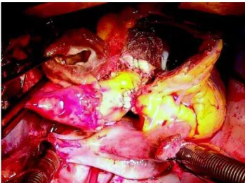
A median transsternal thoracotomy was performed with the establishment of cardiopulmonary bypass. The aorta was clamped and the right superior pulmonary vein was opened for suction of the left atrium. Hypothermic anterograde sanguineous cardioplegia was utilized at 20-minute intervals in the coronary ostia. The aorta was sectioned crosswise displaying a bivalved aortic valve with significant stenosis, justacomissural left coronary ostium and the presence of calcification of the annulus towards the anterior cuspid of the mitral valve. The pulmonary trunk was sectioned directly below the bifurcation of the pulmonary branches. The coronary ostia and aortic valve were dissected and removed (Figure 1). The pulmonary trunk and valve were dissected and removed. The aortic commissural pillars were marked and a pulmonary graft was implanted using individual 3-0 mersilene sutures. The new ring was anchored with a strip of autologous pericardium and reinforced with 5-0 prolene sutures. A window was opened in the graft and the coronary grafts were implanted. A neoaorta was then made followed by implantation of a Nº 28 pulmonary valve in the pulmonary position using 5-0 polypropylene sutures (Figure 2). Thus, the Ross operation was completed utilizing the technique of total replacement of the root [1]. The perfusion time was 185 minutes and myocardial ischemia time 160 minutes. In the postoperative period, the patient evolved with instability of the sternum with the necessity of re-intervention to redo the suture. The patient was released from hospital on the 22nd postoperative day in an excellent clinical state and with an echocardiogram demonstrating there was no significant gradient between the left ventricle and the aorta and that the new aortic valve was competent (Figure 3).
REFERENCE
1. Elkins RC. The Ross operation: a 12-year experience. Ann Thorac Surg. 1999;68(3 Suppl):S14-8.
Fig. 1 – Removed coronary ostia (tweezers) and plain sutures passed through the aortic valvar annulus prior to the implantation of the pulmonary valve
Fig. 2 – Pulmonary valve implanted in the aortic position and the pulmonary homograft (tweezers) ready to be implanted in the pulmonary position