Specific Staining and Increased Neuronal
m
-Receptor
Immunoreactivity at the Injured Nerve Trunk in Mice
Yvonne Schmidt1, Claire Gave´riaux-Ruff2, Halina Machelska1*
1Klinik fu¨r Ana¨sthesiologie und operative Intensivmedizin, Freie Universita¨t Berlin, Charite´- Universita¨tsmedizin Berlin, Campus Benjamin Franklin, Berlin, Germany,
2Institut de Ge´ne´tique et de Biologie Mole´culaire et Cellulaire, UdS Universite´ de Strasbourg, Strasbourg, Inserm, U964; CNRS, UMR7104, Illkirch, France
Abstract
Neuropathic pain is a debilitating chronic disease often resulting from damage to peripheral nerves. Activation of opioid receptors on peripheral sensory neurons can attenuate pain without central nervous system side effects. Here we aimed to analyze the distribution of neuronalm-opioid receptors, the most relevant opioid receptors in the control of clinical pain, along the peripheral neuronal pathways in neuropathy. Hence, following a chronic constriction injury of the sciatic nerve in mice, we used immunohistochemistry to quantify them-receptor protein expression in the dorsal root ganglia (DRG), directly at the injured nerve trunk, and at its peripheral endings in the hind paw skin. We also thoroughly examined them -receptor antibody staining specificity. We found that the antibody specifically labeledm-receptors in human embryonic kidney 293 cells as well as in neuronal processes of the sciatic nerve and hind paw skin dermis, but surprisingly not in the DRG, as judged by the use ofm/d/k-opioid receptor knockout mice. Therefore, a reliable quantitative analysis ofm-receptor expression in the DRG was not possible. However, we demonstrate that them-receptor immunoreactivity was strongly enhanced proximally to the injury at the nerve trunk, but was unaltered in paws, on days 2 and 14 following injury. Thus,m -opioid receptors at the site of axonal damage might be a promising target for the control of painful neuropathies. Furthermore, our findings suggest a rigorous tissue-dependent characterization of antibodies’ specificity, preferably using knockout animals.
Citation:Schmidt Y, Gave´riaux-Ruff C, Machelska H (2013)m-Opioid Receptor Antibody Reveals Tissue-Dependent Specific Staining and Increased Neuronalm -Receptor Immunoreactivity at the Injured Nerve Trunk in Mice. PLoS ONE 8(11): e79099. doi:10.1371/journal.pone.0079099
Editor:Bradley Taylor, University of Kentucky Medical Center, United States of America
ReceivedMay 14, 2013;AcceptedSeptember 19, 2013;PublishedNovember 22, 2013
Copyright:ß2013 Schmidt et al. This is an open-access article distributed under the terms of the Creative Commons Attribution License, which permits unrestricted use, distribution, and reproduction in any medium, provided the original author and source are credited.
Funding:This study was supported by a grant from the Deutsche Forschungsgemeinschaft, Klinische Forschergruppe 100/2 (MA 2437/1-4; HM). The funders had no role in study design, data collection and analysis, decision to publish, or preparation of the manuscript.
Competing Interests:The authors have declared that no competing interests exist.
* E-mail: [email protected]
Introduction
Neuropathic pain can result from peripheral nerve injuries such as amputation, entrapment, or compression. Such neuropathies trigger maladaptive alterations in the nervous system leading to peripheral and central sensitization that underlie transition to chronic pain [1]. Therapy with classical opioids predominantly acting at m-opioid receptors is limited by detrimental effects, including respiratory failure, nausea, dependence, and addiction mediated in the central nervous system [2]. Importantly, these side effects can be avoided by activating opioid receptors on peripheral sensory neurons. Periph-eral analgesic effects of opioids in neuropathic conditions were tested in animal models utilizing ligations of the nerve trunk [3]. Yet, opioids were commonly applied to tissues remote from the nerve lesion site, i.e. to paws innervated by damaged nerves, leading to partial attenuation [4–8] or no improvement of hypersensitivity [9– 12]. Interestingly, opioid peptides derived from immune cells accumulating at the site of nerve injury [13,14] or exogenousm -receptor agonists injected at this site [15] reversed mechanical or thermal hypersensitivity, suggesting that opioid receptors at the nerve injury site are functional.
However, the expression of peripheral m-opioid receptors in neuropathy was mostly assessed in the dorsal root ganglia (DRG).
Depending on the nerve damage type or the DRG level, the number of m-receptor-immunoreactive cells examined with immunohiostochemistry was either unaltered [16,17], increased [15], or decreased [11,17–19]. In addition, the m-receptor immunoreactivity assessed with Western blot was elevated [17,20] or diminished [17]. Nevertheless, the opioid receptor level in the DRG might not be predictive for peripheral opioid analgesia in neuropathy. The m-receptor immunoreactivity was enhanced at the nerve injury site [15], while it was either increased [20,21] or decreased [17] in the hind paw skin innervated by the damaged nerve. Still, the receptor cellular sources in these tissues were so far not identified.
performed detailed control experiments to ensure a specific identification ofm-receptors.
Methods
Transfection of HEK 293 cells
HEK 293 cells (Leibniz Institute DSMZ, Germany) were transiently transfected with plasmids containing the full-length cDNA (approximately 2mg) of the mousem-opioid receptor or the
mouse d-opioid receptor fused with enhanced green fluorescent protein (eGFP) [24]. Transfection was done with X-tremeGENE HP DNA transfection reagent, following the protocol of the manufacturer (Roche, Basel, Switzerland).
Animals
Experiments were performed according to the guidelines of the International Association for the Study of Pain [25] and were approved by the State animal care committee (Landesamt fu¨r Gesundheit und Soziales, Berlin). They were carried out in male mice (25–30 g) that were either C57BL/6J wild type (Harlan Laboratories, Horst, Netherlands), or triplem/d/k-opioid receptor knockout on a 129 (50%)/C57BL/6J (50%) genetic background, and the corresponding 129 (50%)/C57BL/6J (50%) wild type [26]. All mice were bred at the Charite´, Berlin, and were kept in groups of 3–5 per cage, with free access to food and water, in environmentally controlled conditions (12 h light/dark schedule; 2260.5uC; humidity 60–65%).
Staining specificity ofm-opioid receptor antibody
After transfection with m- or d-opioid receptor cDNA (see above), HEK 293 cells were washed in phosphate buffered saline (PBS), fixed in 4% paraformaldehyde and 4% sucrose in PBS for 15 min at room temperature, again washed, and permeabilized in 0.25% TritonX-100 in PBS for 5 min. After washing, cells were blocked with 10% bovine serum albumin (BSA) in PBS for 30 min at 37uC, and incubated with a rabbit m-receptor polyclonal primary antibody (1:800; Ab10275, Abcam, Cambridge, UK) in 3% BSA/PBS for 2 h at 37uC. The antibody was designed to recognize the C terminal amino acids 384–398 (i.e. NHQLEN-LEAETAPLP) of the ratm-receptor 1, but it also reacts with the identical sequence present in the C terminus of the mouse m -receptor. Of all three opioid receptors (m, d,k), this sequence is unique for the m-receptor. After washing, the sections were incubated with a goat anti-rabbit texas red-conjugated secondary antibody (Vector Laboratories, Burlingame, CA) in 3% BSA/PBS
for 45 min at 37uC, again washed, and mounted in Mowiol (Merck, Darmstadt, Germany).
m/d/k-Opioid receptor knockout (n = 4) and the corresponding genetic background wild type mice (n = 4) were genotyped by PCR on genomic DNA samples obtained from tails, using specific primers (Table 1). All mice were deeply anesthetized with isoflurane (Abbott, Wiesbaden, Germany) and perfused transcar-dially with 0.1 M PBS (pH 7.4), followed by ice-cold 0.1 M PBS containing 4% paraformaldehyde (pH 7.4) (fixative solution). The lumbar 4 and 5 DRG, the sciatic nerve parts (8–10 mm-long), the skin with subcutaneous tissue from the plantar surface of hind paws, and the lumbar enlargement of the spinal cord were isolated and postfixed in the fixative solution for 2 h at 4uC, cryoprotected in 30% sucrose at 4uC overnight, embedded in OCT compound (Miles Inc. Elkhart, IN), and frozen at280uC. The DRG were additionally incubated (approximately 30 s) in ice-cold 2-methyl-butane before embedding. Tenmm-thick sections were prepared from the DRG and longitudinally cut sciatic nerves, while 12m
m-thick sections were prepared from longitudinally cut paw tissue and transversely cut spinal cord, using a cryostat. The sections were mounted on gelatin-coated slides.
To verify the expression ofm-receptors in neurons, the sections were stained with them-receptor antibody (1:800; described above) and an antibody to pan-neuronal marker protein gene product 9.5 (PGP 9.5; 1:800; Abcam). The sections were exposed to the blocking solution for 1 h. They were subsequently incubated overnight with the primary antibody solution. After washing, the sections were incubated for 1 h with texas red-conjugated goat anti-rabbit (1:200; Vector Laboratories) or Alexa FlourH donkey anti-rabbit (1:1000; Invitrogen, Carlsbad, USA), and fluorescein isothiocyanate (FITC)-conjugated goat anti-chicken (1:200; Gene-Tex, Irvine, USA) or Alexa FlourH donkey anti-chicken (1:1000; Jackson ImmunoResearch, Pennsylvania, USA) secondary anti-bodies, washed in PBS, and mounted in Mowiol, as previously [13].
Additional DRG sections were incubated for 45 min in PBS with 0.5% H2O2 and 45% methanol to block endogenous
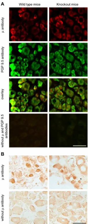
peroxidase. To prevent nonspecific binding, the sections were incubated for 60 min in PBS containing 0.3% Triton X-100, 1% BSA, and 5% goat serum (blocking solution). The sections were then incubated overnight with them-receptor antibody (1:1500), washed, and stained with a vectastain avidin-biotin peroxidase complex using goat anti-rabbit biotinylated secondary antibody (Vector Laboratories). After washing, the sections were stained with 39, 39-diaminobenzidine (DAB) tetrahydrochloride (Vector
Table 1.Primer sequences used for PCR to genotypem/d/k-opioid receptor knockout (KO) and the corresponding genetic background wild type (WT) mice.
Receptor Product size Primer Sequence (59- 39)
m WT: 648 bp Forward GAGTTAGGAGAATCAGGAGTTCAAG
KO: 422 bp Reverse TGCCATGAACATTACGGGCAGAC
Forward middle ACCGCTTCCTCGTGCTTTACGGTA
d WT: 1035 bp Forward GACCACGTGGTGCGCGCAGC
KO: 591 bp Reverse AGAACACGCAGCACAAAGACTGG
Forward middle ACCGCTTCCTCGTGCTTTACGGTA
k WT: 290 bp Forward TTCTCGCTTTCCAGCTGCAGC
KO: 580 bp Reverse CCTGAACTCACCGGATGATGACA
Forward middle ACCGCTTCCTCGTGCTTTACGGTA
Laboratories) for 30–60 s, washed in tap water, dehydrated in alcohol, cleared in xylene, and mounted in Entellan (Merck). Additional staining was performed after omission of the primary antibody.
Nerve injury
CCI was induced in deeply isoflurane-anesthetized C57BL/6J mice by exposing the sciatic nerve at the level of the right mid-thigh and placing three loose silk ligatures (4/0) around the nerve. The wound was closed with silk sutures. Sham operation was performed in a similar manner, but without nerve ligation [13]. The following experiments were performed on days 2 and 14 after surgeries, which respectively represent early and later stages of CCI-induced neuropathic pain, examined in our previous studies [13,14].
Immunostaining ofm-opioid receptors following nerve injury
On days 2 and 14 after CCI or sham operation, animals were deeply anesthetized and perfused transcardially, as described above. The lumbar 4 and 5 DRG from ipsi- and contralateral sides to the CCI, parts (8–10 mm-long) of the injured and contralateral sciatic nerves, and skin with subcutaneous tissue from both hind paws were isolated. Parts of injured nerves included the ligation site and sites proximal and distal to it. Corresponding tissues were also obtained from sham-operated and naı¨ve mice. All tissues were postfixed, cryoprotected, embedded in OCT compound, and frozen at -80uC, as described above (see ‘‘Staining specificity ofm -opioid receptor antibody’’).
m-Receptor staining using DAB in the DRG as well as using immunofluorescence in the nerve trunk and the hind paw skin was performed as described above (see ‘‘Staining specificity ofm-opioid receptor antibody’’), except that PGP 9.5 was not stained. The sections following these staining procedures were used for quantitative analysis of m-receptor expression described below. Additional staining was performed following omission of the m -receptor primary antibody or preabsorption of the primary antibody with the m-receptor immunizing peptide (5–10-fold excess, preincubation for 3 h).
Additionally, to verify the neuronal expression of m -recep-tors, the sections of DRG ipsilateral to the CCI (on days 2 and 14 after injury; n = 3 mice per time point) were incubated overnight with the m-receptor antibody (1:800) alone and in combination with isolectin B4 (IB4) FITC-conjugated (1:150; Sigma-Aldrich, St. Louis, USA), chicken neurofilament 200 (NF200; 1:500; Chemicon, Billerica, USA), or guinea pig a -calcitonin gene-related peptide (CGRP; 1:800; Bachem, Bubendorf, Switzerland) antibodies. After washing, the sections were incubated with texas red-conjugated goat anti-rabbit and goat anti-guinea pig, or FITC-conjugated goat anti-chicken secondary antibodies (1:200; Vector Laboratories and Gene-Tex). Thereafter, the sections were washed in PBS, mounted in Mowiol, and viewed under a fluorescence microscope (Zeiss) with appropriate filters.
Quantification ofm-opioid receptor immunostaining following nerve injury
Tissues from 5–6 mice were used per each experimental condition, i.e. naı¨ve, sham operation, and CCI (as described under ‘‘Immunostaining ofm-opioid receptors following nerve injury’’). Images were taken using light (m-receptors in DRG) or fluorescent microscope with appropriate filters (m-receptors in nerves and paws) and 206objectives (Zeiss Axioskop 2), and the AxioVision
program. Quantification was performed using the ImageJ graphic program (http://rsb.info.nih.gov/ij/).
Every second section of each serially cut DRG was stained form -receptors. The total number of all DRG neurons and of m -receptor-immunoreactive neurons was counted, and the data expressed as percent of the total number of neurons per section. To assess the labeling intensity ofm-receptors in DRG neurons the images were converted to grayscale. For each image, the background intensity was assessed in three random areas not covered by neurons, averaged, and subtracted. Then, the m -receptor-immunoreactive neurons were marked with a freehand selection tool. Their labeling intensity (in arbitrary units) was acquired and expressed as a mean staining intensity per section. For each group, six sections per animal were analyzed. The examiner was unaware of the experimental groups.
Every third section of each serially cut sciatic nerve was stained for m-receptors. In injured nerves, the images were taken from three different areas in relation to ligatures: directly proximally (0– 700mm; proximal I), further proximally (700–1400mm; proximal
II), and directly distally (0–700mm). Images of 700mm-areas (corresponding to the injury site) in contralateral nerves of CCI mice, both sciatic nerves of sham-operated and naı¨ve mice were also obtained. To measure the staining intensity, the upper and lower threshold density ranges were adjusted to encompass and match the immunoreactivity (red fluorescence) to provide an image with positive staining appearing in white pixels, and background staining in black pixels, for all images. A standardized box (0.2 mm2) was positioned over each 700mm-area and the number of positively-stained (white) pixels per section was calculated. In parallel, the number of m-receptor-stained fibers was quantified in each area, except for the proximal I region because strong immunoreactivity made the distinction of fibers difficult. For each animal, four sections from each nerve were analyzed. The examiner was unaware of the identity of nerve images from naı¨ve and sham-operated mice, and of contralateral nerves from the CCI animals. It was not possible to fully blind injured nerves because the ligation site was visible. However, to minimize a possible bias, the images of the three areas (proximal I, proximal II, and distal) of injured nerves were blinded for quantification.
Every second section of each serially cut paw tissue was stained form-receptors. A rectangular box of constant size was placed over the immunostained area. The box size was based on the averaged area from four images showingm-receptor-immunoreactive fibers, and calculated as 0.19 mm2. The total number of m -receptor-immunoreactive fibers was counted per section. Additionally, to assess the labeling intensity ofm-receptors in sensory fibers, the images were converted to grayscale. For each image, the background intensity was assessed in three random areas not covered by fibers, averaged, and subtracted. Then, them -receptor-immunoreactive fibers were marked with a freehand selection tool, their labeling intensity (in arbitrary units) was acquired and expressed as a mean staining intensity per section. For each group, four sections per animal were analyzed by the examiner unaware of experimental groups. For all tissues, the data were first averaged for each animal and these values were used for statistical evaluations.
Statistical analysis
Results
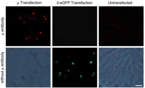
m-Opioid receptor antibody reveals specific staining in HEK 293 cells
Staining with the m-receptor antibody of HEK 293 cells transfected with the mousem-receptor revealed positively stained cells. In contrast, no positively stained cells were found in untransfected HEK 293 cells or HEK 293 cells transfected with the moused-opioid receptor fused with eGFP, which appeared in green in the absence of them-receptor antibody (Fig. 1).
m-Opioid receptor antibody staining in DRG neurons
Specific staining of them-receptor antibody in HEK 293 cells prompted us to analyze the impact of nerve damage onm-receptor expression in peripheral sensory pathways. On days 2 (Fig. S1) and 14 (data not shown) following nerve injury, we found numerousm -receptor antibody-stained small- and medium-size DRG cells co-expressing CGRP which labels peptidergic C and A neurons, whereas few DRG cells co-expressed IB4 which binds non-peptidergic C neurons, or NF200 which marks myelinated A neurons, ipsilaterally to the nerve injury. Of all DRG neurons, 3662% were positively stained withm-receptor antibody in naı¨ve animals, in agreement with previous studies [11,15,27]. Neither sham surgery nor CCI significantly changed the percentage ofm -receptor antibody-stained cells (p.0.05; Fig. S2A and B). The intensity ofm-receptor antibody labeling in DRG neurons was also not altered by the surgeries on days 2 and 14 (p.0.05; Fig. S2A and C). Preabsorption of the m-receptor antibody with the m -receptor immunizing peptide showed a lack ofm-receptor specific staining in DRG (Fig. S3); some background staining following preabsorption in the DAB staining image is similar to that seen in DAB experiments with the omission of the m-receptor antibody (see Fig. 2B).
After completion of these experiments, we had access to opioid receptor knockout mice and decided to verify the m-receptor antibody staining specificity. To ensure the targeting of neurons we also stained for the pan-neuronal marker PGP 9.5 with double
immunofluorescence. Surprisingly, we found no difference in the
m-receptor antibody staining of DRG cells (co-labeled with PGP 9.5) between wild type andm/d/k-opioid receptor knockout mice (Fig. 2A). Similarly, there was no difference between the two genotypes in them-receptor antibody labeling using DAB staining (Fig. 2B). Omission of antibodies to m-receptors and PGP 9.5 showed no immunofluorescent staining (Fig. 2A), while some background staining was seen in the absence of m-receptor antibody in experiments using DAB (Fig. 2B), both in wild type andm/d/k-receptor knockout mice. Together, despite the positive outcome of the control experiments in HEK 293 cells (Fig. 1) and of the preabsorption experiments in DRG (Fig. S3), the staining in opioid receptor knockout mice clearly shows that the antibody did not specifically label m-opioid receptors in the DRG, in our experimental conditions. Consequently, the analysis ofm-receptor expression in DRG neurons appears invalid (Fig. S1 and S2).
m-Opioid receptor antibody specifically labels peripheral neuronal processes in the sciatic nerve and the paw skin
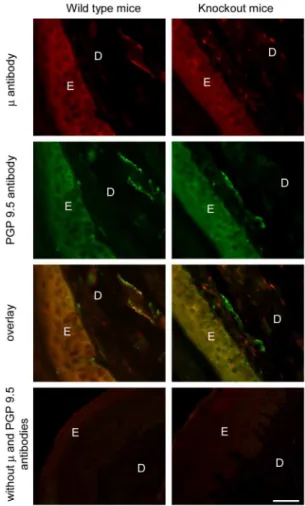
Interestingly, the m-receptor antibody staining of neuronal processes in the sciatic nerve (Fig. 3) and in the hind paw skin dermis (Fig. 4) was present in wild type mice, but it was absent in
m/d/k-opioid receptor knockout mice. Thus,m-receptor labeling overlaid the PGP 9.5 labeling in wild type mouse sciatic nerves and the paw skin dermis, whereas inm/d/k-receptor knockout mice, them-receptor antibody staining was absent, but that of PGP 9.5 remained (Fig. 3 and 4). The skin epidermis was similarly labeled by both antibodies in both genotypes (Fig. 4). Omission of antibodies tom-receptors and PGP 9.5 showed no staining in the nerve and paw skin of both genotypes (Fig. 3 and 4).
Because of this unexpected tissue-dependent staining specificity ofm-receptor antibody, we additionally tested spinal cord, which is known to be rich inm-receptors. We found thatm-receptor and PGP 9.5 antibodies co-labeled the dorsal horn of the spinal cord in wild type mice, while inm/d/k-receptor knockout mice only PGP 9.5 antibody staining was preserved (Fig. S4). Thus, it appears that them-receptor antibody specifically stains spinal cord, similarly to the sciatic nerve and paw skin dermis.
Figure 1. Specific staining ofm-opioid receptors in HEK 293 cells.Representative immunofluorescence images showing that them-receptor antibody only stains HEK 293 cells transfected with the mousem-opioid receptors, but not HEK 293 cells transfected with the moused-opioid receptors coupled to eGFP, or in untransfected HEK 293 cells (upper panel: left, middle and right images, respectively). In the absence ofm-receptor antibody there was no staining in HEK 293 cells transfected with them-receptors or eGFP-coupledd-receptors (showing only eGFP staining in green), or in untransfected HEK 293 cells (lower panel: left, middle and right images, respectively). Scale bar = 50mm.
Nerve injury enhancesm-opioid receptor immunoreactivity at the injured nerve trunk
Specific m-receptor labeling in peripheral neuronal processes prompted us to examine m-receptor expression following nerve damage. In the sciatic nerve, representative immunofluorescence images show strong m-receptor immunorectivity directly proxi-mally to the nerve injury (Fig. 5A). Quantitative analysis revealed that there were no significant differences in the number of m -receptor-immunoreactive sciatic nerve fibers among naı¨ve, sham-operated, and CCI animals, on days 2 and 14 following surgeries (p.0.05; Fig. 5B). In contrast, we found a robust and significantly higher intensity of m-receptor staining directly proximally to the ligatures on days 2 and 14 after CCI as compared to all other conditions (i.e. to areas located further proximally and distally to CCI, or to nerves of naı¨ve and sham-operated mice) (p,0.05; Fig. 5C). There were no significant differences in them-receptor staining intensity between the two time points after surgeries (p.0.05; Fig. 5C). Preabsorption control experiments showed a lack ofm-receptor specific staining in the sciatic nerve and paw skin (Fig. S3).
Figure 2. Non-specific staining of m-opioid receptors in DRG
neurons. (A) Representative double immunofluorescence images showing similarm-receptor and PGP 9.5 staining in DRG neurons of wild type mice (left panel) andm/d/k-opioid receptor knockout mice (right panel). Omission of antibodies to m-receptors and PGP 9.5 resulted in no staining in both genotypes (bottom panel). (B) Representative DAB staining images showing similar m-receptor labeling in DRG neurons of wild type mice (left image in the upper panel) andm/d/k-opioid receptor knockout mice (right image in the upper panel). In the absence ofm-receptor antibody, some background DAB staining was visible in both genotypes (lower panel). Scale bars = 50mm.
doi:10.1371/journal.pone.0079099.g002
Figure 3. Specific staining ofm-opioid receptors in the sciatic
nerve.Representative double immunofluorescence images showing m-receptor and PGP 9.5 staining in the sciatic nerve of wild type mice (left panel), but only PGP 9.5 and nom-receptor labeling in them/d/k-opioid receptor knockout mice (right panel). Omission of antibodies to m-receptors and PGP 9.5 resulted in no staining in both genotypes (bottom panel). Scale bar = 50mm.
Nerve injury does not alter them-opioid receptor immunoreactivity in paws innervated by injured nerves
In the hind paw skin from naı¨ve and CCI animals,m -receptor-immunoreactive nerve fibers were found predominately within the dermis (Fig. 6A), in agreement with the labeling in Fig. 4. The number ofm-receptor-immunoreactive fibers was not significantly changed on days 2 and 14 after CCI or sham surgery (p.0.05; Fig. 6B). Likewise, there were no significant alterations in the labeling intensity of m-receptors in sensory fibers after surgeries (p.0.05; Fig. 6C).
Discussion
In this study, we focused on the impact of nerve injury on them -opioid receptor protein expression along the relevant peripheral neuronal pathways. We found that neuronalm-receptor immuno-reactivity was strongly enhanced at the nerve lesion site, while it was unaltered in the hind paw skin, at early (2 days) and later (14 days) neuropathy stages. In previous studies, m-receptor agonists applied to paws innervated by injured nerves often moderately attenuated neuropathy-induced hypersensitivity [4–8] or were
ineffective [9–12]. Notably, we have previously shown that activation of opioid receptors at the nerve damage site by opioid peptides derived from local immune cells inhibited CCI-induced mechanical hypersensitivity on days 2 and 14 [13,14]. Similar time-course analgesia was also observed after exogenous m -receptor agonist application at this site [15]. These findings suggest that opioid receptors at the site of axonal damage might be a promising target for neuropathic pain treatment.
The current debate about the lack of specificity of antibodies to opioid receptors and G protein-coupled receptors in general [22,23,28–30] prompted us to perform a detailed analysis of them -receptor antibody staining specificity. The antibody we used (Abcam; Ab10275) specifically stained m-receptors in HEK 293 cells since only cells transfected withm-receptors, but not withd -receptors or untransfected cells, were positively labeled. Addition-ally, the lack ofm-receptor specific staining in the DRG, sciatic nerve, and paw skin in preabsorption control experiments suggests that the antibody selectively binds to its commercial immunizing peptide. However, the antibody did not specifically recognize nativem-receptors in the DRG, as it equally labeled DRG cells from wild type andm/d/k-receptor knockout mice. This was true regardless whether we used immunofluorescence or DAB staining, in several independent experiments. Hence, although in our experiments the m-receptor antibody stained a comparable percentage of DRG neurons (36%) and the same neuronal subpopulations (mostly CGRP- and some IB4- or NF200-positve cells), as in previous immunohistochemical studies assessing m -receptor expression [11,15,27,31], the labeled DRG proteins were apparently notm-receptors or any other opioid receptors in our study.
Interestingly, however, the m-receptor antibody specifically labeledm-receptors in the neuronal processes of the sciatic nerve and paw skin dermis, and the spinal cord dorsal horn. This is supported by a co-staining ofm-receptors and the pan-neuronal marker PGP 9.5 in wild type mice, but only single PGP 9.5 staining in the nerve, skin dermis, and spinal cord of m/d/k -receptor knockout animals. It seems unlikely that the antibody recognizedd- ork-opioid receptors in sciatic nerve, paw skin, and spinal cord, because it is directed against an amino acid sequence that is absent ind- ork-receptors (see methods). Additionally, the lack of staining in d-receptor transfected HEK cells and the unspecific staining in the DRG of triplem/d/k-receptor knockout mice support the notion that the antibody does not cross-react withd- ork-receptors.
On the other hand, both m-receptor and PGP 9.5 antibodies stained the skin epidermis in wild type and m/d/k-receptor knockout animals. PGP 9.5 is abundantly present in the nervous system and is commonly used as an immunohistochemical marker for nerves [32], although melanocytes and Merkel cells also revealed PGP 9.5 immunoreactivity in human skin biopsies [33]. This might be a possible explanation for the immunoreactivity we found in the paw epidermis. Nevertheless, we cannot exclude non-specific PGP 9.5 labeling of the epidermis because this structure was non-specifically stained by the m-receptor antibody in our study. Clearly, rigorous control experiments are needed when using commercial antibodies, despite the companies’ claims on their specificity. Apparently, single bands of expected sizes on Western blots, provided on datasheets of commercial antibodies, or the disappearance of staining after preabsorption with immunizing peptides, are insufficient indicators for a specific labeling in immunohistochemistry [34,35]. Our results support the use of animals genetically lacking the proteins of interest as the first choice criterion for specificity controls, in agreement with other studies [29,30,35,36], since also data obtained from experiments
Figure 4. Specific staining of m-opioid receptors in the hind
paw skin dermis.Representative double immunofluorescence images showingm-receptor and PGP 9.5 staining in the hind paw skin dermis of wild type mice (left panel), but only PGP 9.5 and nom-receptor labeling in them/d/k-opioid receptor knockout mice (right panel). In contrast, the epidermis appears similarly stained by both antibodies in both genotypes. Omission of antibodies tom-receptors and PGP 9.5 resulted in no staining in both genotypes (bottom panel). Scale bar = 50mm. E, epidermis; D, dermis.
using cell lines might not always be predictive for post-in vivo antibody staining. Moreover, our results indicate that antibodies might be even tissue- or tissue structure-selective. Interestingly, similar observations were made in another study, which examined antibodies to muscarinic receptors [34]. Thus, of 24 antibodies tested, only two antibodies were specific for muscarinic 2 receptors, as judged by the use of muscarinic 2 receptor knockout mice. Of these two antibodies, one was specific in 11 tissues but not in one (of 12 tissues examined), while the specificity of the other antibody depended on the batches [34]. Although there is no clear explanation for these differences, the results in our study and in that by Jositsch et al. [34] favor the examination of antibodies’ specificity in each tissue of interest.
Thus, unfortunately, our detailed analysis aiming at the quantification of m-receptor protein expression in the DRG following nerve damage appears inconclusive. Nevertheless, regardless of the opioid receptor expression in DRG cell bodies, the net protein level in peripheral sensory pathways might depend on injury-induced alterations in the receptor expression along the neuronal processes. Only one previous study analyzedm-receptors directly at the nerve injury site, and reported an elevation of its immunoreactivity distally to the CCI [15]. Another study found decreasedm-receptor immunoreactivity proximally to the ligature in the sciatic nerve in animals with spinal nerve ligation (SNL), and suggested a reduced receptor anterograde transport in the sciatic nerve [17]. However, it is conceivable thatm-receptors accumu-lated at the SNL site (located proximally to the sciatic nerve
Figure 5. Elevation ofm-opioid receptor immunoreactivity at the injured nerve trunk.(A) Representative immunofluorescence images showing enhancedm-receptor immunoreactivity directly proximally (0–700mm; proximal I) to the CCI compared to regions more distant proximally (700–1400mm; proximal II) and directly distally (0–700mm) to the CCI in injured nerves. Corresponding region (700mm-long) from the naı¨ve nerve is also shown. Scale bar = 50mm. (B) Quantitative analysis showing no significant alterations in the number ofm-receptor-immunoreactive neuronal fibers following surgeries (p.0.05; one-way RM ANOVA). (C) Quantitative analysis showing significantly increased intensity ofm-receptor staining (expressed as the number of positively-stained pixels) directly proximally (prox I) to the CCI (*p,0.05, versus all other conditions; one-way RM ANOVA, Bonferroni test). Experiments were performed in naı¨ve mice and in mice on days 2 and 14 following CCI or sham surgery. Ipsi, ipsilateral; contra, contralateral; nd, not determined. Data are means6SEM. N = 5–6 mice per group.
ligation), but the receptor expression was not analyzed at the SNL site [17]. Moreover, since both studies used Western blot, the cellular sources of opioid receptors remain enigmatic [15,17]. Using immunofluorescence and an antibody specifically staining
m-receptors in peripheral neuronal processes, we detected strongly enhancedm-receptor immunoreactivity proximally to the CCI in the sciatic nerve. The colocalization of thesem-receptors with PGP 9.5 (this study) or with CGRP [13], suggests we labeled sensory fibers. The lack of changes in the number of fibers expressingm -receptors indicates that the immunoreactivtiy increased only in fibers expressingm-receptors already before nerve injury.
Explanation of the origin of the increasedm-receptor immuno-reactivity at the nerve injury site is complicated by the lack of specific m-receptor labeling in the DRG in our study and, therefore, can only be speculated. Thus, the increasedm-receptor immunoreactivity proximally to the nerve injury site could result
from: (i) concomitantly increased (pre-existing or de novo) m -receptor synthesis and anterograde transport leading to the receptor accumulation at the CCI site, ifm-receptor immunore-activity in the DRG was enhanced, or (ii) a stronger rate of the receptor transport relative to its synthesis, ifm-receptor immuno-reactivity in the DRG was unchanged or decreased. Alternatively, since mRNA of various proteins, including opioid receptors, have been found in axons [37,38], the increasedm-receptor immuno-reactivity at the nerve damage site could result from its locally enhanced synthesis. Additionally, a combination of several mechanisms cannot be excluded.
In paws innervated by injured nerves, former studies reported thatm-receptor immunoreactivity was elevated after CCI or partial nerve ligation, but decreased following SNL, using Western blot, without specifying cell types [17,20,21]. In contrast, we observed no changes in the number and the labeling intensity of sensory
Figure 6. Unalteredm-opioid receptor immunoreactivity in hind paws following nerve injury.(A) Representative immunofluorescence images showingm-receptor-immunoreactive neuronal fibers (marked with arrows) in paws of naı¨ve animals and paws innervated by injured nerves. Scale bar = 50mm. E, epidermis; D, dermis. (B) Quantitative analysis showing no significant alterations in the number ofm-receptor-immunoreactive fibers following surgeries (p.0.05; one-way RM ANOVA). (C) Quantitative analysis showing no alterations in the intensity ofm-receptor staining (expressed in arbitrary units per section following surgeries (p.0.05, one-way RM ANOVA, Bonferroni test). Experiments were performed in naı¨ve mice and in mice on days 2 and 14 following CCI or sham surgery. Ipsi, ipsilateral; contra, contralateral. Data are means6SEM. N = 5–6 mice per group.
fibers expressingm-receptors in the paw skin. Thus, methodolog-ical targeting of different cellular sources and/or nerve injury type might account for the variations among the studies.
Conclusions
Because the antibody we used did not specifically stain m -receptors in the DRG and it is unclear whether antibodies used to detect these receptors in other studies were rigorously assessed for the specificity [11,15–17,19,20], it is currently difficult to judge whether the DRGm-receptor protein levels are predictive for the peripheralm-receptor-mediated analgesia in neuropathy. On the other hand, the specific labeling of m-receptors in peripheral neuronal processes in our study suggests that the lack of increased neuronalm-receptor immunoreactivity in the peripheral terminals might account for the moderate [4–8] or lacking [9–12] analgesic effects ofm-receptor agonists in paws. In contrast, sincem-receptor immunoreactivity was elevated at the site of axonal injury, targeting of these receptors might be more important for the control of neuropathic pain. Supporting this notion we have recently reported a stronger analgesic efficacy of opioids at the CCI site than in injured nerve-innervated paws [39].
Future studies should elucidate other mechanisms of peripheral opioid analgesia in neuropathy (e.g. ligand accessibility and affinity, receptor coupling and signaling). Notably, animal studies, which so far concentrated onm-receptors on peripheral terminals of sensory neurons, have shown that these receptors can mediate a substantial portion of analgesia produced by systemically (intrave-nously, subcutaneously) injected m-receptor-preferring agonists (morphine, loperamide) in neuropathic pain models [40,41]. To strengthen the clinical application of these findings, a technology-oriented research is needed to find novel ways of drug delivery to the most relevant injured tissue [42]. For example, a recent study has shown that liposomes loaded with loperamide and conjugated with an antibody to intercellular adhesion molecule-1, injected intravenously, exclusively targeted damaged tissue and produced local analgesia in a model of inflammatory pain [43]. Clearly, opioid analgesics selectively acting in the most relevant injured peripheral tissue would be preferred for the lack of central and systemic adverse effects [2].
Supporting Information
Figure S1 Staining of m-opioid receptor antibody and
sensory neuron markers in the DRG.Representative double
immunofluorescence images showing that m-receptor antibody predominantly stained DRG cells expressing CGRP (upper panel) and, to a lesser extend, cells expressing IB4 (middle panel) or NF200 (lower panel). Staining was performed in DRG ipsilateral to the injured nerve, at 2 days after CCI. Arrows indicate double-stained cells. Scale bar = 50mm.
(TIF)
Figure S2 Unaltered m-opioid receptor antibody
stain-ing in the DRG followstain-ing nerve injury.(A) Representative
DAB staining images showing m-receptor antibody-stained neu-rons (marked with arrows) in DRG of naı¨ve mice and in DRG ipsilateral to the nerve injury. Scale bar = 50mm. (B) Quantitative
analysis depicting no significant differences in the percentage ofm -receptor antibody-labeled DRG neurons following surgeries (p.0.05; one-way RM ANOVA). (C) Quantitative analysis showing no alterations in the intensity of m-receptor antibody staining (expressed in arbitrary units per section in positively-stained DRG neurons) following surgeries (p.0.05, one-way RM ANOVA). Experiments were performed in naı¨ve mice and in mice on days 2 and 14 following CCI or sham surgery. Ipsi, ipsilateral; contra, contralateral; nd, not determined. Data are means 6
SEM. N = 5–6 mice per group. (TIF)
Figure S3 Preabsorption of m-opioid receptor antibody
with m-receptor immunizing peptide in DRG, sciatic
nerve, and hind paw skin.(Upper panel) Representative DAB
staining image (first from the left) and immunofluorescence images (second to fourth) showingm-receptor staining in the DRG (first two images), the sciatic nerve (third image), and the paw skin (last image) in the presence of m-receptor antibody. (Lower panel) Corresponding images showing the lack of m-receptor staining following preabsorption of them-receptor antibody withm-receptor immunizing peptide. Some background staining was visible in DAB staining image (see also Fig. 2B). Experiments were performed in tissues ipsilateral to nerve injury, at 2 days after CCI. Scale bars = 50mm. E, epidermis; D, dermis.
(TIF)
Figure S4 Specific staining ofm-opioid receptors in the
spinal cord.Representative double immunofluorescence images
showingm-receptor and PGP 9.5 staining in the spinal cord dorsal horn of wild type mice (left panel), but only PGP 9.5 and nom -receptor labeling in the m/d/k-opioid receptor knockout mice (right panel). Omission of antibodies tom-receptors and PGP 9.5 resulted in no staining in both genotypes (bottom panel). Scale bar = 50mm.
(TIF)
Acknowledgments
We thank Brigitte L. Kieffer for providing the m/d/k-opioid receptor knockout and the corresponding wild type mice, and for helpful discussions on the manuscript. We also thank Melih O. Celik for genotyping the mice.
Author Contributions
Conceived and designed the experiments: HM. Performed the experi-ments: YS. Analyzed the data: YS CGR HM. Contributed reagents/ materials/analysis tools: CGR. Wrote the paper: YS HM. Revised the article: CGR.
References
1. Costigan M, Scholz J, Woolf CJ (2009) Neuropathic pain: a maladaptive response of the nervous system to damage. Annu Rev Neurosci 32: 1–32. 2. Stein C, Reinecke H, Sorgatz H (2010) Opioid use in chronic noncancer pain:
guidelines revisited. Curr Opin Anaesthesiol 23: 598–601.
3. Machelska H (2011) Control of neuropathic pain by immune cells and opioids. CNS Neurol Disord Drug Targets 10: 559–570.
4. Pertovaara A, Wei H (2001) Peripheral effects of morphine in neuropathic rats: role of sympathetic postganglionic nerve fibers. Eur J Pharmacol 429: 139–145. 5. Martinez V, Christensen D, Kayser V (2002) The glycine/NMDA receptor antagonist (+)-HA966 enhances the peripheral effect of morphine in neuropathic rats. Pain 99: 537–545.
6. Obara I, Makuch W, Spetea M, Schutz J, Schmidhammer H, et al. (2007) Local peripheral antinociceptive effects of 14-O-methyloxymorphone
deriv-atives in inflammatory and neuropathic pain in the rat. Eur J Pharmacol 558: 60–67.
7. Hervera A, Negrete R, Leanez S, Martin-Campos JM, Pol O (2011) Peripheral effects of morphine and expression of mu-opioid receptors in the dorsal root ganglia during neuropathic pain: nitric oxide signaling. Mol Pain 7: 25.
8. Kabli N, Cahill CM (2007) Anti-allodynic effects of peripheral delta opioid receptors in neuropathic pain. Pain 127: 84–93.
9. Aley KO, Levine JD (2002) Different peripheral mechanisms mediate enhanced nociception in metabolic/toxic and traumatic painful peripheral neuropathies in the rat. Neuroscience 111: 389–397.
PI3K-gamma/AKT/nNOS/NO/KATP signaling pathway. Proc Natl Acad Sci U S A 107: 4442–4447.
11. Rashid MH, Inoue M, Toda K, Ueda H (2004) Loss of peripheral morphine analgesia contributes to the reduced effectiveness of systemic morphine in neuropathic pain. J Pharmacol Exp Ther 309: 380–387.
12. Uchida H, Ma L, Ueda H (2010) Epigenetic gene silencing underlies C-fiber dysfunctions in neuropathic pain. J Neurosci 30: 4806–4814.
13. Labuz D, Schmidt Y, Schreiter A, Rittner HL, Mousa SA, et al. (2009) Immune cell-derived opioids protect against neuropathic pain in mice. J Clin Invest 119: 278–286.
14. Labuz D, Schreiter A, Schmidt Y, Brack A, Machelska H (2010) T lymphocytes containing beta-endorphin ameliorate mechanical hypersensitivity following nerve injury. Brain Behav Immun 24: 1045–1053.
15. Truong W, Cheng C, Xu QG, Li XQ, Zochodne DW (2003) Mu opioid receptors and analgesia at the site of a peripheral nerve injury. Ann Neurol 53: 366–375.
16. Kolesnikov Y, El-Maarouf A, Rutishauser U, Pasternak G (2007) Reorganiza-tion of dorsal root ganglion neurons following chronic sciatic nerve constricReorganiza-tion injury: correlation with morphine and lidocaine analgesia. Eur J Pharmacol 568: 124–133.
17. Lee CY, Perez FM, Wang W, Guan X, Zhao X, et al. (2011) Dynamic temporal and spatial regulation of mu opioid receptor expression in primary afferent neurons following spinal nerve injury. Eur J Pain 15: 669–675.
18. Zhang X, Bao L, Shi TJ, Ju G, Elde R, et al. (1998) Down-regulation of mu-opioid receptors in rat and monkey dorsal root ganglion neurons and spinal cord after peripheral axotomy. Neuroscience 82: 223–240.
19. Kohno T, Ji RR, Ito N, Allchorne AJ, Befort K, et al. (2005) Peripheral axonal injury results in reduced mu opioid receptor pre- and post-synaptic action in the spinal cord. Pain 117: 77–87.
20. Walczak JS, Pichette V, Leblond F, Desbiens K, Beaulieu P (2005) Behavioral, pharmacological and molecular characterization of the saphenous nerve partial ligation: a new model of neuropathic pain. Neuroscience 132: 1093–1102. 21. Walczak JS, Pichette V, Leblond F, Desbiens K, Beaulieu P (2006)
Characterization of chronic constriction of the saphenous nerve, a model of neuropathic pain in mice showing rapid molecular and electrophysiological changes. J Neurosci Res 83: 1310–1322.
22. Scherrer G, Imamachi N, Cao YQ, Contet C, Mennicken F, et al. (2009) Dissociation of the opioid receptor mechanisms that control mechanical and heat pain. Cell 137: 1148–1159.
23. Niwa H, Rowbotham DJ, Lambert DG (2012) Evaluation of primary opioid receptor antibodies for use in western blotting. Br J Anaesth 108: 530–532. 24. Scherrer G, Tryoen-Toth P, Filliol D, Matifas A, Laustriat D, et al. (2006)
Knockin mice expressing fluorescent delta-opioid receptors uncover G protein-coupled receptor dynamics in vivo. Proc Natl Acad Sci U S A 103: 9691–9696. 25. Zimmermann M (1983) Ethical guidelines for investigations of experimental
pain in conscious animals. Pain 16: 109–110.
26. Martin M, Matifas A, Maldonado R, Kieffer BL (2003) Acute antinociceptive responses in single and combinatorial opioid receptor knockout mice: distinct mu, delta and kappa tones. Eur J Neurosci 17: 701–708.
27. Yamamoto J, Kawamata T, Niiyama Y, Omote K, Namiki A (2008) Down-regulation of mu opioid receptor expression within distinct subpopulations of dorsal root ganglion neurons in a murine model of bone cancer pain. Neuroscience 151: 843–853.
28. Pradidarcheep W, Stallen J, Labruyere WT, Dabhoiwala NF, Michel MC, et al. (2009) Lack of specificity of commercially available antisera against muscar-inergic and adrenergic receptors. Naunyn Schmiedebergs Arch Pharmacol 379: 397–402.
29. Couchman JR (2009) Commercial antibodies: the good, bad, and really ugly. J Histochem Cytochem 57: 7–8.
30. Michel MC, Wieland T, Tsujimoto G (2009) How reliable are G-protein-coupled receptor antibodies? Naunyn Schmiedebergs Arch Pharmacol 379: 385–388.
31. Li JL, Ding YQ, Li YQ, Li JS, Nomura S, et al. (1998) Immunocytochemical localization of mu-opioid receptor in primary afferent neurons containing substance P or calcitonin gene-related peptide. A light and electron microscope study in the rat. Brain Res 794: 347–352.
32. Wilson PO, Barber PC, Hamid QA, Power BF, Dhillon AP, et al. (1988) The immunolocalization of protein gene product 9.5 using rabbit polyclonal and mouse monoclonal antibodies. Br J Exp Pathol 69: 91–104.
33. Wang L, Hilliges M, Jernberg T, Wiegleb-Edstrom D, Johansson O (1990) Protein gene product 9.5-immunoreactive nerve fibres and cells in human skin. Cell Tissue Res 261: 25–33.
34. Jositsch G, Papadakis T, Haberberger RV, Wolff M, Wess J, et al. (2009) Suitability of muscarinic acetylcholine receptor antibodies for immunohisto-chemistry evaluated on tissue sections of receptor gene-deficient mice. Naunyn Schmiedebergs Arch Pharmacol 379: 389–395.
35. Saper CB (2005) An open letter to our readers on the use of antibodies. J Comp Neurol 493: 477–478.
36. Burry RW (2011) Controls for immunocytochemistry: an update. J Histochem Cytochem 59: 6–12.
37. Wei LN (2011) The RNA superhighway: axonal RNA trafficking of kappa opioid receptor mRNA for neurite growth. Integr Biol (Camb) 3: 10–16. 38. Jung H, Yoon BC, Holt CE (2012) Axonal mRNA localization and local protein
synthesis in nervous system assembly, maintenance and repair. Nat Rev Neurosci 13: 308–324.
39. Labuz D, Machelska H (2013) Stronger antinociceptive efficacy of opioids at the injured nerve trunk than at its peripheral terminals in neuropathic pain. J Pharmacol Exp Ther 346: 535–544.
40. Guan Y, Johanek LM, Hartke TV, Shim B, Tao YX, et al. (2008) Peripherally acting mu-opioid receptor agonist attenuates neuropathic pain in rats after L5 spinal nerve injury. Pain 138: 318–329.
41. Kayser V, Lee SH, Guilbaud G (1995) Evidence for a peripheral component in the enhanced antinociceptive effect of a low dose of systemic morphine in rats with peripheral mononeuropathy. Neuroscience 64: 537–545.
42. Rosen H, Abribat T (2005) The rise and rise of drug delivery. Nat Rev Drug Discov 4: 381–385.