FACULDADE DE FARMÁCIA, ODONTOLOGIA E ENFERMAGEM PROGRAMA DE PÓS-GRADUAÇÃO EM ODONTOLOGIA
MESTRADO EM ODONTOLOGIA
NADINE LUÍSA GUIMARÃES ALBUQUERQUE
EFEITO DA INCORPORAÇÃO DE MICROPARTÍCULAS POLIMÉRICAS CARREGADAS COM CATEQUINA NAS PROPRIEDADES FÍSICO-QUÍMICAS DE
UM SISTEMA ADESIVO
EFEITO DA INCORPORAÇÃO DE MICROPARTÍCULAS POLIMÉRICAS CARREGADAS COM CATEQUINA NAS PROPRIEDADES FÍSICO-QUÍMICAS DE
UM SISTEMA ADESIVO
Dissertação de Mestrado apresentada ao Programa de Pós-Graduação em Odontologia da Faculdade de Farmácia, Odontologia e Enfermagem da Universidade Federal do Ceará, como requisito parcial para obtenção do título de Mestre em Odontologia. Área de concentração: Clínica Odontológica.
Orientador: Prof. Dr. Sérgio Lima Santiago. Coorientadores: Profa. Dra. Monica Yamauti e Prof. Dr. Francisco Fábio Oliveira de Sousa.
Dados Internacionais de Catalogação na Publicação Universidade Federal do Ceará
Biblioteca de Ciências da Saúde
A298e Albuquerque, Nadine Luísa Guimarães.
Efeito da incorporação de micropartículas poliméricas carregadas com catequina nas propriedades físico-químicas de um sistema adesivo / Nadine Luísa Guimarães Albuquerque. – 2015.
57 f. : il. color.
Dissertação (Mestrado) – Universidade Federal do Ceará, Faculdade de Farmácia, Odontologia e Enfermagem, Departamento de Clínica Odontológica, Programa de Pós-Graduação em Odontologia,
Mestrado em Odontologia, Fortaleza, 2015. Área de Concentração: Clínica Odontológica.
Orientação: Prof. Dr. Sérgio Lima Santiago.
Coorientação: Profa. Dra. Monica Yamauti e Prof. Dr. Francisco Fábio Oliveira de Sousa.
1. Adesivos. 2. Camellia sinensis. 3. Polímeros. 4. Catequina. 5. Metaloproteinases da Matriz. I. Título.
EFEITO DA INCORPORAÇÃO DE MICROPARTÍCULAS POLIMÉRICAS CARREGADAS COM CATEQUINA NAS PROPRIEDADES FÍSICO-QUÍMICAS DE
UM SISTEMA ADESIVO
Dissertação de Mestrado apresentada ao Programa de Pós-Graduação em Odontologia da Faculdade de Farmácia, Odontologia e Enfermagem da Universidade Federal do Ceará, como requisito parcial para obtenção do título de Mestre em Odontologia. Área de concentração: Clínica Odontológica.
Aprovada em: ___/___/______.
BANCA EXAMINADORA
________________________________________ Prof. Dr. Sérgio Lima Santiago (Orientador)
Universidade Federal do Ceará (UFC) _________________________________________
Prof. Dr. Ricardo Marins de Carvalho University of British Columbia (UBC) _________________________________________
À Universidade Federal do Ceará, na pessoa do Magnífico Reitor Prof. Dr. Jesualdo Pereira Farias.
À Faculdade de Farmácia, Odontologia e Enfermagem, representada pela sua Diretora, Profa. Dra. Maria Goretti Rodrigues de Queiroz.
Ao Curso de Odontologia da Universidade Federal do Ceará, na pessoa do Coordenador Prof. Dr. Fabrício Bitu Sousa.
Ao Programa de Pós-Graduação em Odontologia, em nome da Coordenadora Profa. Dra. Lidiany Karla Azevedo Rodrigues.
Aos meus pais, Luis Alberto e Mirtes, pela compreensão nos meus momentos ausentes, pela força e ajuda sempre que precisei. Amo vocês! Obrigada.
Ao meu esposo, Ivo, pelo amor e pela força que me deu em todos os momentos. Sem ele teria sido muito mais difícil. Eu te amo muito.
Aos meus sogros, Josué e Lúcia, que sempre vibraram com cada conquista minha como se fosse dos seus filhos. Obrigada.
À minha cunhada Karine, sempre presente e torcendo pelo meu sucesso.
Ao meu orientador, Prof. Dr. Sérgio Lima Santiago, pela eficiente orientação e pela ajuda e incentivo que me foram dados. Muito obrigada mesmo!
Aos coorientadores, Prof. Dr. Francisco Fábio Oliveira e Profª Drª Monica Yamauti, pela disponibilidade e ajuda mesmo a distância.
Ao pós-doutorando Victor Feitosa, por sua constante disponibilidade para esclarecer minhas dúvidas e pela simplicidade com a que repassava seus valiosos conhecimentos.
Aos companheiros de laboratório e colegas de turma, pela amizade e companheirismo nessa jornada.
À amiga Cecília Atem Gonçalves Costa, por me acolher tão bem e compartilhar os seus conhecimentos. Muito obrigada!
Ao técnico David Queiroz de Freitas, pela inestimável ajuda no laboratório. Obrigada por sua gentileza sempre!
À discente de Química (IFCE) Amanda Pontes Maia Pires, por ter ajudado na utilização dos equipamentos no Laboratório de Pesquisas Analíticas, fundamentais para a realização do experimento.
À Central Analítica da UFC, pela oportunidade de utilizar os equipamentos que enriqueceram nosso estudo.
Ao Prof. Alejandro Pedro Ayala, por facilitar o acesso ao Laboratório (LabRam) do Departamento de Física (UFC).
À técnica do Laboratório (LabRam) do Departamento de Física (UFC), Silmara Alves, pelo acompanhamento durante os experimentos.
Às funcionárias da coordenação do Programa de Pós-Graduação, Janaine Marques Leal e Lúcia Ribeiro Marques Lustosa, pela eficiência na resolução de todas as questões burocráticas referentes à pós-graduação.
The aim of this in vitro study was to evaluate the performance of polymeric microparticles loaded with Epigallocatechin-3-gallate (EGCG) on the physicochemical properties of a two-step etch-and-rinse adhesive system. First, the degree of conversion (%DC) was evaluated by FT-IR spectrophotometry and release assay of adhesives to evaluate the performance of EGCG loaded PLGA microparticles was realized (Experiment 1). For release assay, aliquots were collected of each samples and quantified in terms of EGCG release at pre-defined times by means of UV-Vis Spectrophotometer. In Experiment 2, forty-five molars were divided into 5 groups (n=9) according to the rewetting solution used (distilled water, 0.1% EGCG aqueous solution and 1.0% microparticles aqueous solution (PLGA50:50/EGCG)) and the Adper Single Bond 2 adhesive system used (containing 0.1% free EGCG, 1.0% EGCG loaded PLGA microparticles or in original form as control). Five 1-mm-thick increments of composite resin were build up and light-cured for 40 s individually. The teeth were stored in distilled water at 37ºC for 24 h. After storage, they were longitudinally sectioned in both directions to obtain bonded sticks with a cross-sectional area of approximately 1.0 mm². Each bonded stick was testing to a tensile force of 0.5 mm/min in the universal testing machine. %DC and µTBS values were statistically analyzed with ANOVA, with significance level of 5%. There was no statistically significant difference between the DC means after PLGA-microparticles loaded with EGCG incorporated (p>0.05). In relation to release assay, the 1.0% PLGA50:50/EGCG group presented better results, achieving the highest release in quantitative terms, being the elect to be used in bond strength test (Experiment 2). After 24 h of storage, there was no statistically significant difference between the mean bond strength values of the tested groups (p>0.05). The incorporation of the polymeric microparticles loaded with EGCG did not interfere in the adhesive degree of conversion. The adhesive system loaded microparticles EGCG incorporated in its composition was able to release EGCG. However, the flavonoid epigallocatechin-3-gallate (EGCG) had no effect when incorporated into etch-and-rinse adhesive system or applied as dentin pretreatment, on free and microencapsulated forms, in the immediate bond strength.
1 INTRODUÇÃO GERAL ………. 11
2 PROPOSIÇÃO ... 15
2.1. Objetivo Geral ... 15
2.2. Objetivos Específicos ... 15
3 CAPÍTULO ... 16
3.1. Capítulo 1 ... 17
4 CONCLUSÃO GERAL ... 38
REFERÊNCIAS GERAIS... 39
APÊNDICE ... 46
ANEXOS ... 47
ANEXO A – Norma do Periódico Dental Materials ... 47
1 INTRODUÇÃO GERAL
Os sistemas adesivos têm demonstrado resultados promissores na prática clínica diária. Entretanto, apesar de todo o arsenal que temos à disposição para uma adequada adesão, ainda existe uma dificuldade para se conseguir união entre os materiais restauradores e as estruturas dentárias em virtude da heterogeneidade dos substratos envolvidos (PASHLEY, TAY, IMAZATO, 2011). O esmalte é considerado um substrato com maior conteúdo mineral e de morfologia homogênea possibilitando uma adesão efetiva, enquanto a dentina é mais complexa, apresentando menor conteúdo mineral, de natureza heterogênea e intrinsecamente úmida (VAN MEERBEECK et al., 2003). Essa umidade tem papel importante na degradação da interface adesiva e na redução das propriedades mecânicas da adesão (PEREIRA et al., 1999; CARRILHO et al., 2004). Além disso, ainda existem outros desafios para uma boa adesão como a nanoinfiltração que é a diferença entre a zona de dentina desmineralizada pelo ácido e a zona infiltrada pelo adesivo durante a formação da camada híbrida, gerando fibras colágenas expostas e desprotegidas tornando-as susceptíveis à ação dos fluidos orais (SANO et al., 1995; WANG e SPENCER, 2003; DE MUNCK et al., 2005); e a permeabilidade dos adesivos dentinários (TAY, PASHLEY, YOSHIYAMA, 2002).
Essa degradação das fibrilas colágenas expostas na interface adesiva se dá pelas metaloproteinases de matriz (MMPs), enzimas endógenas presentes na dentina, responsáveis pela organização e mineralização da matriz dentinária (CHAUSSAIN-MILLER et al., 2006; CARRILHO et al., 2007; STANISLAWCZUK et al., 2009; BRESCHI et al., 2010a; YIU et al., 2012). Estas se encontram inativas e são ativadas em baixo pH, ou seja, quando é realizado o condicionamento ácido dos sistemas adesivos convencionais ou na aplicação dos adesivos autocondicionantes (TURK et al., 1995; TJÄDERHANE et al., 1998; VAN STRIJP et al., 2003; VISSE, NAGASE, 2003; PASHLEY et al., 2004; NISHITANI et al., 2006; TERSARIOL et al., 2010; LIU et al., 2011; TJÄDERHANE et al., 2013). Alguns estudos confirmaram a presença da MMP-2, MMP-3, MMP-8 e MMP-9 em dentina humana desmineralizada (PASHLEY et al., 2004; NISHITANI et al., 2006; MAZZONI et al., 2007; SULKALA et al., 2007 STANISLAWCZUK et al., 2009; BRESCHI et al., 2010b). A MMP-2, também conhecida como Gelatinase A, degrada colágeno tipo I, principal colágeno encontrado na dentina e a MMP-9, ou Gelatinase B, degrada o colágeno tipo IV, que é o principal componente do colágeno desnaturado (MORGUNOVA et al., 1999; CHAUSSAIN-MILLE et al., 2006). Já a MMP-8, também denominada de colagenase-2, foi identificada por Sulkala et al., em 2007, como a principal enzima colagenolítica da dentina humana. Mais recentemente, outro grupo de enzimas proteolíticas foi identificado na dentina humana indicando que a atividade colagenolítica da dentina não se dá apenas pela presença das MMPs, mas também pela atividade das cisteínas catepsinas (TERSARIOL et al., 2010; NASCIMENTO et al., 2011).
Portanto, durante os últimos anos, o entendimento dos mecanismos que envolvem a degradação proteolítica da interface adesiva tem ganhado imensa atenção e rapidamente vêm crescendo o interesse da comunidade científica em desenvolver estratégias para aumentar a longevidade clínica das restaurações adesivas. Assim, o uso de inibidores da atividade enzimática tem sido aceito como uma estratégia eficaz para melhorar a longevidade das restaurações adesivas (LOGUERCIO, STANISLAWCZUK, POLLI, 2009; OSORIO et al., 2011; YIU et al., 2012; SANTIAGO et al., 2013).
et al., 2007; LOGUERCIO, STANISLAWCZUK, POLLI, 2009; OSORIO et al., 2011). A clorexidina é uma molécula sintética que se liga a várias proteínas por meio de um mecanismo quelante e pode inibir a ação dessas enzimas mesmo em baixas concentrações, desacelerando assim o processo de degradação da interface de união (CARRILHO et al., 2007; LOGUERCIO, STANISLAWCZUK, POLLI, 2009). O primeiro estudo que constatou a benéfica ação do digluconato de clorexidina usado como pré-tratamento da dentina foi realizado por Hebling et al., em 2005. Seus resultados mostraram uma diminuição da degradação de colágeno da camada híbrida se comparado à técnica original de condicionamento ácido e posterior aplicação do adesivo. Em seguida, diversos estudos apontaram a clorexidina como potente inibidor das metaloproteinases seja como solução para pré-tratamento da dentina (CARRILHO et al., 2007; LOGUERCIO, STANISLAWCZUK, POLLI, 2009) ou incorporada ao sistema adesivo (YIU et al., 2012). No entanto, há uma preocupação na aplicação de digluconato de clorexidina em tecidos humanos no que se refere à segurança biológica, sendo necessária a procura por inibidores mais biocompatíveis (FARIA, CARDOSO, LARSON, 2009).
(abaixo de 0,2%), não promoveu alterações significativas no grau de conversão dos monômeros resinosos (DU et al., 2012; PALLAN et al., 2012). Embora, a incorporação de EGCG aos sistemas adesivos tenha demonstrado resultados promissores, há uma preocupação em relação à disponibilidade da catequina dentro da camada híbrida. Sendo o EGCG uma molécula solúvel em água (solubilidade 5mg/ml) (Sigma-Aldrich, St. Louis, MO, USA) foi observada uma alta taxa de liberação nas primeiras 24 horas de exposição à agua destilada, seguida de uma redução significativa na taxa de liberação até o 28° dia (PALLAN et al., 2012). Portanto, o tempo de permanência do EGCG na camada híbrida pode ser curto devido à ação da água (PALLAN et al., 2012). Contudo, esse inconveniente pode ser contornado a partir de métodos de liberação controlada de fármacos (LIANG, WONG, BURT, 2005; GAIGNAUX et al., 2012).
Para isso, buscou-se a utilização de micropartículas poliméricas, produzidas a partir de alguns polímeros, como o ácido poli láctico-co-glicólico (PLGA), padrão-ouro na liberação controlada de fármacos, devido sua incomparável biodegradação e biocompatibilidade (PRIOR et al., 2000; BLANCO-PRIETO et al., 2002; GRAVES et al., 2004; SCHNIEDERS et al., 2006). A incorporação de micropartículas poliméricas carregadas com EGCG ao sistema adesivo convencional pode ser uma estratégia eficaz se comparado à incorporação de EGCG puro ao sistema adesivo. Além de uma liberação mais prolongada e controlada, apresenta vantagem em relação à proteção do fármaco frente aos processos fisiológicos degradativos e à bioabsorção, visto que a ocorrência desses fenômenos acarretaria na perda da atividade farmacológica (PRIOR et al., 2000; BLANCO-PRIETO et al., 2002; GRAVES et al., 2004; SCHNIEDERS et al., 2006).
Diante do exposto, faz-se necessário que as técnicas adesivas sejam reformuladas ao longo do tempo justificando-se o estudo do flavonóide Epigalocatequina-3-galato como agente inibidor da atividade enzimática conseguindo assim, a preservação da camada híbrida e consequentemente, maior durabilidade das restaurações adesivas.
2 PROPOSIÇÃO
2.1 Objetivo Geral
Avaliar o efeito da incorporação de micropartículas poliméricas carregadas com Epigalocatequina-3-galato (EGCG) nas propriedades físico-químicas de sistema adesivo convencional de 2 passos.
2.2 Objetivos Específicos
- Avaliar o grau de conversão dos monômeros em polímeros dos adesivos em sua composição original e nas formulações contendo as micropartículas carregadas com EGCG.
- Analisar a liberação do princípio ativo a partir do adesivo convencional contendo micropartículas carregadas com EGCG.
3 CAPÍTULO
Esta dissertação está baseada no Artigo 46 do Regimento Interno do Programa de Pós-Graduação em Odontologia da Universidade Federal do Ceará que regulamenta o formato alternativo para dissertações de Mestrado e teses de Doutorado e permite a inserção de artigos científicos de autoria ou coautoria do candidato. Por se tratar de um estudo envolvendo dentes humanos, o projeto de pesquisa deste trabalho foi submetido à apreciação do Comitê de Ética em pesquisa da Universidade Federal do Ceará, tendo sido aprovado, conforme o parecer consubstanciado no 459.659 de 14 de novembro de 2013 (ANEXO B).
Assim sendo, esta dissertação é composta de um capítulo contendo um artigo científico que será submetido ao periódico Dental Materials (ANEXO A) conforme descrito na sequência:
Adhesive system containing polymeric microparticles loaded with catechin: Physicochemical characterisation.
CAPÍTULO 1
Adhesive system containing polymeric microparticles loaded with catechin: Physicochemical characterisation.
Nadine Luísa Guimarães Albuquerquea
Jiovanne Rabelo Neria
Monica Yamautib
Francisco Fábio Oliveira de Sousac
Sérgio Lima Santiagoa
a
Department of Restorative Dentistry, Federal University of Ceará, Fortaleza, Ceará,
Brazil. Rua Monsenhor Furtado s/n, 60430-655, Fortaleza, Ceará, Brazil.
b
Department of Dentistry, Federal University of Minas Gerais, Belo Horizonte, Minas
Gerais, Brazil. Av. Presidente Antônio Carlos 6627, 31270-902, Pampulha, Belo
Horizonte, Minas Gerais, Brazil.
c
Department of Pharmaceutical Sciences, Federal University of Amapá, Macapá,
Amapá, Brazil. Rod. Juscelino Kubitschek km 2, 68902-280, Macapá, Amapá, Brazil.
Corresponding Author:
Dr. Sérgio Lima Santiago
Rua Monsenhor Furtado s/n, Rodolfo Teófilo, CEP: 60430-355, Fortaleza, CE- Brasil
Tel.: 55 (85) 3366 8030
ABSTRACT
Objective. Evaluate the performance of polymeric microparticles loaded with Epigallocatechin-3-gallate (EGCG) on degree of conversion and release assay of adhesives and examine resin–dentin bond strength with different EGCG application modes.
Methods. The degree of conversion (%DC) was evaluated by FT-IR spectrophotometry and release assay of adhesives containing different proportions of EGCG loaded PLGA microparticles was performed. Aliquots were collected of each samples and quantified in terms of EGCG release at pre-defined times by means of UV-Vis Spectrophotometer (Experiment 1). After that, forty-five molars were divided into 5 groups (n=9) according to EGCG different application modes. The teeth were prepared to bonding and testing in the universal testing machine (Experiment 2). %DC and µTBS values were statistically analyzed with ANOVA.
Results. There was no statistically significant difference between the DC means after PLGA-microparticles loaded with EGCG incorporated (p>0.05). The 1.0% PLGA50:50/EGCG group presented better results, achieving the highest release in quantitative terms, being the elect to be used in bond strength test. After 24 h of storage, there was no statistically significant difference among the mean bond strength values of the tested groups (p>0.05).
Significance. EGCG can be effective in improving the longevity of adhesive restorations therefore, their permanence time in the hybrid layer can be short due to its solubility in water. Hence, the strategy to produce polymeric microparticles loaded with EGCG to achieve a more controlled and prolonged release.
1. Introduction
Studies have shown that adhesive systems lose their bond to dentin over time, and there is a consensus that the hybrid layer created by current adhesive systems is imperfect, susceptible to degradation and, could negatively affect the bond strength [1,2]. The decrease of bond strength is related to a hydrolytic degradation of the polymers of the adhesive systems and the proteolysis of collagen matrix of the hybrid layer [3]. Transmission electron microscopy analyses showed that almost 70% of collagen from the adhesive interface disappears after 44 months water storage [4]. Host-derived proteases (matrix metalloproteinases and cysteine cathepsins) with collagenolytic activity in hybrid layers are the mainly responsible by degradation of collagen fibrils reducing the longevity of clinically applied resin-based restorations [2,5,6].
Therefore, studies have focused their research on the modification of dental adhesives to improve the durability of bonding to dentin of resin-based restorations [1,2]. The use of protease inhibitors on dentin surface after acid etching or incorporation into the adhesive system has been well accepted [7-11]. Chlorhexidine (CHX) was the first protease inhibitor proposed for preserve the hybrid layer through the inhinition of MMPs [12] and cysteine cathepsins [13] but recently other inhibitors has received increased attention from researchers.
flavonoid is effective in improving the longevity of adhesive procedures independent of adhesive system.
In attempt to achieve a release of EGCG in a more controlled and prolonged as possible, sought the use of polymeric microparticles produced by polymers as Poly (D-L lactide-co-glycolide) acid (P(D-LGA), one of the main polymers used in the development of release systems. It considered gold standard in controlled drug delivery, mostly due to its superior biocompatibility [16-19]. Besides be used in the development of release systems, this polymer has others applications as: prevention of postsurgical adhesions through of application of nanofibers of poly (lactic-co-glycolic acid) (PLGA) loaded with epigallocatechin-3-O-gallate (EGCG) [20] and enhance wound healing by accelerating cell infiltration, re-epithelialization and angiogenesis by eletroctrospum membranes composed of PLGA containing 1 wt% EGCG [21]. Thus, the use of polymeric microparticles loaded with EGCG can be an effective strategy in comparison to free EGCG due to the protection of the drug from the physiological degradation and bioadsorption, avoiding the loss of pharmalogical activity. In addition, the controlled drug delivery systems could contribute to a prolonged effect of the drug in the specific therapeutic site.
Therefore, the aim of this study was to evaluate the effect of different EGCG application modes on the resin-dentin bonds. To achieve this objective, the study was divided into two experiments. The objective of the first experiment was to evaluate the performance of polymeric microparticles loaded with EGCG on degree of conversion and release assay of adhesives and the second experiment was to examine early resin– dentin bond strength with two EGCG formulations applied as pretreatment or incorporated into etch-and-rinse adhesive system. The null hypotheses were: (1) incorporation of polymeric microparticles loaded with EGCG have no effect on adhesive degree of conversion; (2) the incorporation of PLGA-microparticles loaded with EGCG into adhesive will not be able to release EGCG; (3) the use of EGCG in free form or loaded into PLGA-microparticles as dentin pretreatment or incorporated into the adhesive does not affect the immediate bond strength.
2. Materials and Methods
2.1. Epigallocatechin-3-gallate microparticles used
The materials and chemicals used in this study are described in Table 1.
Epigallocatechin-3-gallate (EGCG) (Sigma Aldrich, St. Louis, MO, USA) was added to different formulations of microparticles using two types of Poly (D-L lactide-co-glycolide) acid (PLGA, Sigma Aldrich, St. Louis, MO, USA): Resomer® RG502H (PLGA 50:50) and Resomer® RG756S (PLGA 75:25) by means of spray-drying technique [22].
Formulations were prepared in the ratio PLGA:EGCG 16:1. A blank formulation (PLGA only) from each polymer was obtained and used as a reference. Due to differences in solubility among the drugs and polymers, an emulsification process was proposed. Briefly, PLGA was dissolved in dichloromethane (DCM) and EGCG was dissolved in ethyl acetate in another vial. The solutions were mixed in a disperser system (Ultraturrax IKA T10B; IKA/Works, Inc. NC, USA) and immediately spray dried in a laboratory spray drier (Mini Spray Drier Buchi 290, Buchi, Flawil, Switzerland).
Table 1 – Materials and chemicals used in the study.
Material (/Manufacturer) Batch number Basic formulation
AdperSingle Bond 2* (3M/ESPE®, St. Paul, MN,
EUA)
1312201025
Etchant: 35% phosphoric acid (batch #1219600378).
Bis-GMA, HEMA, dimethacrylates, silica nanofiller
(5 nm), polyalquenoic acid copolymer, initiators, water and
ethanol.
Resin Filtek Z250 XT
(3M/ESPE®,St. Paul, MN, EUA) 37277
Bis-EMA, Bis-GMA and UDMA. Filled to 60% by volume with zirconia silica filler,
average.particle size = 0.6 μm
Poly (D-L lactide – co – glycolide acid) Resomer® RG502H / Sigma Aldrich, St.
Louis, MO, USA
STBD2887V ---
Poly (D-L lactide – co – glycolide acid) Resomer® RG756S / Sigma Aldrich, St.
Louis, MO, USA
STBC6378V ---
Epigallocatechin-3-gallate (Sigma Aldrich, St. Louis, MO,
USA)
SLBL1959V ---
2.2. Preparation of adhesive formulations
The adhesive formulations were prepared by incorporating PLGA microparticles loaded with EGCG into Adper Single Bond 2 adhesive system (3M/ESPE®, St. Paul, MN, EUA) by manual blending. The amount of microparticles incorporated into the adhesives ranged between 0.5, 1.0 and 2.0% (w/w). Each adhesive formulation (Table 2) briefly mixed in a vortex (Biomixer QL-901, SP, Brazil) for 1 min at reduced ambient light.
Table 2 – Description of the experimental groups of Experiment 1 (Degree of conversion and Release assay of adhesives containing microencapsulated EGCG).
GROUPS MATERIAL MICROENCAPSULATED
FORMS OF EGCG Control
Adper Single Bond 2
Control
0.5% PLGA50:50/EGCG Containing 0.5% (w/w) of microparticles
(PLGA50:50) of EGCG
0.5% PLGA75:25/EGCG Containing 0.5% (w/w) of microparticles
(PLGA75:25) of EGCG
1.0% PLGA50:50/EGCG Containing 1.0% (w/w) of microparticles
(PLGA50:50) of EGCG
1.0% PLGA75:25/EGCG Containing 1.0% (w/w) of microparticles
(PLGA75:25) of EGCG
2.0% PLGA50:50/EGCG Containing 2.0% (w/w) of microparticles
(PLGA50:50) of EGCG
2.0% PLGA75:25/EGCG Containing 2.0% (w/w) of microparticles
(PLGA75:25) of EGCG
Experiment 1
2.3. Characterization of the formulations
2.3.1. Degree of conversion
The degree of conversion (DC) of the adhesive resins was assessed by Fourier Transform Infrared Spectroscopy (FTIR) (Perkin-Elmer Spectrum 100, Perkin Elmer, Shelton, CT, USA). Each adhesive system was dispensed into a small agate mortar and thoroughly mixed with potassium bromide (KBr), at a ratio of 4:100. The pellets of KBr/adhesive solution were prepared with a hand press (Hand Press Kit 161-1100, PIKE Technologies, Madison, WI, USA). FTIR spectrum of the uncured adhesive was obtained from each sample using 32 scans in a range of 4000-400 cm-1, at 4 cm-1 resolution in transmission mode.
The adhesive system was light-activated for 40 s using the light source (Ellipar Freelight 2, 3M ESPE, St. Paul, MN, USA). Additional FTIR spectra were obtained immediately after light curing. The analyses were performed at 25 °C with 70% relative humidity. Tree specimens per group (n=3) were tested. The rate of unreacted carbon-carbon double bonds (C=C) was determined from the ratio of absorbance intensities of aliphatic C=C (peak at 1636 cm-1) against an internal standard (aromatic carbon-carbon bond peak at 1608 cm-1) before and after curing. Degree of conversion was determined by subtracting the C=C from 100%.
2.3.2. Release assay of adhesives containing microencapsulated EGCG
In view of the difference in hydrophobicity and also the biodegradation rates of the two PLGA (PLGA 50:50 and PLGA 75:25) used in this study, there was a release assay in order to observe their performance into the adhesive system (Adper Single Bond 2).
A calibration curve was used to quantify the drug from a series of reference solutions ranging from 2.5 to 40 ppm, resulting in a linear relationship between absorbance peak height and drug concentration. A UV-Vis Spectrophotometer (DU-730; Beckman Coulter, Fullerton, CA, USA) was used to evaluate and confirm the absorbance peak of EGCG at 275 nm.
with each adhesive formulation, a mylar strip was placed in the top of it and glass slide was placed to perform the light-curing process. The material was light-cured (Ellipar Freelight 2, 3M ESPE, St. Paul, MN, USA) for 40 s at 600 mW/cm2. Samples were stored in individual vials containing 1 mL of distilled water at 37ºC stored until 2904 h.
Experiment 2
2.4. Microtensile Bond Strength Test
For the second experiment, forty-five (45) extracted, caries-free human third molars were used. The teeth were collected after the patient’s informed consent had been obtained under a protocol reviewed and approved by the local Research and Ethics Committee. The selected teeth were stored for about one month after extraction in 0.01% (w/v) thymol solution.
The occlusal enamel of each tooth was removed using a slow-speed diamond saw (IsoMet; Buehler, Lake Bluff, IL, USA) under water-cooling in order to expose a flat coronal dentin surface. The enamel-free, exposed dentin surfaces were further polished on wet #600-grit SiC paper for 60 s to standardize the smear layer.
Table 3 – Description of the experimental groups of Experiment 2 (Bond strength test).
GROUPS DENTIN PRETREATMENT ADHESIVE SYSTEM
Control Distilled water Adper Single Bond 2
SB+0.1% free EGCG Distilled water Containing 0.1% (w/w) of
free EGCG.
SB+1.0% PLGA50:50/
EGCG
Distilled water Containing 1.0% (w/w) of microparticles (PLGA50:50-EGCG)
RS 0.1% free EGCG Epigallocatechin-3-gallate aqueous solutions (0.1% EGCG)
Adper Single Bond 2
RS 1.0% PLGA50:50/
EGCG
Microparticles aqueous solution (1.0% PLGA50:50/EGCG)
Adper Single Bond 2
The etch-and-rinse adhesive system Adper Single Bond 2 (3M ESPE®) was then applied according to the manufacturer’s instructions. After light curing the adhesive (Ellipar Freelight 2, 3M ESPE, St. Paul, MN, USA), five 1-mm-thick increments of composite resin were build up (Filtek Z250 XT - 3M/ESPE®). Each increment was light cured (Ellipar Freelight 2, 3M ESPE, St. Paul, MN, USA) for 40 s at a power density of 600 mW/cm². The bonded teeth were stored in distilled water at 37ºC for 24 h.
After storage, they were longitudinally sectioned in both ‘x’ and ‘y’ directions
The failure modes were evaluated at 80X magnification (StereoZoom Leica S8 APO, Leica Microsystems; Wetzlar, Hesse, Germany) and classified as cohesive (failure exclusively within the dentin or composite; C), adhesive (A) when failure occurred at the dentin/adhesive interface, ou mixed (M) when two modes of failure occurred simultaneously.
2.5. Statistical Analysis
DC and µTBS values were submitted a Shapiro-Wilk test to analyze the normal distribution of errors. As normal distribution was confirmed, data were analyzed using a statistical analysis of variance one-way ANOVA. Statistical procedures were performed with the SigmaStat 3.5 for Windows statistical program software (Systat Software, San Jose, California, USA). The significance level was set at p < 0.05 for all tests.
3. Results
3.1. Degree of conversion
Table 4 - Mean and standard desviation of the tested groups.
EGCG microencapsulated forms incorporated in SB Mean (SD)
Control 58.32 (0.38)
0.5% PLGA50:50/EGCG 57.29 (0.73)
0.5% PLGA75:25/EGCG 57.64 (0.73)
1.0% PLGA50:50/EGCG 58.11 (0.32)
1.0% PLGA75:25/EGCG 58.10 (0.89)
2.0% PLGA50:50/EGCG 59.07 (0.57)
2.0%PLGA75:25/EGCG 57.61 (0.50)
3.2. Release assay of adhesives containing microencapsulated EGCG
EGCG release profiles of adhesive system are shown in Fig. 1. In groups 0.5% PLGA50:50/EGCG and 1.0% PLGA50:50/EGCG a controlled release was observed reaching the total release (100%) during the assayed period. In contrast, among the PLGA75:25 groups, the highest release obtained did not reach more than 60% of the overall drug content (0.5% PLGA75:25/EGCG).
In all groups, except from 1.0% PLGA75:25/EGCG and 2.0% PLGA75:25/EGCG was observed a pulsatile release profile, characterized by moments of cessation and subsequent rapid increase of release at 300, 600 and 900 h. In 1.0% PLGA75:25/EGCG and 2.0% PLGA75:25/EGCG groups this perfomance was more discreet and less perceived in Fig. 1.
Fig. 1 - Cumulative EGCG release (%) from adhesive system in aqueous medium during the entire evaluation period (2904 h).
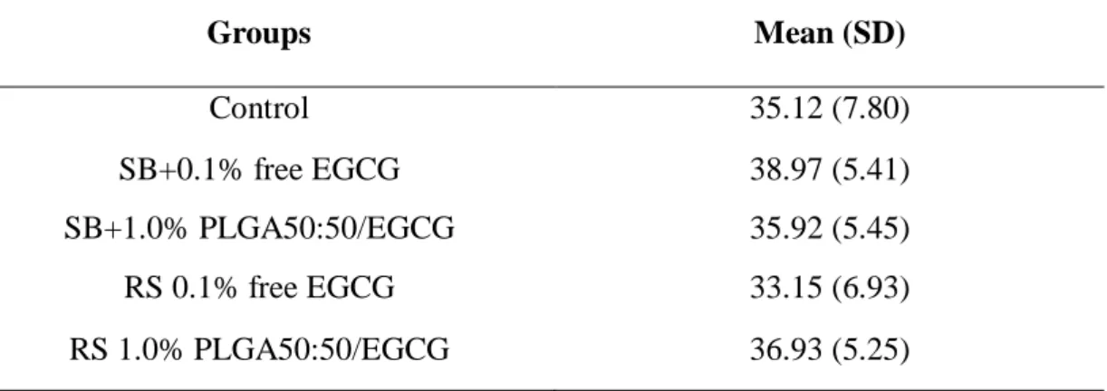
3.3. Bond strength
Mean µTBS values were calculated and are expressed in Table 5. After 24 h of storage, there was no statistically significant difference between the mean bond strength values of the tested groups (p > 0.05).
Table 5 - Mean µTBS values and standard desviation (MPa) of the tested groups.
Groups Mean (SD)
Control 35.12 (7.80)
SB+0.1% free EGCG 38.97 (5.41)
SB+1.0% PLGA50:50/EGCG 35.92 (5.45)
RS 0.1% free EGCG 33.15 (6.93)
4. Discussion
One of the main properties of epigallocatechin-3-galate related to adhesive dentistry is prevent dentin collagen degradation by MMP inhibition [23] differing from others MMP-inhibitors to be a natural product, extracted from green tea (Camellia sinensis). Thus, it can be used in cavities of any depth because of its low toxicity and anti-inflammatory properties [14].
Dentin treatment using EGCG significantly improved the mechanical properties of demineralized dentin, which suggests potential collagen cross-linking [24]. These positive data support the introduction of EGCG in dental practice. However, drugs are released more quickly when uncoated [25-27] for that reason, polymeric microparticles produced by polymers as Poly (D-L lactide-co-glycolide) acid (PLGA) has been extensively used in several applications concerning the controlled release of drugs [22,28,29] include in Endodontics and Periodontics [22,30].
The association between EGCG and polymeric materials aimed to obtain a long-acting drug delivery system, which could be applied in dental therapeutics. It has not been studied, especially when it concerns the physical and/or chemical properties of the molecules and the resulting effect on the release ability from the material. The present study evaluated the effect of two types EGCG solution, free and microencapsuled, applied as dentin pretreatment or incorporated into etch-and-rinse adhesive system on dentin bond strength. In order to establish the better PLGA microparticles loaded with EGCG formulations was conducted degree of conversion and release assay of adhesives containing microencapsulated EGCG (Experiment 1) and after, teeth were prepared to subsequent bond strength test (Experiment 2).
demonstrated that the incorporation of EGCG in low concentrations (0.01 and 0.1%) into a specific one-step self-etch adhesive did not cause any detrimental effect on the DC.
Concerning the EGCG release, the results showed different cumulative release profile between the groups (Fig. 1). Thus, the second null hypothesis, the incorporation of PLGA-microparticles loaded with EGCG into adhesive will not be able to release EGCG was rejected. It was observed a controlled release profile in all the groups but, PLGA 50:50 groups reached a total release (100%) during the assay period while, among PLGA75:25 groups which released more just reached 60% of the total (0.5% PLGA75:25/EGCG). We speculated that this behavior could be explained by polymer hydrophobicity that suits better to hydrophobic adhesive jeopardizing the drug diffusion. Futhermore, in all PLGA 50:50 groups was observed a pulsatile release profile characterized by forming a plateau momentary immediately followed by an increase in release. This support the cases where there are required concentration peaks, such as metabolic disease and/or in which the effect is directly dependent on the minimum plasma level to achieve the pharmacological effect.
In this study, the objective is in situ controlled release with the drug remain covered by the polymer, securing their efficacy over time. The pulsatile release profile enable that higher doses are reached in individual and gradual moments, encouraging the effects related to inhibition enzymatic. Therefore, considering all factors, the 1.0% PLGA50:50/EGCG group was presented better results, being the elect to be used in Experiment 2.
The results of this study revealed that EGCG has not effect on the immediate bond strength independent of the application mode since that there was no statistically significant difference between the mean bond strength values of the tested groups (p > 0.05). Thus, the third null hypothesis, that the use of EGCG in free form or loaded into PLGA-microparticles as dentin pretreatment or incorporated into the adhesive does not affect the immediate bond strength, must be accepted.
bond strength was lower than Control Group unlike the findings of Santiago and others [11] at the same EGCG concentration (0.1%) (Table 5).
In the present study, we speculate that only immediate bond strength test may not have been sufficient to detect the effects of hydrolytic degradation of the adhesive interface. A study by Kiyomura et al. [31] reported that storage times between 2 and 4 years were required to detect the effects of hydrolytic degradation. Therefore, complementary studies are being conducted to confirm the potential of this catechin in preservation of collagen and maintenance of bond strength and evaluate the influence of the polymeric microparticles loaded with EGCG on the physicochemical properties of a commercial etch-and-rinse adhesive system.
5. Conclusion
- The addition of microencapsulated EGCG did not affect the degree of conversion of etch-and-rinse adhesive system, independent of concentration.
- Adhesive system incorporated with PLGA-microparticles loaded with EGCG were able to release EGCG, making these systems viable for dental applications.
REFERENCES
[1] Hashimoto M, Ohno H and Kaga M. In vivo degradation of resin-dentin bonds in
humans over 1 to 3 years. J Dent Res 2000;79:1385-1391.
[2] Tjäderhane L, Nascimento FD, Breschi L, Mazzoni A, Tersariol IL, Geraldeli S, et
al. Optimizing dentin bond durability: Control of collagen degradation by matrix
metalloproteinases and cysteine cathepsins. Dent Mater 2013;29:116-135.
[3] Manso AP, Bredan-Russo AK, Suh B, Pashley DH, Carvalho RM. Mechanical
stability of adhesives under water storage. Dent Mater 2009;25:744-749.
[4] Armstrong SR, Vargas MA, Chung I, Pashley DH, Campbell JA, Laffoon JE, et al.
Resin-dentin interfacial ultrastructure and microtensile dentin bond strength after
five-year water storage. Oper Dent 2004;29:705-712.
[5] Chaussain-Miller C, Fioretti F, Goldberg M and Menashi S. The role of matrix
metalloproteinases (MMPs) in human caries. J Dent Res 2006;85:22-32.
[6] Reis A, Carrilho M, Breschi L and Loguercio AD. Overview of clinical alternatives
to minimize the degradation of the resin-dentin bonds. Oper Dent 2013;38:1-25
[7] Carrilho MR, Carvalho RM, De Goes MF, Di Hipólito V, Geraldeli S, Tay FR, et al.
[8] Loguercio AD, Stanislawczuk R, Polli LG, Costa JA, Michel MD, Reis A. Influence of chlorhexidine digluconate concentration and application time on resin-dentin bond
strength durability. Eur J Oral Sci 2009;117:587-596.
[9] De Munck J, Mine A, Van Den Steen PE, Van Landuyt KL, Poitevin A,
Opdenakker G, et al. Enzymatic degradation of adhesive–dentin interfaces produced by
mild self-etch adhesives. Eur J Oral Sci 2010;118:494–501.
[10] Du X, Huang X, Huang C, Wang Y and Zhang Y. Epigallocatechin-3-gallate
(EGCG) enhances the therapeutic activity of a dental adhesive. J Dent 2012;40:485-492.
[11] Santiago SL, Osorio R, Neri JR, Carvalho RM and Toledano M. Effect of the
Flavonoid Epigallocatechin-3-Gallate on Resin-Dentin Bond Strength. J Adhes Dent
2013;15:535-540.
[12] Pashley DH, Tay FR, Yiu C, Hashimoto M, Breschi L, Carvalho RM, et al.
Collagen degradation by host-derived enzymes during aging. J Dent Res
2004;83:216-221.
[13] Scaffa PM, Vidal CM, Barros N, Gesteira TF, Carmona AK, Breschi L, et al.
Chlorhexidine inhibits the activity of dental cysteine cathepsins. J Dent Res
[14] Perdigão J, Reis A and Loguercio AD. Dentin Adhesion and MMPs: A
comprehensive review. J Esthet Rest Dent 2013;25:219:241.
[15] Neri JR, Yamauti M, Feitosa VP, Pires APM, Araújo RS and Santigo SL.
Physicochemical Properties of a methacrylate-based dental adhesive incorporated with
epigallocatechin-3-gallate. Braz Dent J 2014;25:528-531.
[16] Prior S, Gamazo C, Irache JM, Merkle HP and Gander B. Gentamicin
encapsulation in PLA/PLGA microspheres in view of treating Brucella infections. Int J
Pharm 2000;25:115-125.
[17] Blanco-Prieto MJ, Lecaroz C, Renedo MJ, Kunkova J and Gamazo C. In vitro
evaluation of gentamicin released from microparticles. Int J Pharm 2002;21:203-226.
[18] Graves RA, Pamujula S, Moiseyev R, Freeman T, Bostanian LA and Mandal TK.
Effect of different ratios of high and low molecular weight PLGA blend on the
characteristics of pentamidine microcapsules. Int J Pharm 2004;11:251-262.
[19] Schnieders J, Gbureck U, Thull R and Kissel T. Controlled release of gentamicin
from calcium phosphate - poly(lactic acid-co-glycolic acid) composite bone cement.
[20] Shin YC, Yang WJ, Lee JH, Oh JW, Kim TW, Park JC, et al. PLGA nanofiber
membranes loaded with epigallocatechin-3-O-gallate are beneficial to prevention of
postsurgical adhesions. Int J Nanomedicine. 2014;22:4067-4078.
[21] Kim HL, Lee JH, Kwon BJ, Lee MH, Han DW, Hyon SH, et al. Promotion of
full-thickness wound healing using epigallocatechin-3-O-gallate/poly (lactic-co-glycolic
acid) membrane as temporary wound dressing. Artif Organs. 2014;38:411-417.
[22] Sousa FFO, Luzardo-Alvarez A, Pérez-Estévéz A, Seoane-Prado R and
Blanco-Méndez J. Development of a novel AMX-loaded PLGA/zein microsphere for root canal
disinfection. Biomed Mater 2010;5:055008.
[23] Kato MT, Leite AL, Hannas AR, Calabria MP, Magalhães AC, Pereira JC, et al.
Impact of protease inhibitors on dentin matrix degradation by collagenase. J Dent Res
2012;91:1119-1123.
[24] Hiraishi N, Sono R, Sofiqul I, Yiu C, Nakamura H, Otsuki M, et al. In vitro
evaluation of plant-derived agents to preserve dentin collagen. Dent Mater
2013;29:1048-1054.
[25] Huang J, Wong HL, Zhou Y, Wu XY, Grad H and Komorowski R. In vitro studies
and modeling of a controlled-release device for root canal therapy. J Cont Rel
[26] Guse C, Koennings S, Kreye F, Siepmann F, Goepferich A and Siepmann J. Drug
release from lipid-based implants: Elucidation of the underlying mass transport
mechanism. Int J Pharm 2006;314:137-144.
[27] Sousa FFO, Blanco-Méndez J, Pérez-Estévéz A, Seoane-Prado R and
Luzardo-Álvarez A. Effect of zein on biodegradable inserts for the delivery of tetracycline within
periodontal pockets. J Biomater Appl 2011;27:187-200.
[28] Heling I, Sommer M, Steinberg D, Friedman M and Sela MN. Microbiological
evaluation of the efficacy of chlorhexidine in a sustained-release device for dentine
sterilization. Int Endod J 1992;25:15-19.
[29] Lee DY, Spangberg LS, Bok YB, Lee CY and Kum KY. The sustaining effect of
three polymers on the release of chlorhexidine from a controlled release rug device for
root canal disinfection. Oral Surg Oral Med Oral Pathol Oral Radiol Endod
2005;100:105-111.
[30] De Sousa FFO, Blanco-Méndez J, Pérez-Estévez A, Seoane-Prado R and
Luzardo-Álvarez A. Effect of zein on biodegradable inserts for the delivery of tetracycline within
periodontal pockets. J Biomater Appl 2012;27:187-200.
[31] Kiyomura M. Bonding strength to bovine dentin with 4-META/ MMA-TBB resin
4 CONCLUSÃO GERAL
- A incorporação das micropartículas poliméricas carregadas com EGCG não interferiu no grau de conversão dos adesivos.
- O sistema adesivo com micropartículas carregadas com EGCG incorporado em sua composição foi capaz de liberar EGCG.
REFERÊNCIAS GERAIS
BLANCO-PRIETO, M. J.; LECAROZ, C.; RENEDO, M.J; KUNKOVA, J; GAMAZO, C. In vitro evaluation of gentamicin released from microparticles. International Journal of Pharmaceutics, v. 21, n. 242, p. 203-226, 2002.
BRESCHI, L.; MAZZONI, A.; RUGGER, A; CADENARO, M; DI LENARDA, R; DE STEFANO DORIGO, E. Dental adhesion review: Aging and stability of the bonded interface. Dental Materials, v. 24, n. 1, p. 90-101, 2008.
BRESCHI, L.; MARTIN, P.; MAZZONI, A; NATO, F; CARRILHO, M; TJÄDERHANE, L. et al. Use of a specific MMP-inhibitor (galardin) for preservation of hybrid layer. Dental Materials, v. 26, n. 6, p. 571-578, 2010a.
BRESCHI, L.; MAZZONI, A.; NATO, F; CARRILHO, M; VISINTINI, E; TJÄDERHANE, L. et al. Chlorhexidine stabilizes the adhesive interface: a 2-year in vitro study. Dental Materials, v. 26, n. 4, p. 320-325, 2010b.
BURROW, M. F.; TAGAMI, J.; HOSODA, H. The long-term durability of bond strength to dentin. The Bulletin of Tokyo Medical and Dental University, v. 40, n. 4, p. 173-191, 1993.
CARRILHO, M. R.; CARVALHO, R. M.; TAY, F. R; PASHLEY, DH. Effects of storage media on mechanical properties of adhesive systems. American Journal of Dentistry, v. 17, n. 2, p. 104-108, 2004.
CARRILHO, M. R.; CARVALHO, R. M.; DE GOES, M. F; DI HIPÓLITO, V; GERALDELI, S; TAY, F. R. et al. Chlorhexidine preserves dentin bond in vitro. Journal of Dental Research, v. 86, n. 1, p. 90-94, 2007.
CHAUSSAIN-MILLER, C.; FIORETTI, F.; GOLDBERG, M; MENASHI S. The role of matrix metalloproteinases (MMPs) in human caries. Journal of Dental Research, v. 85, n. 1, p. 22-32, 2006.
DE MUNCK, J.; VAN LANDUYT, K.; PEUMANS, M; POITEVIN, A; LAMBRECHTS, P; BRAEM, M. et al. A Critical Review of the durability of adhesion to tooth tissue: Methods and results. Journal of Dental Research, v. 84, n. 2, p. 118-132, 2005.
DE MUNCK, J.; VAN DEN STEEN, P. E.; MINE, A; VAN LANDUYT, K.L.; POITEVIN, A; OPDENAKKER, G. et al. Inhibition of enzymatic degradation of adhesive-dentin interfaces. Journal of Dental Research, v. 88, n. 12, p. 1101-1106, 2009.
interfaces produced by mild self-etch adhesives. European Journal of Oral Science, v. 118, n. 5, p. 494–501, 2010.
DELL'AICA, I.; CANIATO, R.; BIGGIN, S; GARBISA, S. Matrix proteases, green tea, and St. John's wort: Biomedical research catches up with folk medicine. Clinica Chimica Acta, v. 381, n. 1, p. 69–77, 2007.
DEVIKA, P.T.; PRINCE, P.S. Preventive effect of (-)epigallocatechin-gallate (EGCG) on lysosomal enzymes in heart and subcellular fractions in isoproterenol-induced myocardial infarcted Wistar rats. Chemico-Biological Interactions, v. 172, n. 3, p. 245-252, 2008.
DU, X.; HUANG, X.; HUANG, C.; WANG, Y.; ZHANG, Y. Epigallocatechin-3-gallate (EGCG) enhances the therapeutic activity of a dental adhesive. Journal of Dentistry, v. 40, n. 6, p. 485-492, 2012.
ERHARDT, M. C. G.; OSORIO, R.; TOLEDANO, M. Dentin treatment with MMPs inhibitors does not alter bond strengths to caries-affected dentin. Journal of Dentistry, v. 36, n. 1, p. 1068-1073, 2008.
FARIA, G.; CARDOSO, C. R.; LARSON, R. E. Chlorhexidine-induced apoptosis or necrosis in L929 fibroblasts: A role for endoplasmic reticulum stress. Toxicology and Applied Pharmacology, v. 234, n. 2, p. 256-265, 2009.
GAIGNAUX, A.; RÉEFF, J.; SIEPMANN , F.; SIEPMANN, J.; DE VRIESE, C.; GOOLE, J. et al. Development and evaluation of sustained-release clonidine-loaded PLGA microparticles. International Journal of Pharmaceutics, v. 437, n. 1-2, p. 20-28, 2012.
GARBISA, S.; BIGGIN, S.; CAVALLARIN, N. Tumor invasion: molecular shears blunted by green tea. Nature Medicine, v. 5, n. 11, p. 1216, 1999.
GRAVES, R.A.; PAMUJULA, S.; MOISEYEV, R.; FREEMAN, T.; BOSTANIAN, L.A.; MANDAL, T.K. Effect of different ratios of high and low molecular weight PLGA blend on the characteristics of pentamidine microcapsules. International Journal of Pharmaceutics, n. 11, v. 270, p. 251-262, 2004.
HASHIMOTO, M.; OHNO, H.; KAGA M. In vivo degradation of resin-dentin bonds in humans over 1 to 3 years. Journal of Dental Research, v. 79, n. 6, p. 1385-1391, 2000.
HASHIMOTO, M. A review – micromorphological evidence of degradation in resin-dentin bonds and potential preventional solutions. Journal of Biomedical Materials Research: Part B, Applied Biomaterials, v. 92, n. 1, p. 268-280, 2010.
HIRASAWA, M.; TAKADA, K. Multiple effects of green tea catechin on the antifungal activity of antimycotics against Candida albicans. Journal of Antimicrobial Chemotherapy, v. 53, n. 2, p. 225–229, 2003.
ISBRUCKER, R. A.; BAUSCH, J.; EDWARDS, J. A.; WOLZ, E. Safety studies on epigallocatechin gallate (EGCG) preparations. Part 1: genotoxicity. Food and Chemical Toxicology, v. 44, n. 5, p. 626–635, 2006.
KATO, M.T.; LEITE, A.L.; HANNAS, A.R.; CALABRIA, M.P.; MAGALHÃES, A.C.; PEREIRA, J.C. Impact of protease inhibitors on dentin matrix degradation by collagenase. Journal of Dental Research, v. 91, n. 12, p. 1119-1123, 2012.
LIANG, L.S.; WONG, W.; BURT, H.M. Pharmacokinetic study of methotrexate following intra-articular injection of methotrexate loaded poly(L-lactic acid) microspheres in rabbits. Journal of Pharmaceutical Science, v. 94, n. 6, p. 1204-1215, 2005.
LIU, Y.; TJÄDERHANE, L.; BRESCHI, L; MAZZONI, A.; LI, N.; MAO, J. et al. Limitations in bonding to dentin and experimental strategies to prevent bond degradation. Journal of Dental Research, v.90, n. 8, p. 953-968, 2011.
LOGUERCIO, A. D.; STANISLAWCZUK, R.; POLLI, L. G.; COSTA, J.A.; MICHEL, M.D.; REIS, A. Influence of chlorhexidine digluconate concentration and application time on resin-dentin bond strength durability. European Journal of Oral Science, v. 117, n. 5, p. 587-596, 2009.
MAZZONI, A.; MANNELLO, F.; TAY, F. R.; TONTI, G.A.; PAPA, S.; MAZZOTTI, G. et al. Zymographic analysis and characterization of MMP-2 and -9 isoforms in human sound dentin. Journal of Dental Research, v. 86, n. 5, p. 436-440, 2007.
MORGUNOVA, E.; TUUTTILA, A.; BERGMANN, U.; ISUPOV, M.; LINDQVIST, Y.; SCHNEIDER, G. et al. Structure of human pro-matrix metalloproteinase-2: Activation mechanism. Revealed Science, v. 284, n. 4, p. 1667-1670, 1999.
NASCIMENTO, F.D.; MINCIOTTI, C.L.; GERALDELI, S.; CARRILHO, M.R.; PASHLEY, D.H.; TAY, F.R. et al. Cysteine cathepsins in human carious dentin. Journal of Dental Research, v. 90, n. 4, p. 506-11, 2011.
NISHITANI, Y.; YOSHIYAMA, M.; WADGAONKAR, B.; BRESCHI, L.; MANNELLO, F.; MAZZONI, A. et al. Activation of gelatinolytic/collagenolytic activity in dentin by self-etching adhesives. European Journal of Oral Sciences, v.114, n. 2, p. 160-166, 2006.
OSORIO, R.; ERHARDT, M.C.G.; PIMENTA, L.A.F.; OSORIO, E.; TOLEDANO, M. EDTA treatment improves resin-dentin bonds’ resistance to degradation. Journal of Dental Research, v. 84, n. 8, p. 736-740, 2005.
OSORIO, R.; YAMAUTI, M.; OSORIO, E.; ROMÁN, J.S.; TOLEDANO, M. Zinc-doped dentin adhesive for collagen protection at the hybrid layer. European of Journal Oral Science, v.119, n. 5, p. 401-410, 2011.
PALLAN, S.; FURTADO, M.V.A.; CILLI, R.; PRAKKI, A. Mechanical properties and characteristics of developmental copolymers incorporating catechin or chlorhexidine. Dental Materials, v. 28, n. 6, p. 687-694, 2012.
PASHLEY, D. H.; TAY, F.R.; YIU, C.; HASHIMOTO, M.; BRESCHI, L.; CARVALHO, R.M. et al. Collagen degradation by host-derived enzymes during aging. Journal of Dental Research, v. 83, n. 3, p. 216-221, 2004.
PASHLEY, D. H.; TAY, F. R.; IMAZATO S. How to increase the durability of resin-dentin bonds. Compendium of Continuing Education in Dentistry, v. 32, n. 7, p. 60-64, 2011.
PASHLEY, D.H.; TAY, F. R.; BRESCHI, L.; TJÄDERHANE, L.; CARVALHO, R.M.; CARRILHO, M. et al. State of the art etch-and-rinse adhesives. Dental Materials, v. 27, n. 1, p. 1-16, 2011.
PEREIRA, P.N.; OKUDA, M.; SANO, H.; YOSHIKAWA, T.; BURROW, M.F.; TAGAMI, J. Effect of intrinsic wetness and regional difference on dentin bond strength. Dental Materials, v. 15, n. 1, p. 46-53, 1999.
PRIOR, S.; GAMAZO, C.; IRACHE, J. M.; MERKLE, H.P.; GANDER, B. Gentamicin encapsulation in PLA/PLGA microspheres in view of treating Brucella infections. International Journal of Pharmaceutics, v. 25, n. 196, p. 115-125, 2000.
RASHEED, Z.; ANBAZHAGAN, A.N.; AKHTAR, N.; RAMAMURTHY, S.; VOSS, F.R.; HAQQI, T.M. Green tea polyphenol epigallocatechin-3-gallate inhibits advanced glycation end products-induced expression of tumor necrosis factor-alpha and matrixmetalloproteinase-13 in human chondrocytes. Arthritis Research & Therapy, v. 11, n. 3, 2009.
SANO, H.; YOSHIYAMA, M.; EBISU, S.; BURROW, M.F.; TAKATSU, T.; CIUCCHI, B. et al. Comparative SEM and TEM observations of nanoleakage within the hybrid layer. Operative Dentistry, v.20, n. 4, p. 160-167, 1995.
SCHNIEDERS, J.; GBURECK, U.; THULL, R.; KISSEL, T. Controlled release of gentamicin from calcium phosphate - poly(lactic acid-co-glycolic acid) composite bone cement. Biomaterials, v. 27, n. 23, p. 4239-4249, 2006.
SHONO, Y.; TERASHITA, M.; SHIMADA, J.; KOZONO, Y.; CARVALHO, R.M.; RUSSELL, C.M. et al. Durability of resin-dentin bonds. Journal of Adhesive Dentistry, n. 1, v. 3, p. 211-218, 1999.
STANISLAWCZUK, R.; AMARAL, R. C.; ZANDER-GRANDE, C.; GAGLER, D.; REIS, A.; LOGUERCIO, A.D. Chlorhexidine-containing acid conditioner preserves the longevity of resin-dentin bonds. Operative Dentistry, v. 34, n. 4, p. 481-490, 2009.
SULKALA, M.; TERVAHARTIALA, T.; SORSA, T.; LARMAS, M.; SALO, T.; TJÄDERHANE, L. Matrix metalloproteinase-8 (MMP-8) is the major collagenase in human dentin. Archives of Oral Biology, v. 52, n. 2, p. 121-127, 2007.
TAY, F.R.; PASHLEY, D. H.; YOSHIYAMA, M. Two modes of nanoleakage expression in single-step adhesives. Journal of Dental Research, v. 81, n. 7, p. 472-476, 2002.
TERSARIOL, I. L.; GERALDELI, S.; MINCIOTTI, C. L.; NASCIMENTO, F.D.; PÄÄKKÖNEN, V.; MARTINS, M.T. et al. Cysteine cathepsins in human dentin-pulp complex. Journal of Endodontic, v. 36, n. 3, p. 475-481, 2010.
TJÄDERHANE, L.; LARJAVA, H.; SORSA, T.; UITTO, V.J.; LARMAS, M.; SALO, T. The activation and function of host matrix metalloproteinase in dentin matrix during breakdown in carious lesions. Journal of Dental Research, v.77, n. 8, p. 1622-1629, 1998.
TJÄDERHANE, L.; NASCIMENTO, F. D.; BRESCHI, L.; MAZZONI, A.; TERSARIOL, I.L.; GERALDELI, S. et al. Optimizing dentin bond durability: Control of collagen degradation by matrix metalloproteinases and cysteine cathepsins. Dental Materials, v. 29, n. 1, p. 116-135, 2013.
TURK, B.; BIETH, J.G.; BJÖRK, I.; DOLENC, I.; TURK, D.; CIMERMAN, N. et al. Regulation of the activity of lysosomal cysteine proteinases by pH-induced inactivation and/or endogenous protein inhibitors, cystatins. Biological Chemistry Hoppe Seyler, v. 376, n. 4, p. 225-230, 1995.
VAN MEERBEECK, B.; DE MUNCK, J.; YOSHIDA, Y.; INOUE, S.; VARGAS, M.; VIJAY, P. et al. Buonocore memorial lecture. Adhesion to enamel and dentin: current status and future challenges. Operative Dentistry, v. 28, n. 3, p. 215-235, 2003.
VISSE, R.; NAGASE, H. Matrix metalloproteinases and tissue inhibitors of metalloproteinases. Structure, function, and biochemistry. Circulation Research, v. 92, n. 8, p. 827-839, 2003.
WANG, Y.; SPENCER, P. Hibridization efficiency of the adhesives/dentin interface with wet bonding. Journal of Dental Research, v. 82, n. 2, p. 141-145, 2003.
YIU, C.K.; HIRAISHI, N.; TAY, F.R.; KING, N.M. Effect of chlorhexidine incorporation into dental adhesive resin on durability of resin-dentin bond. Journal of Adhesive Dentistry, v. 14, n. 4, p. 355-362, 2012.
APÊNDICE
TERMO DE DOAÇÃO DE DENTES
Pe lo pres e nt e inst ru me nt o que at ende às e xig ê nc ia s le ga is, o Sr (a) _________ ______ ______ ______ ______ __ ______ , apó s t er t o ma do co nhe c ime nt o do proto co lo da p esq u is a “EFEITO DA INCORPORAÇÃO DE MICROPARTÍCULAS POLIMÉRICAS CARREGADAS COM CATEQUINA NAS PROPRIEDADES FÍSICO-QUÍMICAS DE UM SISTEMA ADESIVO.” que t e m co mo o bjet ivo comparar o efeito da aplicação do flavonóide Epigalocatequina-3-galato (EGCG) como pré-tratamento da dentina ou incorporado ao sistema adesivo convencional, de forma pura e microencapsulada, na resistência de união, através de ensaios de microtração, vem na me lho r fo r ma de d ire it o DO AR à c irurg iã-de nt ist a Nad ine Lu ís a Gu imar ãe s Albu querque __ d e nt es (t erce iro s mo lar es), de c lara ndo , so b a s pe nas d a le i, que o s de nt e s o bjet o da prese nt e do ação fo ra m e xt ra ído s po r ind ic aç ão t erapêut ic a, cu jo s hist ó r ico s c ircu nst a nc ia do s faz e m p art e do s pro nt uár io s do s pac ie nt e s d e que m se o r ig ina m.
Dat a:___/___/__ _
ANEXO
ANEXO