Clo ning and characte rizatio n o f
Echinococcus granulosus
(Ce sto de )
Egact
I and
Egact
II actin ge ne pro m o te rs
and the ir functio nal analysis in the
NIH3 T3 m o use ce ll line
Departamento de Biologia Molecular e Biotecnologia and Centro de Biotecnologia, Universidade Federal do Rio Grande do Sul, Porto Alegre, RS, Brasil
E.R.P. Gimba, G. Chemale, S.S. Farias and A. Zaha
Abstract
We report here for the first time the structure and function of a promoter from a cestode. The ability of DNA fragments respectively encompassing the 935-bp and 524-bp regions upstream from the ATG codon from the EgactI and EgactII actin genes of Echinococcus
granulosus to promote transcription was studied in the NIH3T3
mouse cell line. The results of transfection assays showed that both regions have strong promoter activity in these cells. The fragments were tested in both orientations and the 524-bp fragment of EgactII presented a bidirectional promoter activity. Deletion analysis of EgactI
and EgactII promoters indicated the presence of regulatory regions
containing putative silencer elements. These results indicate that both
EgactI and EgactII promoters are functional and that the preliminary
functional evaluation of E. granulosus and possibly of other cestode promoters can be performed in heterologous cell lines.
Co rre spo nde nce E.R.P. Gimba
Departamento de Bioquímica Médica ICB, CCS, UFRJ, Bloco E, Sala 22 21941-590 Rio de Janeiro, RJ Brasil
Fax: + 55-21-270-8647 E-mail: etel@ bioqmed.ufrj.br Research supported by CNPq, PADCT, FAPERGS and EEC.
Received March 15, 2000 Accepted July 27, 2000
Ke y wo rds
·Echinococcus granulosus
·Actin
·Promoters
·Cestode
·Gene expression
·Deletions
Intro ductio n
Echinococcus granulosus (Cestode) is the causative agent of hydatidosis, which is a zoonosis affecting human and animal health all over the world (1). The South of South America includes some of the countries with the most prevalent levels of infections with this parasite. There is a special interest in the study of the molecular mechanisms involved in the development and differentiation pro-cesses during the life cycle of E. granulosus, particularly concerning the unusual ability of protoscolices to differentiate into either
adult worms or hydatid cysts, depending on environmental conditions.
Based on the conservation of actin genes in eukaryotes and on the temporal/spatial regulation of expression of many members of this multigene family in different organ-isms (3), our laboratory has used these genes as a model to study the basic mechanisms of transcription regulation in this parasitic flat-worm. We have focused our attention on the
cis-linked regulatory regions required for transcriptional activity of two actin genes from E. granulosus, namely EgactI and
EgactII, previously described by our group (4). Functional assays for E. granulosus and other cestode promoter sequences are prob-lematic since homologous cell lines are not available, and techniques to efficiently trans-form these organisms have not yet been de-veloped. In spite of these limitations, the activity of promoters from Schistosoma man-soni (Trematode), Artemia franciscana
(Crustacea) and Ephydatia muelleri (Porif-era) genes has been already analyzed with success using mammalian cell lines, sug-gesting conservation of the general machin-ery of transcription (5-8). Based on this con-servation of the basic transcription appara-tus, we also decided to test the activity of E.
granulosus actin gene promoters on the
NIH3T3 mammalian cell line. The present study provides the first report of the struc-ture and function of a promoter from a ces-tode. We describe the promoter analysis of the EgactI and EgactII actin genes, showing their putative regulatory regions and func-tion in driving chloramphenicol-acetyltrans-ferase (CAT)-reporter gene transcription in the NIH3T3 mouse cell line.
Mate rial and Me tho ds
Plasmids and D NA manipulatio n
All DNA manipulations were carried out by standard techniques and plasmid struc-tures were confirmed by restriction diges-tion (9). Sequencing was performed by dideoxynucleotide method using the T7
Se-quencing Kit (Pharmacia, Uppsala, Swe-den) or by cycle sequencing (DNA Thermo Sequenase Radiolabeled Terminator Cycle Sequencing Kit, Amersham, Little Chalfont, England). Nucleotide sequence analysis was performed using SIGNAL SCAN (10), Mat-Inspector (11) and GCG computer programs (12). Vectors pCAT Basic and pCAT En-hancer were purchased from Promega Corp. (Madison, WI, USA). Plasmid pCMVßgal was a gift from Dr. Vilma Martins (Ludwig Institute for Cancer Research, São Paulo, SP, Brazil). Supercoiled plasmid DNAs were purified using Qiagen tips.
Plasm id co nstructs
Actin gene promoter-CAT reporter con-structs containing 5'-flanking regions (Fig-ure 2) were prepared by PCR amplification using as templates genomic DNA fragments encompassing the entire EgactI and EgactII actin genes (4). The 5'-flanking regions from -935 to -14 and -524 to +50 from EgactI and
EgactII, respectively, were cloned into the promoter-less pCAT Basic vector, generat-ing the constructs pEgI-935CAT and pEgII-524CAT or into the pCAT Enhancer vector, generating the plasmids pEgI-935CATE and pEgII-524CATE. We also constructed plas-mids in which these same sequences were cloned into pCAT Basic in the reverse direc-tion (plasmids pEgI-935CATR and pEgII-524CATR). The primers used to amplify the
EgactI promoter region from -935 to -14
Oppo-site Oppo-sites were designed in the oligonucle-otides used to amplify the fragments that were cloned in the reverse direction.
pCAT Basic plasmids containing dele-tions of both 5'-flanking regions encompass-ing the 935- and 524-bp 5' regulatory regions were also constructed (Figure 2). Plasmids pEgI-717CAT and pEgI-525CAT were pre-pared with the -717/-700 (5'-AAAA AAGCTTGTTTACATAAGGGAAGTC-3') or -525/-509 (5'-AAAAAAGCTTTCATTC ACAAAGAGGGAT-3') 5' primers and with the common EgactI 3' oligonucleotide primer -31/-14. Both the 223CAT and pEgI-100CAT constructs were derived from the 525CAT plasmid. Plasmid pEgI-525CAT was hydrolyzed with both HindIII (at -525) and SacI (at -223) or with both
HindIII (at -525) and PstI (at -100), blunted and ligated, generating the constructs pEgI-223CAT and pEgI-100CAT, respectively. Plasmids pEgII-387CAT, pEgII-288CAT and pEgII-128CAT were prepared with the -387/ -365 (5'-AAAAAAGCTTATATGATATC AGGACAGCCCTCT-3'), -288/-271 (5'-AA AAAAGCTTAAGACGTAAAGCATTATT-3') or -128/-111 (5'-AAAAAAGCTTCCCC TTCGATGAGGTTAA-3') primers and with the common EgactII 3' oligonucleotide primer +34/+50. The 5' and 3' PCR primers contained
HindIII and SalI restriction sites (underlined) at their 5' ends. All PCR reactions were per-formed in 1X ThermoPol reaction buffer (New England Biolabs Inc., Beverly, MA, USA) with 200 mM of each nucleotide, 40 pmol of each primer, and 2 units of Deep Vent poly-merase (New England Biolabs) with proof-reading exonuclease activity. The PCR frag-ments were digested with HindIII and SalI and ligated into the corresponding sites of pCAT Basic. Each construct was verified by sequenc-ing at the 5' and 3' end point-plasmid junctions.
Ce ll culture co nditio ns, transfe ctio ns and
CAT assays
NIH3T3 cells (obtained from Dr. Vilma
Martins) were maintained in Dulbeccos modified Eagles medium (DMEM; Gibco BRL, Gaithersburg, MD, USA) containing 10% fetal bovine serum (FBS) supplemented with 3.7 g/l sodium bicarbonate. For tran-sient transfections, 1.9 x 10⁵
cells were plated onto 35-mm dishes and grown to approxi-mately 75% confluence before adding the transfection mixture. Eight microliters of Lipofectamin
reagent (Life Technologies Inc., Rockville, MD, USA) was added to a mixture of 1 µg of CAT reporter gene con-taining plasmids and 1 µg of internal control plasmid pCMVßgal. This mixture was incu-bated at room temperature for 45 min in 200 µl of serum-free medium. Cells were washed once with 2 ml of serum free-medium and the transfection mixture was added. Cells were incubated for 5 h at 37oC in a CO
2
incubator. Following incubation, 1 ml of DMEM/20% FBS was added. The medium was replaced 18 h after the start of transfec-tion. Forty-eight hours after transfection, cells were collected by centrifugation and washed once with phosphate-buffered saline, and lysates were prepared by freezing and thaw-ing cells 4 times followthaw-ing resuspension in lysate buffer (0.25 M Tris, pH 7.8). Cell lysates were centrifuged for 15 min at 13,000 rpm at 4o
C. Subsequently, the supernatant was assayed for ß-galactosidase and CAT activities as previously described (9,13). CAT activities were normalized taking into ac-count the ß-galactosidase activities meas-ured in all experiments. After chromatogra-phy, dried plates were autoradiographed us-ing Hyperfilm ECL X and intensifyus-ing screens (DuPont, Boston, MA, USA). For quantification of acetylated 14C-labeled
Analysis o f D NA-pro te in inte ractio n by ge l
shift assay
Gel retardation assays were performed as previously described (13), with slight modi-fications: 5 µg of NIH3T3 total nuclear ex-tract or bacterial lysate containing the hu-man YY1 recombinant protein was incu-bated with 1 µg of poly (dI-dC) and 0.5 ng of 5' [32
P]-labeled DNA fragment (13) was then added. After 20 min at room temperature, protein-DNA complexes were separated from free probes on a 6% polyacrylamide gel in 0.5X TBE. The gels were dried and exposed to Kodak X-ray films. NIH3T3 nuclear ex-tracts were prepared from 0.5 x 10⁶
to 1 x 10⁶
cells. Cells were washed with Tris-buffered saline (TBS), scraped from the plate with a rubber spatula, and centrifuged at 1,500 g
for 5 min. The pellet was resuspended in 1 ml of TBS and transferred to another tube. Cells were again sedimented and the pellet was resuspended in 400 µl of ice-cold buffer A (10 mM HEPES, pH 7.9, 10 mM KCl, 0.1 mM EDTA, 0.1 mM EGTA, 1 mM DTT, and 0.5 mM PMSF) and incubated for 15 min on ice. Twenty five microliters of 10% Nonidet P-40 was added and the mixture was submit-ted to vigorous vortexing for 10 s. Nuclei were sedimented for 30 s, resuspended in 50 µl of ice-cold buffer C (20 mM HEPES, pH 7.9, 0.4 M NaCl, 1 mM EDTA, 1 mM EGTA, 1 mM DTT, and 1 mM PMSF), and shaken for 15 min at 4oC. The nuclear extract was
centrifuged for 5 min at 4o
C and the superna-tant containing the nuclear extract was stored at -70o
C. All procedures were carried out at 4o
C and protein concentration in the extracts was determined by the method of Bradford (14).
Plasmid pGEX-2T containing the human YY1-coding sequence was kindly provided by Dr. Thomas Shenk (Princeton Univer-sity). To prepare bacterial lysate containing the GST-YY1 fusion protein we used a pro-tocol described elsewhere (15), with slight modifications. The fusion plasmid was
in-troduced into bacterial strain JM101. Usu-ally a liter of bacterial culture was grown to absorbance at 600 nm of 0.6. Synthesis of the fusion protein was induced with 1 mM IPTG. Bacteria were allowed to grow in the presence of IPTG for 4 h at 37o
C and pelleted by centrifugation and the sediment was then resuspended in 10 ml of NETN (100 mM NaCl, 20 mM Tris, pH 8.0, 1 mM EDTA, and 0.5% NP-40). The suspension was soni-cated 2-3 times for 1 min. All the steps were carried out at 4o
C. The insoluble debris was removed by centrifugation at 17,300 g in a Sorvall SS34 rotor for 10 min. The superna-tant containing the fusion protein was col-lected and stored at -70oC with 10%
glycer-ol.
The DNA fragments containing specific promoter regions used in the gel retardation assays were obtained by PCR amplification by Taq DNA polymerase (CenBiot, Porto Alegre, RS, Brazil) using as templates ge-nomic sequences containing EgactI and
obtained were purified on polyacrylamide gels. An oligonucleotide containing the YY1 consensus binding site (5'-CGCTCCGCGG CCATCTTGGCGGCTGGT-3') was used as a positive control for YY1 binding.
Re sults and D iscussio n
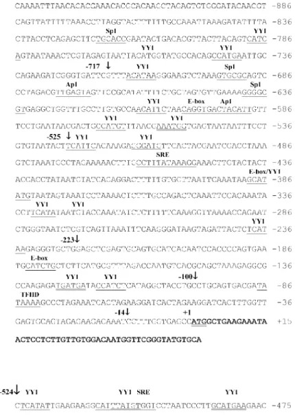
Figure 1 presents the 5'-flanking se-quences of the EgactI and EgactII actin genes. Nucleotide sequences were determined for the 935-bp and 524-bp regions upstream from the ATG (translation start codon), re-spectively (Figure 1A and B). These se-quences did not present significant homol-ogy with each other or with other known promoters whose sequences have been de-posited in the GCG data banks (12).
Computational sequence analysis of the 5'-flanking regions of EgactI and EgactII using SIGNAL SCAN (10) and MatInspector (11) revealed the presence of many binding sites for ubiquitous, developmental, cell type-specific transcription factors and putative elements that are known to contribute to the transcriptional regulation of actin genes. As indicated in Figure 1, E-box (16), SRE (17), and binding sites Sp1 (18), Ap1 (19) MEF-2 (20) and YY1 (21) were identified in both 5'-flanking sequences. However, these se-quences are positioned differentially with respect to their ATG. The EgactI gene pre-sents a canonical TATA box at nucleotide -87, while the EgactII gene promoter shows a TATA box-like sequence at nucleotide
-60. The EgactII 5'-flanking region also pre-sents a poly (dT) element at nucleotide -257. Since most of these putative regulatory ele-ments have been described to be function-ally involved in the control of actin gene regulation, we suggest their putative role in the transcriptional control of EgactI and
EgactII actin genes. Their different struc-tural organization could also suggest a spe-cific pattern of regulation. In spite of the presence of these putative regulatory ele-ments, their role in the transcriptional con-trol of these genes is unknown.
In order to study the functionality of these putative 5' regulatory regions in the tran-scriptional control of the EgactI and EgactII
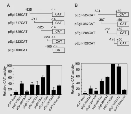
genes, we prepared a series of actin gene promoter-CAT reporter gene constructs (Fig-ure 2). The results of the functional assays are presented in Figure 2A and B. Both pEgI-935CAT and pEgII-524CAT determined a ~4-fold increase in CAT activity relative to pCAT Basic vector activity. These results showed that both 5' regions functioned as active promoters in NIH3T3 cells, suggest-ing that conserved transcription factors pre-sent in these mammalian cells recognize and bind the promoter regions promoting CAT transcription. Their promoter activities are relatively strong, since the levels of CAT expression were similar to those promoted by the SV40 promoter-CAT construct used as a positive control for these experiments (data not shown).
When tested in the presence of the SV40 enhancer sequence, the EgactII promoter activity increased approximately 5-fold, while the activity of the EgactI promoter decreased 1.2-fold when cloned in the same vector (compare pEgII-524CATE and pEgI-935CATE in Figure 2B and A). It has been reported that simultaneous Sp1 and Ap1 bind-ing to the SV40 enhancer sequence and pro-moter elements can have significant effects on gene transcription (22,23). The activation or repression of EgactI and EgactII pro-moter activity could be the result of interac-tions between Sp1 and/or Ap1 NIH3T3 tran-scription factors bound to the promoter re-gions containing the respective binding sites on the SV40 enhancer.
When these promoter sequences were tested in the reverse orientation, we observed that the 524-bp EgactII promoter fragment had activity in either direction (pEgII-524CAT and pEgII-(pEgII-524CATR, Figure 2B), as already observed for enhancers and some promoter elements of many cellular, viral and mitochondrial genes (24). The differ-ence in CAT activity between pEgII-524CATR and pCAT Basic was statistically significant, confirming the EgactII promoter fragment activity in the reverse orientation.
Figure 2 - Transient expression analysis of EgactI and EgactII promoter-CAT reporter constructs in NIH3T3 cells. EgactI (A) and EgactII (B) promoter-CAT constructs are sche-matically represented on the upper part of the figure. The respective transient expression analysis is presented under the promoter-CAT construct schemes. The names of each construct are show n on the left and numbers indicate the relative positions w ith respect to the ATG codon. Also show n is the transcriptional activity of EgactI and EgactII promoters in the reverse orientation and their activity in the presence of the SV40 enhancer, designated by the letters “ R” (reverse) and “ E” (enhancer) in the name of the corresponding con-structs. The level of CAT activity w as expressed as relative activity compared w ith the activity of plasmid constructs pEgI-100CAT or pEgII-128CAT (values set at 100). The data represent the mean and standard deviation of 3-5 independent experiments, each per-formed in duplicate and analyzed by the Student t-test.
R e la ti v e C A T a c ti v it y 100 CAT CAT CAT CAT CAT CAT CAT CAT CAT pEgI-935CAT -935 pEgI-717CAT -717 pEgI-525CAT -525 pEgI-223CAT -223 pEgI-100CAT -100 -14 -14 -14 -14 -14 pEgII-524CAT -524 pEgII-387CAT -387 pEgII-288CAT -288 pEgII-128CAT -128 +50 +50 +50 +50 A B 80 60 40 20 0 R e la ti v e C A T a c ti v it y 100 80 60 40 20 0 pC AT Bas ic pEgI -935 CA T pEgI -935 CA TR pEgI -717 CA T pEgI -525 CA T pEgI -223 CA T pEgI -100 CA T pEgI -935 CA TE pCA T E
nhan cer
pCA T B
asic pEgI I-524 CA T pEgI I-524 CA TR pEgI I-387 CA T pEgI I-288 CA T pEgI I-128 CA T pEgI I-524 CA TE pCA T E
Nevertheless, higher activities were observed when this promoter fragment was tested in the original orientation (compare pEgII-524CAT and pEgII-pEgII-524CATR, Figure 2B). This was also observed for the promoter regions of the Dictyosteliumdiscoideum ac-tin gene (24) and the human hypoxanthine-guanine phosphoribosyl transferase gene (25). The bidirectional activity of the EgactII promoter may be related to the presence of poly (dT) tracts (26), Sp1 binding sites (27), E-box elements (28) and to the lack of a consensus TATA box sequence (29), already described as characteristic promoter elements with bidirectional activity. In marked con-trast, there was no statistically significant difference between pEgI-935CATR and pCAT Basic CAT activities, indicating that the 935-bp EgactI promoter fragment pre-sents no bidirectional activity in this system (Figure 2A). The EgactI promoter sequence, in contrast to the EgactII sequence, has a classical consensus TATA box at position -87, which could have a directional role for transcription, as evidenced by other authors (23).
In order to identify the relevant promoter regions containing regulatory elements in-volved in promoter activity, we constructed a series of actin promoter 5'-deletion mu-tants-CAT reporter constructs. The 5'-dele-tions of the EgactII promoter progressively increased promoter activity to a level 5.7 times higher than that obtained with the in-tact construct (Figure 2B). This increase in promoter activity suggests that the region 524 bp upstream from the ATG contains at least three contiguous regions bearing addi-tive silencer elements: 1) -524 to -388, 2) -387 to -289 and 3) -288 to -129.
5'-Deletions from -935 to -718 and from -717 to -526 of the EgactI promoter did not affect its transcriptional activity, since there was no significant difference between the CAT activities from these constructs and the control (Figure 2), although an E-box and putative binding sites for YY1, Sp1 and Ap1
were present in the deleted regions (see Fig-ure 1). Deletions from -525 to -224 and from -223 to -101 respectively increased CAT activity to a level 1.6 and 2.7 times higher than that obtained with the wild-type pro-moter construct (compare pEgI-223CAT and pEgI-100CAT with pEgI-935CAT in Figure 2A). These results also suggest the presence of additive silencer elements in the region between -525 and -101. An inhibitory sys-tem consisting of additive negative regula-tory regions was also found in the promoters of the actin genes act5C and A3 of Drosophi-la meDrosophi-lanogaster (30) and Bombyx mori (31), respectively. These results suggest that se-quences from -525 to -101 (EgactI) and from -524 to -129 (EgactII) might be involved in the negative regulation of both promoters. Indeed, sequence analysis of both promoter regions (Figure 1) showed the presence of many putative binding sites for the con-served eukaryotic YY1 repressor protein in the deleted regions that contained putative silencer elements. Based on this observa-tion, we considered the possibility that the YY1 repressor protein could be involved in the negative regulation of both promoters. Previous studies have shown that the a-actin gene is regulated by the repressor activity of the YY1 protein (32-35). The YY1 protein seems to present a high degree of evolution-ary conservation, particularly striking in the C-terminal half of the molecule that includes the DNA-binding and repressor domains (21). This repressor protein is an important ponent of the transcription initiation com-plex and is also involved in many aspects of chromatin organization (36). Considering this conservation, we hypothesized that YY1 or a similar DNA-binding activity present in NIH3T3 nuclear extracts could be involved in the negative regulation of both promoters from E. granulosus actin genes.
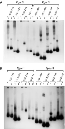
GST/YY1 recombinant protein and the NIH3T3 nuclear extract. Both extracts formed DNA-protein complexes with the YY1 consensus sequence (data not shown). Taken together, these results seem to suggest that the putative binding sites for YY1 pro-tein found in EgactI and EgactII promoter sequences may be required for protein-DNA interaction and may be involved in the tran-scriptional suppression of both actin genes. Further experiments will be necessary to determine whether the factors involved in mediating this repression of EgactI and
EgactII promoters are present in nuclear ex-tracts from E. granulosus cells and to test the specificity and identity of binding of the proteins that are forming the retarded com-plexes with the probes.
The results presented here show, for the first time, the structural and functional char-acterization of cestode promoter sequences. Our study employing mammalian cells rep-resents the first indication that the basic transcriptional control mechanisms of Echi-nococcus genes operate efficiently in a mam-malian context. The successful use of a het-erologous transient expression system to ex-amine the transcriptional activity of these two E. granulosus actin gene promoters can be mostly attributed to the well-known high degree of conservation of basic transcrip-tional mechanisms in eukaryotes, especially those related to transcription initiation (37, 38). Our results concerning the functionality of both 5'-flanking regions indicate that con-served transcription factors present in NIH3T3 nuclear extracts are able to recog-nize, bind and finally activate gene tran-scription from both promoter regions. Al-though very little information is available about gene regulation in E. granulosus, DNA sequences that code for homeobox and GATA transcription factors have already been isolated from cDNA libraries from this worm (39,40). These sequences show a high level of identity to their mammalian counter-parts, indicating that cestode and
mamma-A
-93 5/-7
18
-71 7/-5
26
-52 4/-3
89
-38 7/-2
92
-28 8/-1
31
-12 8/+
50
1 2 1 2 1 2 1 2 1 2 1 2 1 2 1 2
-52 5/-2
24
-22 3/-1
4
EgactI EgactII
B
-93 5/-7
18
-71 7/-5
26
-52 4/-3
89
-38 7/-2
92
-28 8/-1
31
-12 8/+
50
1 2 1 2 1 2 1 2 1 2 1 2 1 2 1 2
-52 5/-2
24
-22 3/-1
4
EgactI EgactII Figure 3 - Binding properties of
NIH3T3 protein nuclear extracts and YY1 recombinant protein in
EgactI and EgactII promoter re-gions. EgactI and EgactII pro-moter regions w ere retarded by NIH3T3 protein nuclear extracts and by YY1 recombinant protein.
EgactI and EgactII [32P]-labeled
promoter fragments (lanes 2) w ere used as probes in a gel mobility shift assay experiment in t he presence of 5 µg of NIH3T3 nuclear extract (panel A, lanes 1) or GST/YY1 recombinant protein (panel B, lanes 1) and resolved on a native 6% poly-acrylamide gel. Four delimited promoter regions (EgactI on the left and EgactII on the right) are show n on the top of the figure. The intense band at the bottom of each lane corresponds to the probe run in the absence of pro-tein.
lian cells present similar transcription fac-tors involved in the basic mechanisms of transcription regulation. For these reasons, the use of a heterologous system to test cestode promoter activity can be of wide interest to elucidate many basic and con-served aspects of transcriptional regulation in these organisms. These studies can also contri-bute to the efforts to better understand the basic molecular mechanisms involved in the control of gene expression in E. granulosus.
Ackno wle dgm e nts
The authors are grateful to Dr. V. Martins for providing NIH3T3 cells and the pCMVßgal plasmid and to Dr. Thomas Shenk for providing the clone containing the hu-man YY1 recombinant protein. We thank Dr. J. Rodrigues for helpful discussions and Dr. Franklin Rumjanek for a critical reading of the manuscript.
Re fe re nce s
1. Thompson RCA (1995). Biology and sys-tematics of Echinococcus granulosus. In: Thompson RCA (Editor), Echinococcus and Hydatid Disease. Cab International, Wallingford, UK.
2. Gustafson TA & Kedes L (1989). Identifi-cation of multiple proteins that interact w ith functional regions of the human car-diac a-actin promoter. M olecular and Cel-lular Biology, 9: 3269-3283.
3. Herman IM (1993). Actin isoforms. Cur-rent Opinion in Cell Biology, 5: 48-55. 4. Silva CM D, Ferreira HB, Picón M ,
Gorfinkiel N, Ehrlich R & Zaha A (1993). M olecular cloning and characterization of actin genes from Echinococcus granulo-sus. M olecular and Biochemical Parasitol-ogy, 60: 209-219.
5. Serra E, Zemzoumi K & Dissous C (1997). Deletion analysis of the Schistosoma mansoni 28-kDa glutathione S-transferase gene promoter in mammalian cells. Im-portance of a proximal activator-protein-1 site. European Journal of Biochemistry, 248: 113-119.
6. Garcia-Saez A, Perona R & Sastre L (1997). Polymorphism and structure of the gene coding for the alpha 1 subunit of the
Artemia franciscana Na/K-ATPase. Bio-chemical Journal, 321: 509-518. 7. Fatyol K, Illes K & Szalay AA (1999). An
alt ernat ive int ronic prom ot er of t he
Bombyx A3 cytoplasmic actin gene exhib-its a high level of transcriptional activity in mammalian cells. M olecular and General Genetics, 261: 337-345.
8. Coutinho CC, Seack J, Van de Vyver G, Bojojevic R & M uller WE (1998). Origin of the metazoa bodyplan: Characterization and functional testing of the promoter of the homeobox gene EmH-3 from the freshw ater sponge Ephydatia muelleri in
mouse 3T3 cells. Biological Chemistry, 379: 1243-1251.
9. Sambrook J, Fritsch EF & M aniatis T (1989). M olecular Cloning. A Laboratory M anual. 2nd edn. Cold Spring Harbor Laboratory Press, Cold Spring Harbor, NY. 10. Prestridge DS (1991). SIGNAL SCAN: a computer program that scans DNA se-quences for eukaryotic transcriptional ele-ments. Computer Applications in the Bio-sciences, 7: 203-206.
11. Quandt K, Frech K, Karas H, Wingender E & Werner T (1995). M atInd and M at-Inspector: new fast and versatile tools for detection of consensus matches in nucle-otide sequence data. Nucleic Acids Re-search, 23: 4878-4884.
12. Gartmann CJ & Grob U (1991). A menu-shell for the GCG programs. Computer Applications in the Biosciences, 7: 457-460.
13. Ausubel F, Brent R, Kingston RE, M oore DD, Seidman JG, Smith JA & Struhl DK (1995). Short Protocols in M olecular Biol-ogy. A Compendium of M ethods from Current Protocols in M olecular Biology. John Wiley and Sons, Inc., New York. 14. Bradford M M (1976). A rapid and
sensi-tive method for the quantitation of micro-gram quantities of protein utilizing the principle of protein-dye binding. Analytical Biochemistry, 72: 248-254.
15. Shi Y, Seto E, Chang LS & Shenk T (1991). Transcriptional repression by YY1, a hu-man GLI-Krüppel-related protein, and re-lief of repression by adenovirus E1A pro-tein. Cell, 67: 377-388.
16. Lem ercier C, To RQ, Carrasco RA & Konieczny SF (1998). The basic helix-loop-helix transcription factor M ist1 functions as a transcriptional repressor of M yoD.
EM BO Journal, 17: 1412-1422.
17. Brow ning CL, Culberson DE, Aragon IV, Fillmore RA, Croissant JD, Schw artz RJ & Zimmer WE (1998). The developmentally regulated expression of serum response factor plays a key role in the control of smooth muscle-specific genes. Develop-mental Biology, 194: 18-37.
18. Pugh BF & Tjian R (1990). M echanism of transcriptional activation by Sp1: evidence for coactivators. Cell, 61: 1187-1197. 19. Paradis P, M acLellan WR, Belaguli NS,
Schw artz RJ & Schneider M D (1996). Se-rum response factor mediates AP-1-de-pendent induction of the skeletal a-actin promoter in ventricular myocytes. Journal of Biological Chem istry, 271: 10827-10833.
20. Akkila WM , Chambers RL, Ornatsky OI & M cDermott JC (1997). M olecular cloning of up-regulated cytoskeletal genes from regenerating skeletal muscle: potential role of myocyte enhancer factor 2 pro-teins in the activation of muscle-regen-eration-associated genes. Biochemical Journal, 325: 87-93.
21. Shi Y, Lee JS & Galvin KM (1997). Every-thing you ever w anted to know about Ying Yang 1. Biochimica et Biophysica Acta, 1332: F49-F66.
22. Lee W, Haslinger A, Karin M & Tjian R (1987). Activation of transcription by tw o factors that bind promoter and enhancer sequences of the human metallothionein gene and SV40. Nature, 325: 368-372. 23. Jin Y, Pasumarthi KBS, Bock M E, Chen Y,
Kardami E & Cattini PA (1995). Effect of “ enhancer” sequences on ventricular myosin light chain-2 promoter activity in heart muscle and nonmuscle cells. Bio-chemical and Biophysical Research Com-munications, 210: 260-266.
(1993). Bi-directional transcription from actin promoters in Dictyostelium. Bio-chimica et Biophysica Acta, 1216: 105-109.
25. Johnson P & Friedmann T (1990). Limited bidirectional activity of tw o housekeeping gene promoters: human HPRT and PGK.
Gene, 88: 207-213.
26. Iyer V & Struhl K (1995). Poly (dA:dT), a ubiquitous promoter element that stimu-lates transcription via its intrinsic DNA structure. EM BO Journal, 14: 2570-2579. 27. Harrison L, Ascione AG, Takiguchi Y, Wil-son DM , Chen DJ & Demple B (1997). Com parison of the prom oters of the mouse (APEX) and human (APE) apurinic endonuclease genes. M utation Research, 385: 159-172.
28. Li N & Seetharam B (1998). A 69-base pair fragment derived from human transcobal-amin II promoter is sufficient for high bi-directional activity in the absence of a TATA box and an initiator element in trans-fected cells. Role of an E-box in transcrip-tional activity. Journal of Biological Chem-istry, 273: 28170-28177.
29. Grichnik JM , French BA & Schw artz RJ (1988). The chicken skeletal alpha-actin gene promoter region exhibits partial dyad symmetry and a capacity to drive bi-direc-tional transcription. M olecular and Cellu-lar Biology, 8: 4587-4597.
30. Chung YT & Keller EB (1990). Positive and negative regulatory elements mediating transcription from the Drosophila melano-gaster actin 5C distal promoter. M olecu-lar and Celluolecu-lar Biology,10: 6172-6180. 31. M angé A, Julien E, Prudhomme JC &
Couble P (1997). A strong inhibitory ele-m ent dow n-regulat es SRE-st iele-m ulat ed transcription of the A3 cytoplasmic actin gene of Bombyx mori. Journal of M olecu-lar Biology, 265: 266-274.
32. Lee T, Shi Y & Schw artz RJ (1992). Dis-placement of BrdUrd-induced YY1 by se-rum response factor activates skeletal a -actin transcription in embryonic myo-blasts. Proceedings of the National Acad-emy of Sciences, USA, 89: 9814-9818. 33. M cLellan WR, Lee T, Schw artz RJ &
Schneider M D (1994). Transf orm ing grow th factor-ß response elements of the skeletal a-actin gene. Combinatorial ac-tion of serum response factor, YY1, and the SV40 enhancer-binding protein, TEF-1. Journal of Biological Chemistry, 269: 16754-16760.
34. Bushel P, Kim JH, Chang W, Catino JJ, Ruley HE & Kumar CC (1995). Tw o serum response elements mediate transcrip-tional repression of human smooth alpha-actin promoter in ras-transformed cells.
Oncogene, 10: 1361-1370.
35. Chen CV & Schw artz RJ (1997).
Competi-tion betw een negative acting YY1 versus positive acting serum response factor and tinman NKx-2.5 regulates cardiac alpha-actin promoter activity. M olecular Endo-crinology, 11: 812-822.
36. Austen M , Luscher B & Luscher-Firzlaff JM (1997). Characterization of the tran-scriptional regulator YY1. The bipartite transactivation domain is independent of interaction w ith the TATA box-binding pro-tein, transcription factor IIB, TAFII55, or cAM P-responsive element-binding pro-tein (CBP)-binding propro-tein. Journal of Bio-logical Chemistry, 272: 1709-1717. 37. Schena M (1989). The evolutionary
con-servation of eukaryotic gene transcription.
Experientia, 45: 972-983.
38. Guarentee L & Bermingham-M cDonogh O (1992). Conservation and evolution of transcriptional mechanisms in eukaryotes.
Trends in Genetics, 8: 27-32.
39. Oliver G, Vispo M , M ailhos A, M artinez C, Sosa-Pineda B, Fielitz W & Ehrlich R (1992). Homeoboxes in flatw orms. Gene, 121: 337-342.