Regulation of gap junctions by
protein phosphorylation
Departamento de Ciencias Fisiológicas,
Pontificia Universidad Católica de Chile, Santiago, Chile J.C. Sáez,
A.D. Martínez, M.C. Brañes and H.E. González
Abstract
Gap junctions are constituted by intercellular channels and provide a pathway for transfer of ions and small molecules between adjacent cells of most tissues. The degree of intercellular coupling mediated by gap junctions depends on the number of gap junction channels and their activity may be a function of the state of phosphorylation of connexins, the structural subunit of gap junction channels. Protein phosphorylation has been proposed to control intercellular gap junc-tional communication at several steps from gene expression to protein degradation, including translational and post-translational modifica-tion of connexins (i.e., phosphorylamodifica-tion of the assembled channel acting as a gating mechanism) and assembly into and removal from the plasma membrane. Several connexins contain sites for phosphoryla-tion for more than one protein kinase. These consensus sites vary between connexins and have been preferentially identified in the C-terminus. Changes in intercellular communication mediated by protein phosphorylation are believed to control various physiological tissue and cell functions as well as to be altered under pathological conditions.
Correspondence J.C. Sáez
Departamento de Ciencias Fisiológicas
Pontificia Universidad Católica de Chile
Alameda 340 Santiago Chile
Fax: (562) 222-5515 E-mail: [email protected]
Presented at the XII Annual Meeting of the Federação de Sociedades de Biologia Experimental, Caxambu, MG, Brasil, August 27-30, 1997.
J.C. Sáez, A.D. Martínez and M.C. Brañes are recipients of FONDECYT grants (Nos. 1960-559, 2960-001 and 2960-002, respectively).
Received September 10, 1997 Accepted September 22, 1997
Key words •Gap junctions •Connexins
•Protein phosphorylation •Sites of phosphorylation •Protein kinases
•Cell-to-cell communication
Introduction
Most cells, with the exception of few types, such as spermatozoids, red blood cells, and skeletal muscle of adult vertebrates, can communicate to adjacent cells by gap junc-tions. These membrane specializations, also referred to as nexuses or macula communi-cans, contain intercellular channels which mediate movement of ions and small mol-ecules (<1.2 kDa) between contacting cells. Each channel is formed by two hemichan-nels or connexons and each one of them is
contributed by one of the two adjacent cells. A connexon is an oligomeric assembly of six polypeptide subunits termed connexins (Cxs) which are highly homologous and are en-coded by a gene family (1,2). While a par-ticular Cx (e.g., Cx43) can be expressed by a wide spectrum of tissues and cell types (3), the expression of some other Cxs (e.g., Cx33, Cx50 and Cx30.1, found in testis, lenses and skin, respectively) is apparently much more restricted (4,5).
tran-script is translated in Xenopus oocytes, usu-ally the expression of a single Cx type is sufficient to establish intercellular gap junc-tional communication (2). Frequently, a single cell can express more than one Cx, which can be localized in the same (6,7) or in different gap junction plaques (8). More-over, functional gap junctions can be estab-lished in an exogenous expression system (4,5,9-17) where the role of protein phos-phorylation in channels formed by a particu-lar Cx can be analyzed at the functional level.
The extent to which cells are functionally coupled by gap junction channels depends on a multiplicity of control mechanisms, including gene transcription, message stabil-ity, translational and post-translational modi-fication of the protein, and assembly into and removal from the membrane. In addition, a number of factors affect gating of assembled channels (1). Analysis of mechanism and regulation of each of these steps has become a key element in the study of gap junctions. Gap junction regulatory mechanisms can lead to an increase or reduction of intercellu-lar coupling with a wide spectrum of time courses (from milliseconds to hours) (18). The turnover of Cx26, Cx32 and Cx43 is between 2 and 5 h (7,19-25). Hence, changes in intercellular coupling that occur within a time course of a few hours in cell types expressing these Cxs could involve any of the steps in Cx biosynthesis from transcrip-tion to degradatranscrip-tion. It has been shown that changes in Cx mRNA transcription rate and mRNA stability can be affected by the acti-vation of intracellular second messenger path-ways and affect intercellular gap junctional communication within a few hours. Although these changes could involve protein phos-phorylation they will not be presented in this article and readers are referred to reviews published elsewhere (1,18).
Formation of gap junctions also requires appropriate cell adhesion mediated by either Ca2+-dependent (NCAMs) or Ca2+-
independ-ent (cadherins) cell adhesion molecules (23,26-30). Cell lines deficient in cell adhe-sion molecules do not assemble gap junc-tions and Cx43 localizes in a perinuclear cytoplasmic compartment (23,29), where it is found preferentially in its unphosphory-lated form (23). Transfection with cDNA encoding a cell adhesion molecule induces at least two changes in Cx43: it promotes i) the insertion of Cx43 into the plasma mem-brane and assembly into gap junctions (23,29) and ii) phosphorylation of Cx43 (23). Al-though phosphorylation might play a role regulating the insertion and/or assembly of a phospho-Cx into the plasma membrane (23), this might not be an absolute requirement. For example, in exogenous expression sys-tems the expression of mRNAs encoding Cx43 or Cx32 mutants with a shortened carboxyl terminal (devoid of all phosphory-latable seryl residues) is able to induce inter-cellular coupling (12,17). In addition, it has been reported that in an MDCK cell line, while formation of gap junctions depends on cell contact, phosphorylation of Cx43 by a protein kinase C-dependent pathway can occur in the absence of Ca2+-dependent cell
adhesion activity (30) and does not correlate with expression of intercellular coupling.
Phosphorylation of Cxs and possible functional roles
Cx26 has been reported not to be phos-phorylated in rat hepatocytes (7) or in iso-lated mouse gap junctions treated with cAMP-dependent kinase (cAMP-dPK) (7,34), protein kinase C (PKC) or Ca2+
/calmodulin-dependent kinase II (Ca2+-CM-dPK II) (34).
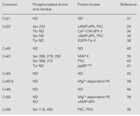
Presently, nine Cxs are known to be phos-phoproteins and their state of phosphoryla-tion can be affected by different protein kinases (Table 1). Nonetheless, only few phosphorylation sites and protein kinases that phosphorylate them have been identi-fied.
Various phospho-forms of Cx43 can be resolved by immunoblotting, making it a useful technique to evaluate its state of phos-phorylation (13-15,20,25,29,30,33,35,37,41, 42,45-56). Nonetheless, not all changes in the state of phosphorylation of Cx43 alter the electrophoretic mobility of the protein (42). In addition, immunoblots do not allow further analysis of the different Cx43 phospho-forms (i.e., amino acid analysis and two-dimensional tryptic maps). Therefore, analysis of the state of phosphorylation of Cxs, including Cx43, requires complemen-tary studies with metabolic radiolabelling followed by immunoprecipitation.
Cyclic nucleotides, diacylglycerol, lyso-phosphatidic acid, tumor promoter phorbol esters, growth factors, and oncogene products (pp60v-src, p130gag-fps and ras
oncogen) alter intercellular gap junctional communication and effect of some these agents depends on cell and Cx type (13,16, 20,30,34-36,38,41-47,51-71). It has also been suggested that phosphorylation of Cx43 in seryl residues might be involved in a number of different processes including its insertion into the plasma membrane (56), increase in its degradation, changes in unitary conduc-tance of single gap junction channels and closure of gap junction channels (13,16, 45,57,61,62). In addition, phosphorylation of tyrosine residues has been associated with reduction in intercellular coupling (10,20).
Effect of Cx32 phosphorylation on intercellular gap junctional communication
Cx32 was one of the first channel-forming proteins shown to be phosphorylated in intact cells (63). Activation of either cAMP-dPK or PKC increases the state of phosphorylation of Cx32 (34,63,70,72) and at least the effect of cAMP-dPK is tempo-rally correlated with an increased junctional conductance (gj). Moreover, Chanson et al.
(71) reported that an increase in intracellular [cAMP] in a human colonic T84 cell line induces a rapid (<20 min) increase in inter-cellular gap junctional communication me-diated by Cx32 gap junctions, that is directly related to an increase in fluid secretion.
Table 1 - Connexins known to be phosphoproteins.
Phosphorylated amino acid residues identified in the intracellular domains of connex-ins are indicated. Isolated gap junctions, fusion proteconnex-ins or synthetic peptides were used as substrates for in vitro phosphorylation assays. Purified protein kinases used in these assays are also indicated. ND: Not determined; Ser: serine; Thr: threonine; Tyr: tyrosine; PK: protein kinase; EGFR-Tyr K: epidermal growth factor receptor Tyr kinase; cAMP-dPK: cAMP-dependent protein kinase; Ca2+-CM-dPK II: Ca2+
/calmodulin-de-pendent protein kinase type II; pp60v-src: Tyr kinase encoded by the oncogene v-src;
MAP K: mitogen-activated protein kinase.
Connexin Phosphorylated amino Protein kinase Reference acid residue
Cx31 ND ND 37
Cx32 Ser 233 cAMP-dPK, PKC 34 Thr ND Ca2+-CM-dPK II 34
Ser ND cAMP-dPK, PKC 34 Tyr ND EGFR-Tyr K 38
Cx40 ND ND 40
Cx43 Ser 266, 279, 282 MAP K 35 Ser 368, 372 PKC 42 Tyr ND pp60v-src 41
Cx45 ND ND 43
Cx45.6 ND Mg2+-dependent PK 39
Cx46 ND ND 44
Cx50 ND Mg2+-dependent PK 39
ND cAMP-dPK
cAMP increases the amount of metaboli-cally labelled Cx32 in primary cell cultures of fetal hepatocytes (70), implying either an increased rate of synthesis or a reduced rate of degradation. The stoichiometry of phos-phorylation of Cx32 in vitro is low; by PKC it approaches 1 mol/mol and by cAMP-dPK it is about 0.1 mol of Pi/mol of protein
(34,63,72). Nevertheless, the stoichiometry of phosphorylation in vivo and the cell com-partment in which Cx32 phosphorylation occurs have not been determined. Moreover, in isolated liver gap junctions previously phosphorylated by PKC, treatment with cAMP-dPK does not increase the incorpora-tion of 32P into Cx32. On the other hand, if
gap junctions are first phosphorylated by cAMP-dPK, treatment with PKC increases the incorporation of 32P into Cx32,
suggest-ing that PKC can phosphorylate other amino acyl residues besides those phosphorylated by cAMP-dPK (72). Using synthetic pep-tides corresponding to regions of the C-terminus of Cx32, it has been demon-strated that Ser 233 is phosphorylated by both cAMP-dPK and PKC (34). Other sites have not yet been identified.
A Cx32 mutant, with serine residues 233 and 240 replaced by asparagine residues, forms gap junctions in Xenopus oocytes with macroscopic gating properties (voltage de-pendence and pH sensitivity) that are indis-tinguishable from those formed by the wild-type Cx (12). These findings suggest that phosphorylation of those serine residues is not required for channel opening or clos-ing by these conditions. Nevertheless, modu-lation of gj due to changes in the assembly or
retrieval of channels into or from the plasma membrane has not been studied. Phospho-rylated Cx32 by PKC but not by cAMP-dPK is less sensitive to degradation by m and mµ-calpains (73), suggesting an alternative mechanism for the regulation of intercellular coupling. Thus, the extent to which intercel-lular coupling between Cx32-containing cells is modulated by phosphorylation requires a
more exhaustive exploration.
Although Cx32 in isolated rat liver gap junctions is also a moderate substrate for Ca2+-CM-dPK II, resulting in serine and
threonine phosphorylation (34), the state of phosphorylation of Cx32 has not been ex-haustively studied in cells treated with agents that selectively activate this kinase. Cx32 is not phosphorylated by pp60v-src in isolated
rat liver gap junctions (Sáez JC, Nairn AC and Hertzberg EL, unpublished observation) or in Xenopus oocytes (10). Nevertheless, tyrosyl phosphorylation of Cx32 can occur in isolated liver gap junctions treated with the epidermal growth factor receptor tyrosine kinase (38). The functional consequence of Cx32 tyrosyl phosphorylation remains un-raveled.
Effect of Cx43 phosphorylation on intercellular gap junctional
communication
In cell lines and in neonatal (25,42) cardiac myocytes (33,50), two phosphorylated forms of Cx43 with slower electrophoretic mobili-ties (43-47 kDa) than the unphosphorylated form (41 kDa) can be identified either by immunoblotting or in immunoprecipitated Cx43 from 32P-labelled cells. Immunoblot
In Rous sarcoma virus-transformed fi-broblasts, where coupling is low, all forms of Cx43 are phosphorylated in both seryl and tyrosyl residues, and threonine phosphoryla-tion of the least mobile species is also noted (20). A mutation replacing tyrosine 265 of Cx43 with phenylalanine does not prevent gap junction formation in Xenopus oocytes but it completely abolishes the inhibition of intercellular gap junctional communication and the tyrosyl phosphorylation induced by pp60v-src; gap junctions formed by wild-type
Cx43 and then exposed to src are phospho-rylated in tyrosine residues and intercellular gap junctional communication is completely inhibited (10).
Activation of cAMP-dPK or PKC leads to various effects on cells of different types, even when cells expressing the same junc-tional proteins are compared (18). Frequently, in cells where the unphosphorylated form of Cx43 (Cx43-NP) predominates under basal conditions, stimulation of PKC with a tumor promoter phorbol ester (e.g., TPA) leads to rapid cell uncoupling and shifts the electro-phoretic mobility of Cx43 forms (30,45,52, 53,55). By contrast, in cells where the phos-phorylated forms of Cx43 predominate (e.g., rat cardiocytes) TPA promotes intercellular communication with no detectable changes in the state of phosphorylation, as evaluated by Western blots (42). Nevertheless, there are exceptions: in rat leptomeningeal cells (8), and in a rat liver epithelial cell line (IRA 20) (54) the phosphorylated forms of Cx43 predominate and TPA induces uncoupling without detectable changes in the state of Cx43 phosphorylation detected by Western blots. In IRA 20 cells TPA did not change the levels of Cx43 or of its mRNA but did result in the loss of Cx43 immunoreactivity by indirect immunofluorescence (54), suggest-ing that the inhibition in intercellular gap junctional communication might involve a post-translational modification that perhaps cannot be detected in denaturing gels. There-fore, Western blot analysis might not be the
appropriate approach to study the correla-tion between all changes in phosphorylacorrela-tion. For example, the state of Cx43 phosphoryla-tion studied by Western blot in rat heart myocytes does not change in response to agents that affect the activity of protein ki-nases or phosphoprotein phosphatases, al-though treatment with some of these agents does affect the incorporation of 32P into
Cx43 (42). The stimulation of cAMP- or cGMP-dPK or PKC does not significantly increase the incorporation of 32P presumably
because Cx43 is maximally phosphorylated under basal conditions. However, the incor-poration of 32 P is greatly reduced after
inhi-bition of protein kinase activities with stau-rosporine, and can then be stimulated by TPA (42). In neonatal rat cardiac myocytes Cx43 is predominantly phosphorylated (25,42,50) and it is localized in the plasma membrane (33). Agents that affect the incor-poration of 32P into Cx43 do not affect the
distribution of the protein tested immunocy-tochemically in rat myocytes, suggesting that changes in the rate of phosphorylation de-tected with 32P
i occur within the plasma
mem-brane compartment. In all the systems men-tioned above it seems that PKC mediates the effects of TPA. In support of this, after PKC is down-regulated cells are coupled and both coupling and state of phosphorylation of Cx43 become insensitive to TPA and phos-phorylated forms of Cx43 are still detected as major components (45). Using synthetic peptides corresponding to the deduced se-quences in the C-terminal region or recom-binant fusion protein of the C-terminus of Cx43 it has been shown that residues 368 and 372 are phosphorylated by PKC but not by cAMP- or cGMP-dPK or Ca2+-CM-dPK
II (42) (Table 1). Both sites have been found mutated in visceroatrial heterotaxia and there-fore implicated in the pathogenesis of this disease (74).
presumably are the same residues phospho-rylated by cdc2 kinase, since a fusion protein of the carboxy terminal of Cx43 phosphoryl-ated either by MAP or cdc2 kinase shows identical tryptic fingerprints (42) (Table 1). Furthermore, EGF-induced cell uncoupling is mediated by MAP kinase Cx43 phosphoryla-tion in seryl residues of proline rich regions (35). A similar mechanism may operate in the PDGF-induced cell uncoupling which is PKC independent (68).
A membrane permeable derivative of cGMP reduces gj in neonatal rat myocytes
(61) and in SKHep1 cells transfected with rat Cx43 but not in SKHep1 cells transfected with human Cx43 (14). The state of phos-phorylation of Cx43 expressed by SKHep1 cells stably transfected with rat Cx43 cDNA is increased by 8Br-cGMP but the site of phosphorylation is unknown as also is its
interaction with other sites of phosphoryla-tion by other protein kinases. In rat but not in human Cx43 the seryl residue 257 located in the carboxy terminal is flanked by proline and lysine, making it a possible site for phos-phorylation by cGMP-dependent protein ki-nase. Although Cx43 does not present an obvious consensus site for cAMP-dPK phos-phorylation, an increase in intracellular [cAMP] increases gj in neonatal rat
myo-cytes (57) and induces phosphorylation of Cx43 in the ovary (47).
Finally, indications that phosphoprotein phosphatases 1 and 2A participate in de-phosphorylation of Cx43 have been obtained. In MDCK cells, treatment with okadaic acid, an inhibitor of these phosphatases, potenti-ates the increase in the relative amount of phosphorylated Cx43 induced by activation of a PKC-dependent pathway (30).
References
1. Bennett MVL, Barrio LC, Bargiello TA, Hertzberg EL & Sáez JC (1991). Gap junc-tions: new tools, new answers, new ques-tions. Neuron, 6: 305-320.
2. Bruzzone R, White TH & Paul DL (1996). Connections with connexins: the molecu-lar basis of direct intercellumolecu-lar signaling.
European Journal of Biochemistry, 238: 1-27.
3. Beyer EC, Kistler J, Paul DL & Goodenough DA (1989). Antisera directed against connexin43 peptides react with a 43-kD protein localized to gap junctions in myocardium and other tissues. Journal of Cell Biology, 108: 595-605.
4. Haefliger JA, Bruzzone R, Jenkins NA, Gilbert DJ, Copeland NG & Paul DL (1992). Four novel members of the connexin fam-ily of gap junction proteins. Molecular cloning, expression, and chromosome mapping. Journal of Biological Chemistry, 267: 2057-2064.
5. White TW, Bruzzone R, Goodenough DA, & Paul DL (1992). Mouse Cx50, a func-tional member of the connexin family of gap junction proteins, is the lens fiber protein MP70. Molecular Biology of the Cell, 3: 711-720.
6. Nicholson BJ, Dermietzel R, Teplow D, Traub O, Willecke K & Revel J-P (1987). Two homologous protein components of hepatic gap junctions. Nature, 329:
732-734.
7. Traub O, Look J, Dermietzel R, Bümmer F, Hülser D & Willecke K (1989). Com-parative characterization of the 21-kD and 26-kD gap junction proteins in murine liver and cultured hepatocytes. Journal of Cell Biology, 108: 1039-1051.
8. Spray DC, Moreno AP, Kessler JA & Dermietzel R (1991). Characterization of gap junctions between cultured leptome-ningeal cells. Brain Research, 568: 1-14. 9. Dahl G, Werner R, Levine E &
Rabadan-Diehl C (1992). Mutational analy-sis of gap junctional formation. Biophysi-cal Journal, 62: 172-182.
10. Swenson KI, Piwnica-Worms H, McNamee H & Paul DL (1990). Tyrosine phosphorylation of the gap junction pro-tein connexin43 is required for the pp60v-src-induced inhibition of communi-cation. Cell Regulation, 1: 989-1002. 11. Eghbali B, Kessler JA & Spray DC (1990).
Expression of gap junction channels in communication-incompetent cells after stable transfection with cDNA encoding connexin 32. Proceedings of the National Academy of Sciences, USA, 87: 1328-1331.
12. Werner R, Levine E, Rabadan-Diehl C & Dahl G (1991). Gating properties of con-nexin32 cell-cell channels and their mu-tants expressed in Xenopus oocytes.
Pro-ceedings of the Royal Society of London, 243: 5-11.
13. Kwak BR, Hermans MMP, De Jonge HR, Lohmann SM, Jongsma H & Chanson M (1995). Differential regulation of distinct types of gap junction channels by similar phosphorylating conditions. Molecular Bi-ology of the Cell, 6: 1707-1719. 14. Kwak BR, Sáez JC, Wilders RW, Chanson
M, Fishman GL, Hertzberg EL, Spray DC & Jongsma HJ (1995). cGMP-dependent phosphorylation of connexin43: influence on gap junction channels conductance and kinetics. Pflügers Archiv. European Jour-nal of Physiology, 430: 770-778. 15. Moreno AP, Fishman GI & Spray DC
(1992). Phosphorylation shifts unitary con-ductance and modifies voltage depend-ent kinetics of human connexin43 gap junction channels. Biophysical Journal, 62: 1-3.
16. Moreno AP, Sáez JC, Fishman GI & Spray DC (1994). Human connexin43 gap junc-tion channels. Regulajunc-tion of unitary con-ductances by phosphorylation. Circulation Research, 74: 1050-1057.
Spray DC (1993). Gap junctions: multiplic-ity of controls in differentiated and undif-ferentiated cells and possible functional implications. In: Shenolikar S & Nairn AC (Editors), Advance in Second Messenger Phosphoprotein Research. Raven Press, New York, 163-198.
19. Fallon RF & Goodenough DA (1981). Five hour half-life of mouse liver gap junction.
Journal of Cell Biology, 90: 521-526. 20. Crow DS, Beyer EC, Paul DL, Kobe SS &
Lau AF (1990). Phosphorylation of con-nexin43 gap junction protein in uninfected and Rous sarcoma virus-transformed mammalian fibroblasts. Molecular and Cellular Biology, 10: 1754-1763. 21. Laird DW, Castillo M & Kasprzak L (1995).
Gap junction turnover, intracellular traf-ficking, and phosphorylation of con-nexin43 in brefeldin A-treated rat mam-mary tumor cells. Journal of Cell Biology, 131: 1191-1203.
22. Musil LS, Beyer EC & Goodenough DA (1990). Expression of the gap junction pro-tein connexin43 in embryonic chick lens: molecular cloning, ultrastructural localiza-tion, and post-translational phosphoryla-tion. Journal of MembraneBiology, 116: 163-175.
23. Musil LS, Cunningham BC, Edelman GM & Goodenough DA (1990). Differential phosphorylation of the gap junction pro-tein connexin43 in junctional communica-tion-competent and -deficient cell lines.
Journal of Cell Biology, 111: 2077-2088. 24. Musil LS & Goodenough DA (1991).
Bio-chemical analysis of connexin43, intracel-lular transport, phosphorylation, and as-sembly into gap junctional plaques. Jour-nalofCellBiology, 115: 1357-1374. 25. Laird DW, Puranam KL & Revel JP (1991).
Turnover and phosphorylation dynamics of connexin43 gap junction protein in cul-tured cardiac myocytes. Biochemical Jour-nal, 273: 67-72.
26. Imhof BA, Vollmers PH, Goodmand SL & Birchmeir W (1983). Cell-cell interaction and polarity of epithelial cells: specific per-turbation using a monoclonal antibody.
Cell, 35: 667-675.
27. Mege RM, Matsuzaki F, Gallin WF, Golber JI, Cunningham BA & Edelman GM (1988). Construction of epithelioid sheets by transfection of mouse sarcoma cells with cDNAs for chicken cell adhesion mol-ecules. Proceedings of the National Acad-emy of Sciences, USA, 85: 7274-7278. 28. Keane RW, Mehta PP, Rose B, Honig LS,
Loewenstein WR & Rutishauser U (1988). Neuronal differentiation, N-CAM-medi-ated adhesion and gap junctional
commu-nication in neuroectoderm. A study in vi-tro. Journal of Cell Biology, 106: 1307-1319.
29. Jongen WMF, Fitzgeral DJ, Asamoto M, Piccoli C, Slaga JT, Gros D, Takeichi M & Yamasaki H (1991). Regulation of con-nexin 43-mediated gap junctional intercel-lular communication by Ca2+ in mouse
epidermal cells is controlled by E-cad-herin. Journal of Cell Biology, 114: 545-555.
30. Berthoud VM, Ledbetter MLS, Hertzberg EL & Sáez JC (1992). Connexin43 in MDCK cells: regulation by a tumor-promoting phorbol ester and Ca2+. Euro-pean Journal of Cell Biology, 57: 40-50. 31. Zimmerman DB, Green CR, Evans WH &
Gilula N (1987). Topology of the 32-kD liver gap junction protein in isolated rat liver gap junctions and gap-derived single membrane structures. Journal of Biologi-cal Chemistry, 262: 7751-7763.
32. Hertzberg EL, Disher RM, Tiller AA, Zhou Y & Cook RG (1988). Topology of the Mr 27,000 liver gap junction protein. Journal of Biological Chemistry, 263: 19105-19111.
33. Yancey SB, John SA, Lal R, Austin BJ & Revel J-P (1989). The 43-kD polypeptide of heart gap junctions: immunolocaliza-tion, topology, and functional domains.
Journal of Cell Biology, 108: 2241-2254. 34. Sáez JC, Nairn AC, Czernik AJ, Spray DC,
Hertzberg EL, Greengard P & Bennett MVL (1990). Phosphorylation of connexin 32, a hepatocyte gap-junction protein, by cAMP-dependent protein kinase, protein kinase C and Ca2+/calmodulin-dependent
protein kinase II. EuropeanJournal of Bio-logical Chemistry, 192: 263-273. 35. Warn-Cramer BJ, Lampe PD, Kurata WE,
Kanemitsu MY, Loo LWM, Eckhart W & Lau AF (1996). Characterization of the mitogen-activated protein kinase phos-phorylation sites on the connexin-43 gap junction protein. Journal of Biological Chemistry, 271: 3779-3786.
36. Berthoud VM, Beyer EC, Kurata WE, Lau AF & Lampe PD (1997). The gap-junction protein connexin 56 is phosphorylated in the intracellular loop and the car-boxy-terminal region. European Journal of Biochemistry, 244: 89-97.
37. Traub O, Butterweck A, Elfgang C, Hertlein B, Balzer K, Gergs U, Hafemann B & Willecke K (1995). Immunochemical characterization of connexin31, -37, -40, -43, and -45 in cultured primary cells, transfected cell lines and murine tissues. In: Kano Y, Katoaka K, Shiba Y, Shibata Y & Shimazu T (Editors), Progress in Cell
Biology. Vol. 4. Elsevier Science Publish-ers B.V., Amsterdam, 343-347.
38. Díez JA, Elvira M & Villalobo A (1995). Phosphorylation of connexin-32 by the epidermal growth factor tyrosine kinase.
Annals of the New York Academy of Sci-ences, 766: 477-480.
39. Arneson ML, Cheng HL & Louis CF (1995). Characterization of the ovine-lens plasma membrane protein kinase sub-strate. European Journal of Biochemistry, 234: 670-679.
40. Traub O, Eckert R, Lichtenberg-Frate H, Elfgang C, Bastide B, Scheidtman KH, Hülser BF & Willecke K (1994). Immu-nochemical and electrophysiological char-acterization of murine connexin 40 and 43 in mouse tissues and transfected human cells. European Journal of Cell Biology, 64: 101-112.
41. Loo LWM, Berestecky JM, Kanemitsu MY & Lau AF (1995). pp60src-mediated
phos-phorylation of connexin 43, a gap junction protein. Journal of Biological Chemistry, 270: 12751-12761.
42. Sáez JC, Nairn AC, Czernik AJ, Fishman GI, Spray DC & Hertzberg EL (1997). The functional state of gap junctions between neonatal rat cardiac myocytes is directly related to changes in the state of phos-phorylation of connexin43. Journal of Mo-lecular and Cellular Cardiology, 29: 2131-2145.
43. Laing JG, Westphale EM, Enelmann GL & Beyer EC (1994). Characterization of the gap junction protein, connexin45. Journal of Membrane Biology, 139: 31-40. 44. Jiang JX, Paul DL & Goodenough DA
(1993). Posttranslational phosphorylation of lens fibre connexin46: a slow occur-rence. Investigative Ophthalmology and Visual Science, 34: 3558-3565.
45. Berthoud VM, Rook M, Hertzberg EL & Sáez JC (1993). On the mechanism of cell uncoupling induced by a tumor promoter phorbol ester in clone 9 cells, a rat liver epithelial cell line. European Journal of Cell Biology, 62: 384-396.
46. Berthoud VM & Sáez JC (1993). Regula-tion of connexin43, the main gap junc-tional protein between astrocytes, during pineal ontogeny. Journal of Pineal Re-search, 14: 67-72.
47. Granot I & Dekel N (1994). Phosphoryla-tion and expression of connexin-43 ova-rian gap junction protein are regulated by luteinizing hormone. Journal of Biological Chemistry, 269: 30502-30509.
development and by sexual hormones.
EuropeanJournalof Cellular Biology (in press).
49. Kadle R, Zhang JT & Nicholson BJ (1991). Tissue-specific distribution of differen-tially phosphorylated forms of Cx 43. Mo-lecular and Cellular Biology, 11: 363-369. 50. Lau AF, Hatch-Pigott V & Crow DS (1991).
Evidence that heart connexin43 is a phos-phoprotein. Journal of Molecular and Cel-lular Cardiology, 23: 659-663.
51. Lau AF, Kanemitsu MY, Kurata WE & Danesh AL (1992). Epidermal growth fac-tor disrupts gap-junctional communication and induces phosphorylation of con-nexin43 on serine. Molecular Biology of the Cell, 3: 865-874.
52. Oh SY, Grupen CG & Murray AW (1991). Phorbol ester induces phosphorylation and down-regulation of connexin 43 in WB cells. Biochimica et Biophysica Acta, 1094: 243-245.
53. Reynout JK, Lampe PD & Johnson RG (1992). An activator of protein kinase C inhibits gap junctional communication be-tween cultured bovine lens cells. Experi-mental Cell Research, 198: 337-342. 54. Asamoto M, Oyamada M, El-Aoumari A,
Gros D & Yamasaki H (1991). Molecular mechanisms of TPA-mediated inhibition of gap-junctional intercellular communica-tion: evidence for action on the assembly or function but not the expression of con-nexin 43 in rat liver epithelial cells. Molec-ularCarcinogenesis, 4: 322-327. 55. Brissette JL, Kumar NM, Gilula NB &
Dotto GP (1991). The tumor promoter 12-O-tetradecanoylphorbol-13-acetate and the ras oncogene modulate expres-sion and phosphorylation of gap junction proteins. Molecular and Cellular Biology, 11: 5364-5371.
56. Lampe P (1994). Analyzing phorbol ester effects on gap junctional communication: a dramatic inhibition of assembly. Journal of Cell Biology, 127: 1895-1905. 57. Burt JM & Spray DC (1988). Ionotropic
agents modulate gap junctional conduc-tance between cardiac myocytes. Ameri-can Journal of Physiology, 254:
H1206-H1210.
58. Chanson M, Bruzzone R, Spray DC, Regazzi R & Meda P (1988). Cell uncou-pling and protein kinase C: correlation in a cell line but not in a differentiated tissue.
American Journal of Physiology, 255: C699-C704.
59. Chanson M, Meda P & Bruzzone R (1989). Increase in pancreatic exocrine secretion during uncoupling: evidence for a protein kinase C-independent effect. Experimen-tal Cell Research, 182: 349-357. 60. Kurata W & Lau AF (1994). p130gag-fps
disrupts gap junctional communication and induces phosphorylation of con-nexin43 in a manner similar to that of pp60v-src. Oncogene, 9: 329-335.
61. Takens-Kwak BR & Jongsma H (1992). Cardiac gap junction: three distinct single channels conductances and their modula-tion by phosphorylating treatments.
Pflügers Archiv, 422: 198-200.
62. Kwak BR, Van Veen TAB, Analbers LJS & Jongsma H (1995). TPA increases con-ductance but decreases permeability in neonatal rat cardiomyocyte gap junction channels. Experimental Cell Research, 220: 456-463.
63. Sáez JC, Spray DC, Nairn AC, Hertzberg EL, Greengard P & Bennett MVL (1986). cAMP increases junctional conductance and stimulates phosphorylation of the 27-kDa principal gap junction polypeptide.
Proceedings of the National Academy of Sciences, USA, 83: 2473-2477.
64. Maldonado PE, Rose B & Loewenstein WR (1988). Growth factors modulate junc-tional cell-to-cell communication. Journal of Membrane Biology, 106: 203-210. 65. Yada T, Rose B & Loewenstein WR
(1985). Diacylglycerol downregulates junctional membrane permeability, TMB blocks this effect. Journal of Membrane Biology, 88: 217-232.
66. Matesic DF, Rupp HL, Bonney WJ, Ruch RJ & Trosko JE (1994). Changes in gap-junction permeability, phosphoryla-tion, and number mediated by phorbol ester and non-phorbol-ester tumor pro-moters in rat liver epithelial cells.
Molecu-lar Carcinogenesis, 10: 226-236. 67. Hii CS, Oh SY, Schmidt SA, Clark KJ &
Murray AW (1994). Lysophosphatidic acid inhibits gap-junctional communication and stimulates the phosphorylation of con-nexin-43 in WB cells: possible involve-ment of the mitogen-activated protein ki-nase cascade. Biochemical Journal, 303: 475-479.
68. Pelletier DB & Boynton AL (1994). Disso-ciation of the PDGF receptor tyrosine ki-nase activity from the PDGF-mediated in-hibition of gap junctional communication.
Journal of Cell Physiology, 158: 427-434. 69. Roseng LE, Redival E & Sanner T (1993). 12-O-tetradecanoylphorbol-13-acetate-induced inhibition of gap junctional com-munication is differentially regulated in a transformation-sensitive Syrian hamster embryo cell line compared to early pas-sage SHE cells. Carcinogenesis, 14: 1851-1855.
70. Traub O, Look J, Paul D & Willecke K (1987). Cyclic adenosine monophosphate stimulates biosynthesis and phosphoryla-tion of the 26 kDa gap juncphosphoryla-tion protein in cultured mouse hepatocytes. European Journal of CellBiology, 43: 48-54. 71. Chanson M, White MM & Garber SS
(1996). cAMP promotes gap junctional coupling in T84 cells. American Journal of Physiology, 271: C533-C539.
72. Takeda A, Saheki S, Yamamura H & Shimazu T (1989). Phosphorylation of the 27-kDa gap junction protein by protein ki-nase C in vitro and in rat hepatocytes.
JournalofBiochemistry, 106: 723-727. 73. Elvira M, Díez JA, Wang KKW & Villalobo
A (1993). Phosphorylation of connexin-32 by protein kinase C prevents its proteoly-sis by mµ-calpain and m-calpain. Journal of Biological Chemistry, 268: 14294-14300.