Control of the phosphorylation of
the astrocyte marker glial fibrillary
acidic protein (GFAP) in the immature
rat hippocampus by glutamate and
calcium ions: possible key factor in
astrocytic plasticity
Departamento de Bioquímica, Instituto de Biociências,
Universidade Federal do Rio Grande do Sul, 90046-900 Porto Alegre, RS, Brasil R. Rodnight, C.A. Gonçalves,
S.T. Wofchuk and R. Leal
Abstract
The present review describes recent research on the regulation by glutamate and Ca2+ of the phosphorylation state of the intermediate
filament protein of the astrocytic cytoskeleton, glial fibrillary acidic protein (GFAP), in immature hippocampal slices. The results of this research are discussed against a background of modern knowledge of the functional importance of astrocytes in the brain and of the structure and dynamic properties of intermediate filament proteins. Astrocytes are now recognized as partners with neurons in many aspects of brain function with important roles in neural plasticity. Site-specific phos-phorylation of intermediate filament proteins, including GFAP, has been shown to regulate the dynamic equilibrium between the polymer-ized and depolymerpolymer-ized state of the filaments and to play a fundamen-tal role in mitosis. Glutamate was found to increase the phosphoryla-tion state of GFAP in hippocampal slices from rats in the post-natal age range of 12-16 days in a reaction that was dependent on external Ca2+. The lack of external Ca2+ in the absence of glutamate also
increased GFAP phosphorylation to the same extent. These effects of glutamate and Ca2+ were absent in adult hippocampal slices, where the
phosphorylation of GFAP was completely Ca2+-dependent. Studies
using specific agonists of glutamate receptors showed that the gluta-mate response was mediated by a G protein-linked group II metabotro-pic glutamate receptor (mGluR). Since group II mGluRs do not act by liberating Ca2+ from internal stores, it is proposed that activation of the
receptor by glutamate inhibits Ca2+ entry into the astrocytes and
consequently down-regulates a Ca2+-dependent dephosphorylation
cascade regulating the phosphorylation state of GFAP. The functional significance of these results may be related to the narrow developmen-tal window when the glutamate response is present. In the rat brain this window corresponds to the period of massive synaptogenesis during which astrocytes are known to proliferate. Possibly, glutamate liber-ated from developing synapses during this period may signal an increase in the phosphorylation state of GFAP and a consequent increase in the number of mitotic astrocytes.
Correspondence
R. Rodnight
Departamento do Bioquímica I.B., UFRGS
Rua Sarmento Leite, 500 90046-900 Porto Alegre, RS Brasil
Fax: 55 (051) 227-1343
Research supported by CNPq, FINEP, FAPERGS, PROPESP and the European Commission (No. CI1*-CT94-0116).
Received November 27, 1996 Accepted January 6, 1997
Key words
•Astrocytes
•Glial fibrillary acidic protein •GFAP
•Glutamate receptors •Protein phosphorylation •Calcium
Introduction
The mammalian brain contains two main groups of cells: the neurons and the neuroglia. Until recently the neuroglia were considered to play only a minor role in brain function, serving merely as a physical framework of support for neurons (the so-called “nerve glue” of Virchow, see Ref. 1). This concept has changed dramatically in the past decade: neuroglia are now recognized as partners with neurons in normal and abnormal brain func-tion (2,3). The neuroglia belong to three main subdivisions: the oligodendrocytes, whose main function is to provide the myelin which insulates the neuronal axons; a morphologi-cally and biochemimorphologi-cally heterogeneous group known as the astrocytes, and the microglia which are the immune cells of the brain. Of these 3 groups the astrocytes are the most numerous and interact functionally with neu-rons. Astrocyte processes envelop synaptic structures forming “perineuronal nets” (4,5), while other processes make intimate contact with the blood capillaries through specialized structures known as “end feet”. Thus, astro-cytes provide a conduit for the transport of energy from the blood stream to the nerve terminals and recent work has demonstrated a tight coupling between synaptic activity and the uptake of glucose by the astrocytic “end feet” (6). Interestingly, astrocytes first me-tabolize glucose to lactate before supplying it as energy to neurons. Further astrocytes modulate the transmission of synaptic signals by the uptake of K+ and neurotransmitters
(7-9). Depending on cell type, developmental stage and brain area, glial cells, and especially astrocytes, express many receptors (10-15) and ion channels (16,17) found in neurons. These cells also secrete factors which regu-late the growth of neurons (18). Conversely, there is evidence that glutamate released from synapses regulates glial proliferation and dif-ferentiation (19). Direct signalling from as-trocytes to neurons via glutamate-mediated calcium waves has been described (20-23) as
also have acute effects of neuronal activity on the morphology of astrocytes (24) or of perisynaptic Schwann cells in the frog neuro-muscular junction (25).
Astrocytes are believed to play a funda-mental role in the modelling of the nervous system during ontogeny (26,27). Experience-dependent plasticity of astrocytes has been studied in experimental approaches that em-brace models involving modification of as-trocyte structure and function such as dark-rearing (28), hippocampal kindling (29,30), spreading depression (31,32), rearing in com-plex environments (33), and long-term poten-tiation (34,35). Astrocytes also exhibit plastic changes in response to cerebral injury (36), electroconvulsive seizures (37) and the ad-ministration of psychotropic drugs (38). Moreover, transplants of astrocytes alleviate memory deficits induced by lesions in cholin-ergic pathways in the rat hippocampus (39).
In this review we focus on factors regulat-ing the phosphorylation state of glial fibril-lary acidic protein (GFAP) in immature astro-cytes. This cytoskeletal glial protein is ex-pressed mainly in astrocytes and is a valuable marker for these cells since it is absent in neurons (40). GFAP is a class III intermediate filament (IF) protein which exhibits dynamic properties similar to those of other members of this class of proteins. These dynamic prop-erties of GFAP are modulated by phosphory-lation and play a fundamental role in astro-cytic plasticity. We will first discuss briefly the background to present knowledge of the structure and general dynamic properties of IF proteins.
Intermediate filament proteins - struc-ture and dynamic properties
at least 70% homology between class III IF proteins (41,42) and consists of four tracts (1A, 1B, 2A and 2B) of repetitive heptads, where residues a and d generally have high hydropathy and form a hydrophobic line along the helical structure that allows a fit between two helical chains. There are three non-heli-cal segments linking these tracts. All type III IF proteins possess a highly charged N-termi-nal segment which, in the case of GFAP, incorporates 5 phosphorylation sites. Between the N-terminal segment and the rod domain there is a well-conserved segment named H1. All IF proteins possess a well-conserved non-helical C-terminal segment which is not
charged in type III IF proteins. In porcine but not in rat GFAP this segment is phosphorylat-ed at a single serine residue.
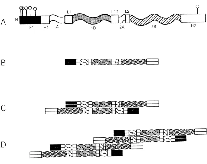
The assembly of IF proteins involves sev-eral steps (Figure 1). First a dimer is formed between two parallel monomers; next anti-parallel dimers interact through residues in their coiled-coil 1B segment forming a tet-ramer or protofilament, and then the protofilaments interact through residues in the coiled-coil 2B segments to form an octamer. These structures were identified in desmin filaments by chemical crosslinking (43). In the case of GFAP, a study of the self-assembly of mutants of this protein showed
A
B
C
D
N
E1 H1 1A
L1
1B
L12
2A L2
2B H2
C
Figure 1 - Schematic illustration of the filamentous structure of GFAP. A, Structure of the monomer. Five phosphorylation sites are shown in
segment E1 in the N-terminal domain and one in segment H2 at the C-terminal (see Figure 2). The rod domain comprises four helicoidal
segments (1A, 1B, 2A and 2B) and three linkers (L1, L2 and L12). B, Structure of the dimer. Parallel monomers interact to form coiled-coil
arrangements in the central domain. C, Structure of the tetramer or protofilament. Anti-parallel dimers interact through residues in the coiled
that the sequence KLLEGEE in tract 2B as well as the entire head domain is essential (44).
In in vitro experiments the assembly state of purified GFAP is influenced by the ionic strength and pH of the medium, the presence of Mg2+ and Ca2+ (45,46) and especially (as
discussed below) by the phoshorylation state of the protein (47). Phosphorylation is also important in the intact cell, particularly in preparing the cytoskeleton for cell division. However, the exact mechanism of IF assem-bly/disassembly in vivo is unknown. Simple self-assembly is unlikely and it is probable that other factors are needed as well as regula-tory phosphorylation. A chaperone-like ac-tivity modulating GFAP and vimentin assem-bly has been described (48). Moreover, S100ß, a Ca2+-binding protein expressed in
astro-cytes, may be involved in glial filament for-mation (49). Another interesting fact is that in immature or injured astrocytes GFAP can co-assemble with vimentin, a process that is dependent on the KLLEGEE sequence men-tioned above (44).
Intermediate filaments, in contrast to mi-crotubules and actin filaments, have no polar-ity, and it is not possible to identify a vectorial array of protofilaments. Filaments are as-sembled or incorporated into preformed fila-ments uniformly throughout the cytoplasm and an apparent polar or vectorial incorpora-tion (from the perinuclear region to the cell periphery) may be the consequence of a non-uniform distribution of these filaments (50,51).
General and functional aspects of GFAP phosphorylation
It has been known for some years that IF proteins, including GFAP, undergo cyclic phosphorylation and dephosphorylation in intact cell preparations (52,53), but until re-cently information on the sites phosphorylat-ed was lacking. These sites have now been described for porcine GFAP phosphorylated
in vitro where six sites were identified by
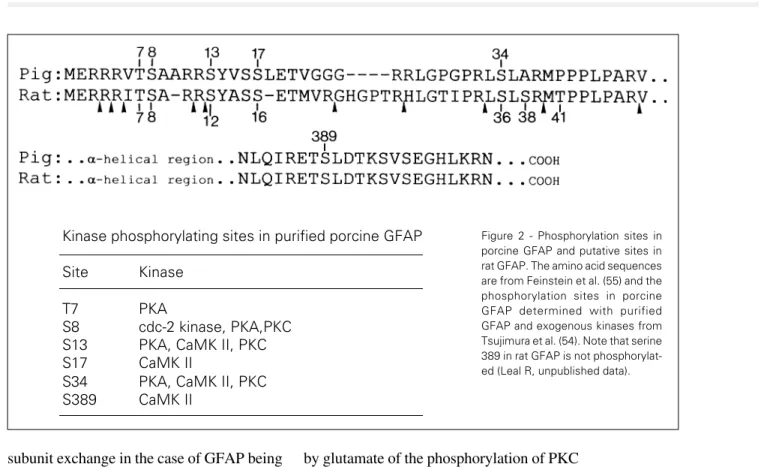
Japanese workers (47,54). Five of these sites are located in the N-terminal head domain (Thr7, Ser8, Ser13, Ser17, Ser34) and one in the tail domain (Ser389) where they are phos-phorylated by cyclic AMP-dependent protein kinase (PKA), Ca2+/calmodulin-dependent
kinase II (CaMK II), protein kinase C (PKC) and the cdc-2 kinase. The phosphorylation sites in rat GFAP are not known but the se-quence homology between porcine and rat GFAP in the N-terminal domain (55) is suffi-cient to assume that the corresponding con-sensus sequences in rat GFAP are potential phosphorylation sites and targets for PKA, PKC, CaMK II and the cdc-2 kinase (Figure 2). Less is known about the sites and kinases involved in GFAP phosphorylation in intact cell systems, but a start has been made. Japa-nese workers used monoclonal antibodies rec-ognizing specific phosphorylation sites and immunofluorescence to demonstrate that one threonine and three serines in the head region of GFAP are phosphorylated in vivo at differ-ent stages of the cell cycle (56-58). With regard to the kinases responsible for these in vivo phosphorylations, present evidence
points to the direct or indirect participation of PKA, CaMK II, the cdc-2 kinase and an un-known kinase (CF kinase) (58). In our labora-tory we have used tryptic phosphopeptide mapping of 32P-labelled GFAP extracted from
incubated hippocampal slices in attempts to identify the kinases involved. Our results sug-gest that CaMK II and PKA are the main kinases involved, either directly or indirectly, in GFAP phosphorylation in this preparation (59 and Leal R, Gonçalves CA and Rodnight R, unpublished data). Phosphorylation of GFAP in primary astrocyte cultures by CaMK II has been reported (60).
subunit exchange in the case of GFAP being suppressed in proportion to the extent of phos-phorylation(62). Moreover, IF protein phos-phorylation plays a crucial role in dividing cells, where evidence from cell cultures has shown that the disassembly of the cytoskel-eton preceding mitosis in some cells is regu-lated by a site-specific increase in the phos-phorylation state of vimentin and GFAP (57,63-65). In interphase cells changes in the phosphorylation state of IF proteins may have profound effects on the structure of the cyto-skeleton, as shown by studies in which hyper-phosphorylation of vimentin induced by pro-tein phosphatase inhibitors (66,67) or the an-titumor drug fostriecin (68) was associated with intermediate filament reorganization. Glutamate agonists and external Ca2+
-lack increase the phosphorylation state of GFAP in immature slices from the rat hippocampus
Our interest in this area arose from a chance observation while using two-dimen-sional electrophoresis to study the regulation
by glutamate of the phosphorylation of PKC substrates in hippocampal slices from imma-ture rats (post-natal days 12-16) labelled with [32P]phosphate. We observed a weak
stimula-tion of the PKC substrates and a much stron-ger stimulation of an unknown protein, desig-nated pp-H47 (69), which was subsequently identified as GFAP (70).
Glutamate receptors are classified into ionotropic (consisting of ligand-gated ion channels) and metabotropic (coupled to sec-ond-messenger systems) (71) receptors. The effect of glutamate on GFAP phosphoryla-tion was shown to occur via a metabotropic receptor since the highly selective metabotro-pic glutamate agonist, (1S,3R)-1-aminocy-clopentane-1,3-dicarboxylic acid (1S,3R-ACPD), stimulated the reaction to the same extent as glutamate, while agonists of ionotropic glutamate receptors were ineffec-tive (72). This effect was shown to occur via a G protein since the stimulation was inhibited by pertussis toxin. Half maximal stimulation was achieved with 20 µM 1S,3R-ACPD. A typical autoradiograph illustrating the effect of glutamate and 1S,3R-ACPD on GFAP
Kinase phosphorylating sites in purified porcine GFAP
Site Kinase
T7 PKA
S8 cdc-2 kinase, PKA,PKC S13 PKA, CaMK II, PKC S17 CaMK II
S34 PKA, CaMK II, PKC S389 CaMK II
phosphorylation is shown in Figure 3. The glutamate response was only observed in the presence of Ca2+ in the medium and was
absent in slices prepared from adult hippo-campus. At first, this need for Ca2+ was not
surprising since we had previously observed that the basal phosphorylation of GFAP in adult slices is completely dependent on exter-nal Ca2+. However, in immature slices we
later found that external Ca2+ partially
inhib-its the incorporation of [32P]phosphate into
GFAP (73). This inhibition starts at a low Ca2+ concentration and reaches a plateau of
about 50% inhibition at 1 mM Ca2+. During
the subsequent ontogenetic development the phosphorylation system gradually becomes dependent on external Ca2+ (Figure 4). For
these contrasting effects of Ca2+ on GFAP
phosphorylation to occur the cation has to cross the cell membrane. This was shown by the use of Ca2+ channel blockers, which in the
presence of external Ca2+ increased GFAP
phosphorylation in immature slices and in-hibited it in adult slices (73). Furthermore, the rates of GFAP phosphorylation in immature slices in the absence of external Ca2+
com-pared with those obtained in the presence of glutamate plus Ca2+ were found to be equal
(Figure 5). Moreover, the effects of Ca2+-lack
and Ca2+-lack plus glutamate were also not
significantly different. These results suggest that glutamate was acting through an unknown mechanism to reverse the inhibitory effect of Ca2+ on the phosphorylation reaction.
Nature of the astrocytic metabotropic glutamate receptor involved in the control of GFAP phosphorylation in immature slices
Both ionotropic (iGluRs) and metabotro-pic glutamate receptors (mGluRs) have been demonstrated in astrocytes in culture and in immature and adult glia in situ (12,74-76). Of
the iGluRs only two of the three main sub-groups, defined by their agonist sensitivity to (RS)-α -amino-3-hydroxy-5-methyl-4-isoxazolepropionic acid (AMPA) and kainic acid (KA), are expressed in astrocytes. Func-tional receptors of the third group, which are activated by N-methyl-D-aspartic acid (NMDA), have not been detected in these
A
kDa 80
50
50
Figure 3 - Autoradiographs illus-trating the stimulation of GFAP phosphorylation by 1 mM gluta-mate (A and B) and 100 µM 1S,3R-ACPD (C and D), where A and C are from control incuba-tions and B and D are from incu-bations in the presence of the agonist. Hippocampal slices from 15-day old rats were labelled
with [32P]phosphate and
ana-lyzed by non-equilibrium pH gra-dient electrophoresis (NEPHGE) for the first dimension and PAGE electrophoresis on 8% gels for the second dimension. To mini-mize intergel variation two first dimension rod gels (control and test) were mounted as mirror images on a single second di-mension slab gel. Arrows point to GFAP. Reproduced, with per-mission, from Ref. 72.
C
B
cells, although they may express non-func-tional subunits of this receptor (77).
The family of mGluRs comprises 8 sub-types divided into three groups according to the extent of amino acid homology, agonist sensitivity and associated signal transduction mechanisms (78). All mGluRs are coupled to G proteins and either regulate the hydrolysis of phosphoinositides (group I, mGluRs1,5) or the synthesis of cyclic AMP (group II, mGluRs2,3, and group III, mGluRs4,6-8) (Table 1). Agonists of mGluRs include the highly selective 1S,3R-ACPD, and L(+)-2-amino-4-phosphonobutyric acid (L-AP4) and the mixed agonists quisqualate and ibotenate. Quisqualate exhibits preference for group I, 1S,3R-ACPD for group II and L-AP4 for group III (Table 1; Refs. 79-81).
Two mGluR subtypes have been un-equivocally detected in astrocytes. One of them is a group I receptor since specific mGluR agonists release Ca2+ from internal
stores in astrocyte cultures (82) and in astro-cytes of hippocampal slices in situ (23,83) through the hydrolysis of phosphatidylinosi-tol and the generation of the Ca2+-releasing
agent inositol trisphosphate (IP3). This re-sponse is apparently due to mGluR5 since high immunoreactivity to this receptor was demonstrated in hippocampal astrocytes in situ (84). The other receptor belongs to group II and has been tentatively identified as mGluR3. Antibody immunocytochemistry and in situ hybridization for this mGluR
showed that it is abundantly expressed in glial cells (85).
Our evidence strongly points to mGluR3 as the receptor responsible for the control of GFAP phosphorylation in immature slices. In a study on the efficacy of a series of mGluR agonists we found that 1S,3R-ACPD was sig-nificantly (P<0.01) more effective than quisqualate in stimulating phosphorylation, suggesting a group II receptor which, from the immunocytochemical evidence cited above, is most likely to be mGluR3 (Wofchuk ST and
Rodnight R, unpublished data). However, this result does not exclude the contribution of a group I receptor since quisqualate was only 22% less effective than 1S,3R-ACPD. We therefore tested the effect of an inhibitor of phosphatidylinositol-specific phospholipase C, the aminosteroid U73122 (86). This com-pound had no effect on the stimulation of GFAP phosphorylation by 1S,3R-ACPD, al-though it effectively inhibited the generation of inositol phosphates from glutamate-stimu-lated phosphatidylinositol hydrolysis in as-trocyte cultures. This result makes less likely the contribution of a group I mGluR to the phosphorylation response. Finally, group III mGluRs were excluded by showing that L-AP4 did not stimulate GFAP phosphoryla-tion.
Peak height (% control)
300
200
100
12-15 20-22 25 40 60
Post-natal days
* *
* *
Peak height (% control)
400 300 200 100 0 Immature Adult +Ca2+
+Ca2+ + glu
-Ca2+
-Ca2+ + glu
1234 1234 1234 1234 1234 1234 1234 1234 1234 1234 1234 1234 1234 1234 1234 1234 1234 1234 1234 1234 1234 1234 1234 12345 12345 12345 12345 12345 12345 12345 12345 12345 12345 12345 12345 12345 12345 12345 12345 12345 12345 12345 12345 12345 12345 12345 1234 1234 1234 1234 1234 1234 1234 1234 1234 1234 1234 1234 12345 12345 12 12 12 123 123 123 * * *
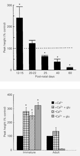
Figure 4 - Developmental profile of the sensitivity of GFAP phos-phorylation in incubated
hippo-campal slices to external Ca2+.
Each bar represents the per-centage change (± SEM) in GFAP phosphorylation in media
lacking Ca2+ from
phosphoryla-tion in media containing 1 mM
Ca2+ (indicated by the dotted
line). *P<0.01 compared to
con-trol (paired t-test). Reproduced,
with permission, from Ref. 73.
Figure 5 - Comparison of the effect of glutamate (glu) and
Ca2+ on GFAP phosphorylation
in hippocampal slices from im-mature and adult tissue. In the immature block the three col-umns marked by an asterisk are significantly different from the 100% control, but do not differ significantly one from another. Each block is the mean of 6-9 observations. Note that in the adult hippocampus glutamate had no effect on GFAP phos-phorylation, which at this age was completely dependent on
external Ca2+. *P<0.001 (paired
t-test) Modified, with
What is the role of external Ca2+?
Membrane exchange of Ca2+in
mamma-lian cells under basal conditions occurs via tonically active voltage-dependentchannels, a plasma membrane Ca2+-ATPase and Na+/
Ca2+ exchange (87).Voltage-dependent Ca2+
channels of the T and L types have been identified in astrocytes (87). Our evidence suggests that in immature hippocampal slices Ca2+ enters astrocytes through tonically
ac-tive L-type channels, since entry (measured by a decrease in GFAP phosphorylation) was blocked by the L-type channel antagonist ni-fedipine (73).
Primary cultures of astrocytes and astro-cytes in situ (e.g., in hippocampal slices) ex-press neurotransmitter receptors that trigger increases in internal Ca2+. These receptors
include glutamate receptors (23,83,88-91) and α1-adrenergic receptors (92). Increases
in internal Ca2+ due to glutamate may be due
to the activation of ionotropic receptors and Ca2+ entry through a receptor channel or
volt-age-dependent channels, or activation of me-tabotropic receptors and the release of Ca2+
from internal stores. In either case the Ca2+
signal is propagated to adjacent cells through gap junctions in the form of waves (90,91,93-95). As already mentioned, these glial cal-cium waves when initiated by glutamate re-leased from neurons may constitute a mech-anism for glia-to-neuron communication (20-22,96).
We initially considered that the Ca2+
de-pendence of the stimulation of GFAP phos-phorylation by glutamate might be due to an activating effect of the cation on the receptor. We viewed the receptor as one that either permitted Ca2+ entry (i.e., ionotropic) or
acti-vated the release of Ca2+ from internal stores
(i.e., metabotropic). The increase in internal Ca2+ was postulated to stimulate a Ca2+
-de-pendent kinase associated with GFAP. This hypothesis was discredited when we discov-ered that 1) external Ca2+ inhibited the basal
incorporation of [32P]phosphate into GFAP
and that 2) the stimulation by glutamate is mediated by a group II mGluR which does not release internal Ca2+. The mechanism by
which glutamate reverses the inhibitory ef-fect of Ca2+ on the reaction is uncertain, but is
probably related to an inhibition of Ca2+ entry
through L-type channels. The inhibition of ion channels by G protein activation is a well-established phenomenon (97,98) and recent studies have demonstrated a G protein-medi-ated inhibition of Ca2+ currents through
neu-ronal L-type channels by stimulation of group II mGluRs (99,100). Channel inhibition of this type is generally considered to be mem-brane delimited and independent of second
Table 1 - Some characteristics of metabotropic glutamate receptors.
*Several splice variants known. **In adult brain slices (115) or expressed
in Chinese hamster ovary cells (81). Activation of these receptors inhibits forskolin-stimulated adenylyl cyclase activity, while in immature brain slices they increase basal and forskolin-stimulated cyclic AMP synthesis (116). PI, Phosphatidylinositol; L-SOP, L-serine-O-phosphate. For com-plete descriptions of mGluRs, see Refs. 71,117-119.
Receptors Characteristics
Group I mGluR1* Increases PI hydrolysis
Quisqualate>glu>>ibotenate>1S,3R-ACPD Expressed in neurons
mGluR5 Increases PI hydrolysis
Quisqualate>glu>>ibotenate>1S,3R-ACPD Expressed in neurons and glia
Group II mGluR2 Regulates** cyclic AMP synthesis
1S,3R-ACPD>ibotenate>quisqualate Expressed in neurons; ? in glia
mGluR3 Regulates** cyclic AMP synthesis
1S,3R-ACPD>ibotenate>quisqualate Expressed in glia and neurons
Group III mGluR4 Regulates cyclic AMP synthesis
L-AP4 = L-SOP>ibotenate Expressed in neurons
mGluR6 Regulates cyclic AMP synthesis
L-AP4
Expressed in retina
mGluR7 Regulates cyclic AMP synthesis
L-AP4
Expressed in neurons
mGluR8 Regulates cyclic AMP synthesis
L-AP4
messenger events involving cyclic AMP or PKC. Recent evidence suggests a direct effect on the channel due to diffusion of ßγ subunits released from the activated G protein hetero-trimer (101).
The exact mechanism of the inhibitory effect of external Ca2+ on basal GFAP
phos-phorylation is unknown, but is unlikely to be due to inhibition of a protein kinase; indeed the main two kinases phosphorylating GFAP in immature slices in the absence of Ca2+
appear to be CaMK and PKA (59 and Leal R, Gonçalves CA and Rodnight R, unpublished data). A more viable hypothesis proposes that external Ca2+ stimulates a Ca2+-dependent
dephosphorylation event associated with GFAP, thus changing the dynamic equilib-rium between phosphorylation and dephos-phorylation and reducing the steady-state level of phosphate in the protein. These con-siderations led us to conduct a study on the enzymes involved in the dephosphorylation of GFAP.
Dephosphorylation of GFAP
Serine/threonine protein phosphatases (PPs) are classified into four groups desig-nated types PP1, PP2A, PP2B and PP2C (102). The activities of PP1 and PP2A are cation-independent, whereas that of PP2B (gener-ally known as calcineurin) is dependent on Ca2+ and calmodulin and that of PP2C on
Mg2+. By using the specific inhibitors of PP1
and PP2A, okadaic acid and microcystin-LR, respectively, we showed that in cytoskeletal preparations and immature hippocampal slices the dephosphorylation of GFAP is cata-lyzed by PP1 (103). No evidence for a direct Ca2+-dependent dephosphorylation of GFAP
was found in this study. However, a highly specific inhibitor of calcineurin, the immuno-suppressant FK506 (104), increased GFAP phosphorylation in immature slices and as-trocyte cultures in the presence of Ca2+, i.e.,
the drug reversed the inhibitory effect of ex-ternal Ca2+ on the phosphorylation reaction
(Vinadé L and Rodnight R, unpublished data). This result strongly suggested the presence of calcineurin associated with GFAP in astro-cytes, a somewhat surprising conclusion since several studies had failed to demonstrate the occurrence of the enzyme in glia by immuno-cytochemistry (105,106). However, we were able to confirm the FK506 result by immuno-blotting using a monoclonal antibody to the ß-subunit of calcineurin. Applied to extracts of primary cultures of astrocytes the antibody revealed a low calcineurin content equivalent to one fiftieth of the content in hippocampal tissue (Vinadé L and Rodnight R, unpub-lished data). These data strongly suggest that calcineurin plays a role in the dephosphoryla-tion of GFAP. Since we found no evidence for a direct Ca2+-dependent dephosphorylation
of GFAP sites, the action of calcineurin prob-ably occurs via the Ca2+-dependent enzyme
cascade which is known to regulate PP1 in many tissues and which is involved in the phenomenon of long-term depression (107). This cascade depends on the fact that in many cells type 1 phosphatases are inhibited by the phosphorylated form of the protein inhibitor-1 (inhibitor-108), which in turn is dephosphorylated (and inactivated) by calcineurin. Thus, down-regulation of calcineurin through inhibition of Ca2+ entry or lack of external Ca2+ would
increase the inhibition by phospho-inhibitor-1 of the type phospho-inhibitor-1 phosphatase associated with GFAP.
Discussion: hypothesis and perspectives
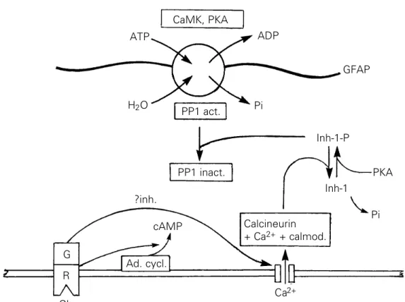
A hypothetical scheme to explain the ac-tion of glutamate agonists on the Ca2+
-de-pendent phosphorylation of GFAP in imma-ture slices is shown in Figure 6. The scheme postulates that the activation of metabotropic glutamate receptors of the mGluR3 subtype inhibits L-type Ca2+ channels and
conse-quently down-regulates the Ca2+-dependent
and the phosphorylation state of GFAP. Note that even though one of the kinases associated with GFAP is a Ca2+/calmodulin-dependent
enzyme, no effect of glutamate stimulation on the phosphorylation of GFAP is postulated since the activation of mGluR3 does not in-crease internal Ca2+. Basal CaMK activity
might be due to a discrete pool of internal Ca2+
or to the activity of autophosphorylated CaMK II which does not require Ca2+ (109). We are
inclined to favor the latter interpretation since autophosphorylated CaMK II has been re-ported to occur in astrocyte cultures (60). It is also necessary to point out that the scheme does not take into account the dynamic nature of the turnover of protein phosphate in GFAP. While phosphorylation of residues is consid-ered to occur on the intact filaments, the substrates of the dephosphorylation reaction are presumably disassembled subunits (dimers or tetramers).
A necessary condition of the mechanism depicted in Figure 6 is the existence of closed intracellular compartments containing group
II receptors, Ca2+ channels and the enzymes
involved in GFAP phosphorylation, to which internal Ca2+ modulated by group I glutamate
and other neurotransmitter receptors is inac-cessible. Indeed membrane-delimited control of Ca2+ entry by G protein subunits would
require the receptors and channels to occur in close proximity. It is tempting to speculate that such compartmentation occurs princi-pally in astrocyte processes where the GFAP filaments are situated close to the plasma membrane. Immature astrocytes in the rat hippocampus possess extensive processes (110) and depolymerization of their filaments can be envisaged as a prerequisite to the round-ing up of the cells prior to mitosis. Localized Ca2+ signalling has been proposed to explain
the spatially separated phosphorylation of vimentin by CaMK II in astrocytes (58).
Future work in this area will involve at-tempts to demonstrate aspects of the scheme in Figure 6 which remain uncertain. These are: 1) the proposed inhibition of Ca2+
chan-nels by a group II mGluR; and 2) the proposed
Figure 6 - Hypothetical scheme
to explain the Ca2+-dependent
action of glutamate in regulating the phosphorylation of GFAP in immature hippocampal slices. Phosphorylation sites in a fila-ment of GFAP are represented by the encircled P and are shown associated with CaMK activity, PKA and protein phos-phatase PP1. The scheme pro-poses that activation of a metab-otropic receptor of subtype mGluR3 by glutamate inhibits
the entry of Ca2+ through Ca2+
channels. The consequent
de-crease in internal Ca2+
down-regulates the protein phos-phatase calcineurin and thus in-creases the phosphorylation state of a putative inhibitor-1 pro-tein. Since phosho-inhibitor-1, in contrast to dephospho-inhibitor-1, inhibits PP1 (108) the phos-phorylation state of GFAP is in-creased.
CaMK, PKA
ATP ADP
H2O Pi
GFAP
PP1 act.
PP1 inact.
Inh-1-P
Inh-1
PKA
Pi Calcineurin
+ Ca2+ + calmod.
Ca2+
Ad. cycl. cAMP
G R
Glu
presence of an inhibitor-1 protein in astro-cytes. With regard to the first aspect, the inhibition of Ca2+ channelshas been
demon-strated in neurons as cited above, but not yet in astrocytes. However, we know that a G protein is necessary for the glutamate-stimu-lated increase in GFAP phosphorylation since the effect was reversed by the G protein an-tagonist, pertussis toxin (72), as was the inhibition of Ca2+ currents in neurons by group
II mGluRs (100). It is unlikely that primary astrocyte cultures will prove to be a useful model to investigate this problem since the expression of Ca2+ channels in these cells is
dependent on a neuronal environment (111). Astrocytes co-cultured with neurons, confo-cal microscopy of astrocytes in situ and elec-trophysiology of acutely isolated astrocytes are possible approaches. In the case of the second aspect, we believe that the question of the occurrence of an inhibitor-1-like protein in hippocampal astrocytes remains open. The neuronal form of inhibitor-1 was not detected by immunocytochemistry in astrocytes (112), but DARPP-32, a protein partly homologous to inhibitor-1 which also inhibits PP1 when phosphorylated, has been found in certain glial cells (113). It is possible that
immuno-blotting (an approach which is more sensitive than immunocytochemistry) will provide more information.
The functional significance of the regula-tion of GFAP phosphorylaregula-tion by glutamate in the immature hippocampus may be related to the narrow developmental window when the glutamate response is present. In the rat brain this window corresponds to the period of massive synaptogenesis during which as-trocytes are known to proliferate. We have therefore speculated that glutamate released from developing synapses during this period may signal an increase in the phosphorylation state of GFAP and a consequent increase in the number of mitotic astrocytes (72). During subsequent development our evidence sug-gests that control by glutamate of GFAP phos-phorylation and expression of the Ca2+
-pendent dephosphorylation mechanism de-clines and that this control is absent in the adult hippocampus when astrocytes normally remain in interphase. A paper by Evans (114) reporting a Ca2+-dependent
dephosphoryla-tion of vimentin by cytosol prepared from mitotically selected cells, which was absent in cytosol from interphase cells, is particu-larly relevant to this hypothesis.
References
1. Tower DB (1988). Development of knowl-edge about astrocytes since Virchow. In: Norenberg MD, Hertz L & Schousboe A
(Editors), The Biochemical Pathology of
Astrocytes. Alan Liss, New York, 3-18. 2. Travis J (1994). Glia: The brains other
cells. Science, 266: 970-972.
3. Kettenmann H & Ransom BR (Editors)
(1995). Neuroglia. Oxford University
Press, Oxford.
4. Spacek J (1985). Three-dimensional anal-ysis of dendritic spines. III. Glial sheath.
Anatomical Embryology, 171: 245-252. 5. Bluemcke I, Eggli P & Celio MR (1995).
Relationship between astrocytic pro-cesses and perineuronal nets in rat
neo-cortex. Glia, 15: 131-140.
6. Tsacopoulus M & Magistretti PJ (1996). Metabolic coupling between glia and
neu-rons. Journal of Neuroscience, 16:
877-885.
7. Keyser DO & Pellmar TC (1994). Synaptic transmission in the hippocampus: Critical
role for glial cells. Glia, 10: 237-243.
8. Newman EA (1995). Glial cell regulation of extracellular potassium. In: Kettenmann H
& Ransom BR (Editors), Neuroglia. Oxford
University Press, Oxford, 717-731. 9. Schousboe A & Westergaard N (1995).
Transport of neuroactive amino acids in astrocytes. In: Kettenmann H & Ransom
BR (Editors), Neuroglia. Oxford University
Press, Oxford, 246-258.
10. Murphy S & Pearce B (1987). Functional receptors for neurotransmitters on
astro-glial cells. Neuroscience, 22: 381-394.
11. McCarthy KD & Salm AK (1991). Pharma-cologically distinct subsets of astrocytes can be identified by their calcium
re-sponse to neuroligands. Neuroscience,
41: 325-333.
12. Teichberg VI (1991). Glial glutamate re-ceptors: likely actors in brain signalling.
FASEB Journal, 5: 3086-3091.
13. Hosli E & Hosli L (1993). Receptors for neurotransmitters on astrocytes in the mammalian central nervous system.
14. Neary JT & Zhu Q (1994). Signalling by
ATP receptors in astrocytes. Neuroreport,
5: 1617-1620.
15. Kimelberg HK (1995). Receptors on
astro-cytes - What possible functions?
Neuro-chemistry International, 26: 27-40. 16. Barres BA, Chun LLC & Corey DP (1990).
Ion channels in vertebrate glia. Annual
Review of Neuroscience, 13: 441-474. 17. Sontheimer H & Ritchie M (1995).
Volt-age-gated sodium and calcium channels in astrocytes. In: Kettenmann H &
Ran-som BR (Editors), Neuroglia. Oxford
Uni-versity Press, Oxford, 202-220.
18. Seil FJ, Eckenstein FP & Reier PJ (1992). Induction of dendrite spine proliferation
by an astrocyte-secreted factor.
Experi-mental Neurology, 60: 85-89.
19. Nicoletti F, Magri G, Ingrao F, Bruno V, Catania MV, DellAlbani P, Condorelli DF & Avola R (1990). Excitatory amino acids stimulate phospholipid hydrolysis and re-duce proliferation in cultured astrocytes.
Journal of Neurochemistry, 54: 771-777. 20. Dani JW & Smith SJ (1995). The
trigger-ing of astrocytic calcium waves by
NMDA-induced neuronal activation. In: Calcium
Waves, Gradients and Oscillations, Ciba Foundation Symposium 188. Wiley, Chichester, UK.
21. Nedergaard M (1994). Direct signalling from astrocytes to neurons in cultures of
mammalian brain cells. Science, 263:
1768-1771.
22. Hassinger TD, Atkinson PB, Strecker GJ, Whalen LR, Dudek FE, Kossel AH & Kater SB (1995). Evidence for glutamate-medi-ated activation of hippocampal neurons
by glial calcium waves. Journal of
Neuro-biology, 28: 159-170.
23. Porter JT & McCarthy KD (1996). Hippo-campal astrocytes respond to glutamate
released from synaptic terminals. Journal
of Neuroscience, 16: 5073-5081. 24. Canady KS & Rubel EW (1992). Rapid and
reversible astrocytic reaction to afferent
blockade in chick cochlear nucleus.
Jour-nal of Neuroscience, 12: 1001-1009. 25. Georgiou J, Robitaille R, Trimble WS &
Charlton MP (1994). Synaptic regulation
of glial protein expression in vivo. Neuron,
12: 443-455.
26. Shao Y & McCarthy KD (1994). Plasticity
of astrocytes. Glia, 11: 147-155.
27. Muller CM (1995). Glial cells and activity-dependent central nervous system plas-ticity. In: Kettenmann H & Ransom BR
(Editors), Neuroglia. Oxford University
Press, Oxford, 805-814.
28. Hawrylak N & Greenough WT (1995). Mo-nocular deprivation alters the morphology of glial fibrillary acidic protein-immunore-active astrocytes in the rat visual cortex.
Brain Research, 683: 187-199.
29. Hansen A, Jorgensen OS, Bolwig TG & Barry DI (1991). Hippocampal kindling in the rat is associated with time-dependent increases in the concentration of glial
fibrillary acidic protein. Journal of
Neuro-chemistry, 57: 1716-1720.
30. Dalby NO, Rondouin G & Lerner-Natoli M (1995). Increase in GAP-43 and GFAP im-munoreactivity in the rat hippocampus subsequent to perforant path kindling.
Journal of Neuroscience Research, 41: 613-619.
31. Kraig RP, Dong L, Thisted R & Jaeger CB (1991). Spreading depression increases immunohistochemical staining of glial
fibrillary acidic protein. Journal of
Neuro-science, 11: 2187-2198.
32. Bonthius DJ, Stringer JL, Lothman EW & Steward O (1994). Spreading depression and reverberatory seizures induce the up regulation of mRNA for glial fibrillary acidic
protein. Brain Research, 645: 215-224.
33. Sirevaag AM & Greenough WT (1991). Plasticity of GFAP-immunoreactive astro-cytes: size and number in visual cortex of rats reared in complex environments.
Brain Research, 540: 273-278.
34. Wenzel J, Lammert G, Meyer U & Krug M (1991). The influence of long-term poten-tiation on the spatial relationship between astrocyte processes and potentiated syn-apses in the dentate gyrus neuropil of rat
brain. Brain Research, 560: 122-131.
35. McCall MA, Gregg RG, Behringer RR, Brenner M, Delaney CL, Galbreath EJ, Zhang CL, Pearce RA, Chiu SY & Messing A (1996). Targeted deletion in astrocyte intermediate filament (Gfap) alters
neu-ronal physiology. Proceedings of the
Na-tional Academy of Sciences, USA, 93: 6361-6366.
36. Hatten ME, Liem RKH, Shelanski ML & Mason CA (1991). Astroglia in CNS injury.
Glia, 4: 233-243.
37. Steward O (1994). Electroconvulsive sei-zures upregulate astroglial gene
expres-sion selectively in the dentate gyrus.
Mo-lecular Brain Research, 25: 217-224. 38. Rocha E & Rodnight R (1994). Chronic
administration of lithium chloride in-creases immunodetectable glial fibrillary acidic protein in the rat hippocampus.
Journal of Neurochemistry, 63: 1582-1584.
39. Bradbury EJ, Kershaw TR, Marchbanks RM & Sinden JD (1995). Astrocyte trans-plants alleviate lesion induced memory deficits independently of cholinergic
re-covery. Neuroscience, 65: 955-972.
40. Eng LF (1985). Glial fibrillary acidic protein (GFAP): the major protein of glial interme-diate filaments in differentiated
astro-cytes. Journal of Neuroimmunology, 8:
203-214.
41. Steinert PM & Roop DR (1988). Molecular and cellular biology of intermediate
fila-ments. Annual Review of Biochemistry,
57: 593-625.
42. Shoeman RL & Traub P (1993). Assembly
of intermediate filaments. Bioessays, 15:
605-611.
43. Geisler N, Schunemann J & Weber K (1992). Chemical cross-linking indicates a staggered and antiparallel protofilament of desmin intermediate filaments and characterizes one higher-level complex
between protofilaments. European
Jour-nal of Biochemistry, 206: 841-852. 44. Chen W-J & Liem RKH (1994). The
end-less story of the glial fibrillary acidic
pro-tein. Journal of Cell Biology, 107:
2299-2311.
45. Eriksson JE, Brautigan DL, Vallee R, Olmstead J, Fujiki H & Goldman RD (1992). Cytoskeletal integrity in interphase cells requires protein phosphatase
activ-ity. Proceedings of the National Academy
of Sciences, USA, 89: 11093-11097. 46. Nakamura Y, Takeda M, Angelides KJ,
Tada K, Hariguchi S & Nishimura T (1991). Assembly, disassembly, and exchange of
glial fibrillary acidic protein. Glia, 4:
101-110.
47. Inagaki M, Nakamura Y, Takeda M, Nishimura T & Inagaki N (1994). Glial fibril-lary acidic protein: Dynamic property and
regulation by phosphorylation. Brain
Pa-thology, 4: 239-243.
48. Nichol ID & Quinlan RA (1994).
Chaper-one activity of α-crystallins modulates
in-termediate filament assembly. EMBO
Journal, 13: 945-953.
49. Bianchi R, Giambanco I & Donato R (1993). S100 protein but not calmodulin binds to the glial fibrillary acidic protein
and inhibits its polymerization in a Ca2+
-dependent manner. Journal of Biological
Chemistry, 268: 12669-12674.
50. Sarria AJ, Nordeen SK & Evans RM (1990). Regulated expression of vimentin cDNA in cells in the presence and absence of a pre-existing vimentin filament network.
51. Vikstrom KL, Lim S-S, Goldman RD & Borisy GG (1992). Steady state dynamics
of intermediate filament networks.
Jour-nal of Cell Biology, 118: 121-129. 52. McCarthy KD, Salm A & Lerea LS (1988).
Astroglial receptors and their regulation of intermediate filament protein
phospho-rylation. In: Kimelberg HK (Editor), Glial
Cell Receptors. Raven Press, New York, 1-22.
53. Harrison B & Mobley PL (1989). Protein phosphorylation in astrocytes mediated by protein kinase C. Comparison with phosphorylation by cyclic
AMP-depend-ent protein kinase. Journal of
Neurochem-istry, 53: 1245-1251.
54. Tsujimura K, Tanaka J, Ando S, Matsuoka Y, Kusubata M, Sugiura H, Yamauchi T & Inagaki M (1994). Identification of phos-phorylation sites on glial fibrillary acidic
protein for cdc2 kinase and Ca2+
-calmod-ulin-dependent protein kinase II. Journal
of Biochemistry, 116: 426-434.
55. Feinstein DL, Weinmaster GA & Milner RJ (1992). Isolation of cDNA clones en-coding glial fibrillary acidic protein: expres-sion in astrocytes and in Schwann cells.
Journal of Neuroscience Research, 32: 1-14.
56. Nishizawa K, Yano T, Shibata M, Ando S, Takahashi T & Inagaki M (1991). Specific localization of phosphointermediate fila-ment protein in the constricted area of
dividing cells. Journal of Biological
Chem-istry, 266: 3074-3079.
57. Sekimata M, Tsujimura K, Tanaka J, Takeuchi Y, Inagaki N & Inagaki M (1996). Detection of protein kinase activity spe-cifically activated at metaphase anaphase
transition. Journal of Cell Biology, 132:
635-641.
58. Inagaki M, Matsuoka Y, Tsujimura K, Ando S, Tokui T, Takahashi T & Inagaki N (1996). Dynamic property of intermediate fila-ments: Regulation by phosphorylation.
Bioessays, 18: 481-487.
59. Leal RB (1995). Estudo do sistema fosforilante da ppH47/GFAP em cérebro de ratos: distribução regional, identificação das quinases envolvidas e mapeamento fosfopeptidico. Doctoral thesis, Universi-dade Federal do Paraná.
60. Yano S, Fukunaga K, Ushio Y & Miyamoto
E (1994). Activation of Ca2+
/calmodulin-dependent protein kinase II and phospho-rylation of intermediate filament proteins by stimulation of glutamate receptors in
cultured rat cortical astrocytes. Journal of
Biological Chemistry, 269: 5428-5439.
61. Ando S, Tanabe K, Gona Y, Sato C & Inagaki M (1989). Domain- and sequence-specific phosphorylation of vimentin in-duces disassembly of the filament
struc-ture. Biochemistry, 28: 2974-2979.
62. Nakamura Y, Takeda M & Nishimura T (1996). Dynamics of bovine glial fibrillary
acidic protein phosphorylation.
Neurosci-ence Letters, 205: 91-94.
63. Chou Y-H, Bischoff JR, Beach D & Goldman RD (1990). Intermediate fila-ment reorganization during mitosis is me-diated by p34cdc2 phosphorylation of
vimentin. Cell, 62: 1063-1071.
64. Chou YH, Opal P, Quinlan RA & Goldman RD (1996). The relative roles of specific N-and C-terminal phosphorylation sites in the disassembly of intermediate filament
in mitotic BHK-21 cells. Journal of Cell
Science, 109: 817-826.
65. Matsuoka Y, Nishizawa K, Yano T, Shibata M, Ando S, Takahashi T & Inagaki M (1993). Two different protein kinases act on a different time schedule as glial
fila-ment kinases during mitosis. EMBO
Jour-nal, 11: 2895-2902.
66. Eriksson JE, Opal P & Goldman RD (1992).
Intermediate filament dynamics. Current
Opinion in Cell Biology, 4: 99-104. 67. Lee W-C, Yu J-S, Yang S-D & Lai Y-K
(1992). Reversible hyperphosphorylation and reorganisation of vimentin intermedi-ate filaments by okadaic acid in 9L rat
brain tumour cells. Journal of Cellular
Bio-chemistry, 49: 378-393.
68. Ho DT & Roberge M (1996). The antitu-mor drug fostriecin induces vimentin hyperphosphorylation and intermediate
filament reorganization. Carcinogenesis,
17: 967-972.
69. Wofchuk ST & Rodnight R (1990). Stimu-lation by glutamate of the phosphoryla-tion of two substrates of protein kinase C, B-50/GAP and MARCKS, and of ppH-47, a protein highly labelled in incubated slices
from the hippocampus. Neuroscience
Re-search Communications, 6: 135-140. 70. Gonçalves CA & Rodnight R (1992).
Ap-parent identity of ppH-47, a protein highly phosphorylated in the hippocampus with
a form of glial fibrillary acidic protein.
Neu-roscience Research Communications, 11: 109-117.
71. Nakanishi S & Masu M (1994). Molecular diversity and functions of glutamate
re-ceptors. Annual Review of Biophysics and
Biomolecular Structure, 23: 319-348.
72. Wofchuk ST & Rodnight R (1994). Gluta-mate stimulates the phosphorylation of glial fibrillary acidic protein in slices of immature hippocampus via a
metabotro-pic receptor. Neurochemistry
Interna-tional, 24: 517-523.
73. Wofchuk ST & Rodnight R (1995). Age-dependent changes in the regulation by external calcium ions of the phosphoryla-tion of glial fibrillary acidic protein in slices
of rat hippocampus. Developmental Brain
Research, 85: 181-186.
74. Cull-Candy SG & Wyllie DJA (1991). Glu-tamate-receptor channels in mammalian
glial cells. Annals of the New York
Acade-my of Sciences, 633: 458-474.
75. Gallo V & Russell JT (1995). Excitatory amino acid receptors in glia: Different
sub-types for distinct functions? Journal of
Neuroscience Research, 42: 1-8. 76. Hansson E & Ronnback L (1995).
Astro-cytes in glutamate transmission. FASEB
Journal, 9: 343-350.
77. Conti F, DeBiasi S, Minelli A & Melone M (1996). Expression of NR1 and NR2A/B subunits of the NMDA receptor in cortical
astrocytes. Glia, 17: 254-258.
78. Steinhäuser C & Gallo V (1996). News of
glutamate receptors in glial cells. Trends
in Neurosciences, 19: 339-345.
79. Aramori I & Nakashini S (1992). Signal transduction and pharmacological charac-teristics of a metabotropic glutamate re-ceptor, mGluR1, in transfected CHO cells.
Neuron, 8: 757-765.
80. Abe T, Sugihara H, Nawa H, Shigemoto R, Mizuno N & Nakanishi S (1992). Molecu-lar characterization of a novel metabotro-pic glutamate receptor mGluR5 coupled
to inositol phosphate/Ca2+ signal
trans-duction. Journal of Biological Chemistry,
267: 13361-13368.
81. Tanabe Y, Nomura A, Masu M, Shigemoto R, Mizuno N & Nakashini S (1993). Signal transduction, pharmacological properties, and expression of patterns of two rat me-tabotropic glutamate receptors, mGluR3
and mGluR4. Journal of Neuroscience, 13:
1372-1378.
82. Kim WT, Rioult MG & Cornell-Bell AH (1994). Glutamate-induced calcium
signal-ing in astrocytes. Glia, 11: 173-184.
83. Porter JT & McCarthy KD (1995).
GFAP-positive hippocampal astrocytes in situ
respond to glutamatergic neuroligands
with increases in [Ca2+]i. Glia, 13:
84. Romano C, Sesma MA, MacDonald C, OMalley K, van den Pol AN & Olney JW (1995). Distribution of metabotropic re-ceptor mGluR5 immunoreactivity in the
rat brain. Journal of Comparative
Neurol-ogy, 355: 455-469.
85. Ohishi H, Shigemoto R, Nakanishi S & Mizuno N (1993). Distribution of the mes-senger RNA for a metabotropic glutamate receptor (mGluR3) in the rat brain -an in situ hybridization study. Journal of Comparative Neurology, 335: 252-266. 86. Thompson AK, Mostafapour SP,
Denlinger LC, Bleasdale JE & Fisher SK (1991). The aminosteroid U-73122 inhib-its muscarinic receptor sequestration and phosphoinositide hydrolysis in SK-N-SH
neuroblastoma cells. Journal of
Biologi-cal Chemistry, 266: 23856-23862. 87. Verkhratsky A & Kettenmann H (1996).
Calcium signalling in glial cells. Trends in
Neurosciences, 19: 346-352.
88. Glaum SR, Holzwarth JA & Miller RJ (1990). Glutamate receptors activate
Ca2+ mobilization and Ca2+ influx into
astrocytes. Proceedings of the National
Academy of Sciences, USA, 87: 3454-3458.
89. Ahmed Z, Lewis CA & Faber DS (1990).
Glutamate stimulates release of Ca2+
from internal stores in astroglia. Brain
Research, 516: 165-169.
90. Cornell-Bell AH & Finkbeiner SM (1991).
Ca2+ waves in astrocytes. Cell Calcium,
12: 185-204.
91. Finkbeiner SM (1993). Glial calcium. Glia,
9: 83-104.
92. Duffy S & MacVicar BA (1995). Adrener-gic calcium signaling in astrocyte
net-works within the hippocampal slice.
Jour-nal of Neuroscience, 15: 5535-5550. 93. Charles AC, Merrill JE, Dirksen ER &
Sanderson MJ (1991). Intracellular sig-nalling in glial cells: calcium waves and oscillations in response to mechanical
stimulation and glutamate. Neuron, 6:
983-992.
94. Sanderson MJ, Charles AC, Boitano S & Dirksen ER (1994). Mechanisms and function of intercellular calcium
signal-ling. Molecular and Cellular
Endocrinol-ogy, 98: 173-187.
95. Glaume C & McCarthy KD (1996). Con-trol of gap-junctional communication in
astrocytic networks. Trends in
Neuro-sciences, 19: 319-325.
96. Parpura V, Basarsky TA, Liu F, Jeftinija K, Jeftinija S & Haydon PG (1994). Gluta-mate-mediated astrocyte-neuron
signal-ling. Nature, 369: 744-747.
97. Hille B (1994). Modulation of ion-channel function by G-protein-coupled receptors.
Trends in Neurosciences, 17: 531-536. 98. Wickham KD & Clapham DE (1995).
G-protein regulation of ion channels.
Cur-rent Opinion in Neurobiology, 5: 278-285. 99. Sayer RJ, Schwindt PC & Crill WE (1992). Metabotropic glutamate receptor-medi-ated suppression of L-type calcium cur-rent in acutely isolated neocortical
neu-rons. Journal of Neurophysiology, 68:
833-842.
100. Chavis P, Shinozaki H, Bockaert J & Fagni L (1994). The metabotropic receptor types 2/3 inhibit L-type calcium channels via a pertussis toxin-sensitive G-protein
in cultured cerebellar granule cells.
Jour-nal of Neuroscience, 14: 7067-7076. 101. Herlitze S, Garcia DE, Mackie K, Hille B,
Scheuer T & Catterall WA (1996).
Modu-lation of Ca2+ channels by G protein ßγ
subunits. Nature,380: 258-262.
102. Stemmer P & Klee CB (1991). Serine/ threonine phosphatases in the nervous
system. Current Opinion in
Neurobiol-ogy, 1: 53-64.
103. Vinadé L & Rodnight R (1996). The de-phosphorylation of glial fibrillary acidic protein (GFAP) in the immature rat hip-pocampus is catalyzed mainly by a type 1
protein phosphatase. Brain Research,
732: 195-200.
104. Liu J, Farmer Jr JD, Lane WS, Friedman J, Weissman I & Schreiber SL (1991). Calcineurin is a common target of cyclo-phylin-cyclosporin A and FKBP-FK506
complexes. Cell, 66: 807-815.
105. Matsui H, Doi A, Itano T, Shimada M, Wang JH & Hatase O (1987). Immuno-histochemical localization of calcineurin, a calmodulin-stimulated phosphatase, in the rat hippocampus using a monoclonal
antibody. Brain Research, 402: 193-196.
106. Goto S, Yamada K, Oyama T, Korematsu K, Nagahiro S, Ushio Y, Fukunaga K, Miyamoto E & Hofer W (1994). Cellular
localization of type II Ca2+
/calmodulin-de-pendent protein kinase in the rat basal ganglia and intrastriatal grafts derived from fetal striatal primordia, in
compari-son with that of Ca2+
/calmodulin-regu-lated protein phosphatase, calcineurin.
Neuroscience, 62: 695-705.
107. Mulkey RM, Endo S, Shenolikar S & Malenka RC (1994). Involvement of a calcineurin/inhibitor-1 phosphatase cas-cade in hippocampal long-term
depres-sion. Nature, 369: 486-488.
108. Cohen P (1989). The structure and
regu-lation of protein phosphatases. Annual
Review of Biochemistry, 58: 453-508.
109. Mukherji S & Soderling TR (1994).
Regu-lation of Ca2+/calmodulin-dependent
pro-tein kinase II by inter- and
intrasubunit-catalyzed autophosphorylations. Journal
of Biological Chemistry, 269: 13744-13747.
110. Nixdorf-Bergweiler BE, Albrecht D & Heinemann U (1994). Developmental changes in the number, size, and orienta-tion of GFAP-positive cells in the CA1
region of rat hippocampus. Glia, 12:
180-195.
111. Corvalan V, Cole R, de Vellis J & Hagiwara S (1990). Neuronal modulation of calcium channel activity in cultured rat
astrocytes. Proceedings of the National
Academy of Sciences, USA, 87: 4345-4348.
112. Gustafson EL, Girault J-A, Hemmings Jr HC, Nairn AC & Greengard P (1991). Im-munocytochemical localization of
phos-phatase inhibitor-1 in rat brain. Journal of
Comparative Neurology, 310: 170-188. 113. Ouimet CC, Miller PE, Hemmings HC,
Walaas SI & Greengard P (1984). DARPP-32, a dopamine- and adenosine 3:5-monophosphate-regulated phosphopro-tein enriched in dopamine-innervated brain regions. III. Immunocytochemical
localization. Journal of Neuroscience, 4:
111-124.
114. Evans RM (1989). Phosphorylation of
vimentin in mitotically selected cells. In
vitro cyclic AMP-dependent and
calcium-stimulated phosphatase activities.
Jour-nal of Cell Biology, 108: 67-78. 115. Schoepp DD & Johnson BG (1993).
Phar-macology of metabotropic glutamate re-ceptor inhibition of cyclic AMP formation
in the adult rat hippocampus.
Neuro-chemistry International, 22: 277-283. 116. Schoepp DD & Johnson BG (1993).
Me-tabotropic glutamate receptor modula-tion of cAMP accumulamodula-tion in the
neona-tal rat hippocampus.
Neuropharmacol-ogy, 32: 1359-1365.
117. Schoepp DD & Conn PJ (1993). Metabo-tropic glutamate receptors in brain
func-tion and pathology. Trends in
Pharmaco-logical Sciences, 14: 13-20.
118. Pin J-P & Duvoisin R (1995). The metabo-tropic glutamate receptors: Structure and
functions. Neuropharmacology, 34: 1-26.
119. Nicoletti F, Bruno V, Copani A, Casabona G & Knopfel T (1996). Metabotropic glu-tamate receptors: a new target for the therapy of neurodegenerative disorders.