Measurement of epidermal thickness
in a patient with psoriasis by
computer-supported image analysis
Departments of 1Pathology, 2Dermatology, and 3Public Health,
Faculty of Medicine, and
4Technical Education Faculty, University of Abant Izzet Baysal,
Konuralp, Düzce, Turkey M. Alper1,A. Kavak2,
A.H. Parlak2,R. Demirci4,
I. Belenli4
andN. Yesildal3
Abstract
The aim of the present study was to measure full epidermal thickness, stratum corneum thickness, rete length, dermal papilla widening and suprapapillary epidermal thickness in psoriasis patients using a light microscope and computer-supported image analysis. The data ob-tained were analyzed in terms of patient age, type of psoriasis, total body surface area involvement, scalp and nail involvement, duration of psoriasis, and family history of the disease. The study was con-ducted on 64 patients and 57 controls whose skin biopsies were examined by light microscopy. The acquired microscopic images were transferred to a computer and measurements were made using image analysis. The skin biopsies, taken from different body areas, were examined for different parameters such as epidermal, corneal and suprapapillary epidermal thickness. The most prominent increase in thickness was detected in the palmar region. Corneal thickness was more pronounced in patients with scalp involvement than in patients without scalp involvement (t = -2.651, P = 0.008). The most prominent increase in rete length was observed in the knees (median: 491 µm, t = 10.117, P = 0.000). The difference in rete length between patients with a positive and a negative family history was significant (t = -3.334, P = 0.03), being 27% greater in psoriasis patients without a family history. The differences in dermal papilla distances among patients were very small. We conclude that microscope-supported thickness measurements provide objective results.
Correspondence
M. Alper
Aibü Düzce Tip Fakültesi Patoloji Bölümü Konuralp, Düzce Turkey
Fax: +90-380-541-4213 E-mail: muratalper@tusdata.com
Received March 17, 2003 Accepted September 19, 2003
Key words
•Psoriasis •Epidermis •Thickness
•Computer-supported image analysis
Introduction
Psoriasis is a noninfectious, inflamma-tory, and hyperproliferative skin disease. Histologically, fully developed psoriasis le-sions are characterized by acanthosis with regular elongation of rete ridges, thickening in their lower portion, thinning of the supra-papillary epidermis, reduced to absent
capil-laries, evaluation of skin surface hydration, and nitric oxide production (1-3).
Our objective was to perform the follow-ing sensitive measurements of epidermal parameters in psoriasis patients: stratum cor-neum thickness, rete length, epidermal thick-ness, suprapapillary epidermal thickthick-ness, and dermal papilla distances by computer-sup-ported image analysis and to correlate them with properties such as type of psoriasis, percent of total body surface area (%TBSA) involvement, scalp and nail involvement, and a family history of psoriasis.
Material and Methods
One hundred and twenty-one skin biop-sies performed at the Abant Izzet Baysal University Medical Faculty, Düzce, Turkey between January 1999 and June 2002 were examined. This series consisted of 64 pa-tients with a clinical and histopathological diagnosis of psoriasis and 57 control sub-jects. Psoriasis patients with active dermato-logical complaints who had come to the Dermatology Clinic were screened and those who fulfilled the inclusion criteria were in-cluded in the study. Inclusion criteria were: psoriasis patients who had not received sys-temic steroids within the last 3 months, pa-tients who had not used topical steroids, calcipotriol or moisturizing agents within the last 6 months, and patients who had not received psoralen + ultraviolet A treatment within the last 6 months. The control group was selected from our archives and con-sisted of patients with unexpected epidermal hyperplasia and hypoplasia, lesions affect-ing deep regions of the skin (panniculitis, small cystic lesions located in the deep der-mis, etc.), and mild lesions. Age, sex, dis-ease duration, extent of disdis-ease, family his-tory, nail involvement, and scalp involve-ment were determined and evaluated by the same dermatologist. Biopsies were per-formed after inper-formed consent was obtained from the patients or their parents. The study
was evaluated and approved by the Ethics Committee of Düzce Medical Faculty.
Histopathological images of hematoxy-lin-eosin-stained vertical 5-µm thick cross-sections taken with an Olympus BX50 mi-croscope were transferred to a computer with a Panasonic GP-KR222 camera and con-verted to BMP files. A PathPic image analy-sis software developed at the Abant Izzet Baysal University Technical Education Fac-ulty was used to examine the images and perform the measurements. Full epidermal thickness, stratum corneum thickness, rete length, dermal papilla distances, and supra-papillary epidermal thickness were meas-ured. First, the distance between the top and the bottom of the rete was measured and full epidermal thickness was determined. Next, the stratum corneum (from the granular layer to the tip), rete (from the upper part of the granular layer to the bottom of the epimis), suprapapillary epidermis (from the der-mal papilla to the top of the granular layer), and dermal papillae (the area where vessels are dilated beneath the epidermis) were meas-ured. For each biopsy specimen, five differ-ent areas represdiffer-enting the lesion were meas-ured and the average was calculated. Psoria-sis patients with a positive family history, nail involvement and scalp involvement were compared to patients without these condi-tions and to the control group.
The psoriasis patient group consisted of 32 females and 32 males, and the control group consisted of 28 females and 29 males. Disease types were classified as plaque, palmar, guttate, erythrodermic, and inverse and were detected at the following rates in psoriasis patients: plaque type in 54 (84.3%), palmar type in 4 (6.2%), guttate type in 4 (6.2%), erythrodermic type in 1 (1.5%), and inverse type in 1 (1.5%).
group, the biopsies were taken from the knees in 16 (28.1%) cases, from the palmar region in 15 (26.3%), from the legs in 10 (17.5%), from the elbows in 9 (15.8%), and from the trunk in 7 (12.3%).
The subjects were divided into three age groups: 2-34 years, 35-54 years, and 55 years or more. Patient age ranged from 2 to 95 years (mean: 39 ± 2.09 years) and control subject age ranged from 10 to 69 years (mean: 37.8 ± 1.89 years). The 2- to 34-year age group consisted of 25 patients and 20 con-trols, the 35- to 54-year age group of 29 patients and 31 controls, and the above 55-year age group of 10 patients and 6 controls. %TBSA was calculated according to the rule of nines and was divided into two groups: 1-5% and 6% and above. %TBSA ranged from 1 to 80% (median: 9.93%), with 36 cases in the first group and 28 cases in the second.
Disease duration ranged from 1 to 420 months (mean: 51 ± 11 months) and was divided into two groups: 1-59 and 60 months or more, with 33 cases in the first group and 31 cases in the second group.
The relationships between these groups and the control group were examined. The biopsy types and sites of the case groups were compared with each other and with the control group.
Data were analyzed statistically using the SPSS statistical program. The Student t-test
was used to compare the averages of con-tinuous variables. When parametric assump-tions could not be satisfied, the Mann-Whitney U-test was used.
Results
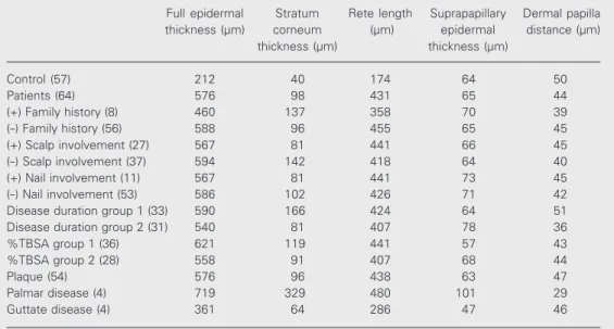
The epidermal measurements for the case and control groups are presented in Table 1. Compared to control, the full epidermal thick-ness of psoriasis patients was 2.71 times greater (t = 10.45, P = 0.000), corneal thick-ness was 2.41 times greater (t = 4.15, P = 0.000) and rete length was 2.47 times greater (t = 12.95, P = 0.000). Suprapapillary epider-mal thickness was similar for the case and control groups (t = 0.67, P = 0.503). How-ever, dermal papillae were 13% narrower in the case group than in the control group (t =
Table 1. Epidermal thickness measurements of psoriasis patients and controls.
Full epidermal Stratum Rete length Suprapapillary Dermal papilla
thickness (µm) corneum (µm) epidermal distance (µm)
thickness (µm) thickness (µm)
Control (57) 212 40 174 64 50
Patients (64) 576 98 431 65 44
(+) Family history (8) 460 137 358 70 39
(-) Family history (56) 588 96 455 65 45
(+) Scalp involvement (27) 567 81 441 66 45
(-) Scalp involvement (37) 594 142 418 64 40
(+) Nail involvement (11) 567 81 441 73 45
(-) Nail involvement (53) 586 102 426 71 42
Disease duration group 1 (33) 590 166 424 64 51
Disease duration group 2 (31) 540 81 407 78 36
%TBSA group 1 (36) 621 119 441 57 43
%TBSA group 2 (28) 558 91 407 68 44
Plaque (54) 576 96 438 63 47
Palmar disease (4) 719 329 480 101 29
Guttate disease (4) 361 64 286 47 46
-2.42, P = 0.018). The difference in rete length between patients having a positive and a negative family history was significant (t = -3.334, P = 0.03), being 27% greater in psoriasis patients without a family history. The differences in the thickness of epider-mis, stratum corneum and suprapapillary epidermis, and also in dermal papilla dis-tance between case and control groups were not statistically significant (P > 0.05).
The epidermal measurements according to type of psoriasis are given in Table 1. When the measurements for the patients with plaque type psoriasis were compared with those for the control group, there were sig-nificant differences in epidermal thickness (t = 10.471, P = 0.000), corneal thickness (t = 3.819, P = 0.000), rete length (t = 12.111, P = 0.000), and dermal papilla distance (t = -1.977, P = 0.042) but not in suprapapillary epidermal thickness (t = 0.652, P = 0.516).
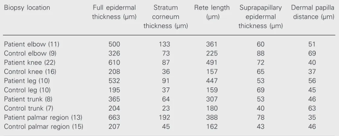
The results concerning the biopsy loca-tion in patients and controls are presented in Table 2. When the measurements in the el-bows of patients and controls were com-pared, the differences in epidermal thick-ness (t = 3.564, P = 0.002) and rete length (t = 5.588, P = 0.000) were statistically sig-nificant while there was no sigsig-nificant dif-ference in stratum corneum (t = 1.348, P =
0.194), suprapapillary epidermal thickness (t = -0.468, P = 0.645), or dermal papilla distances (t = -1.021, P = 0.321). Again, when the lesion areas measured in the knees of the patients and the control group were compared, the differences in epidermal thick-ness (t = 7.113, P = 0.000) and rete length (t = 10.117, P = 0.000) were statistically significant, whereas no significant differ-ences in stratum corneum (t = 1.494, P = 0.144), suprapapillary epidermal thickness (t = 0.518, P = 0.608), or dermal papilla distances (t = -1.170, P = 0.250) were ob-served. When the lesion areas measured in the legs of the patients and the control group were compared, the differences in epidermal thickness (t = 6.002, P = 0.000), rete length (t = 6.187, P = 0.000), and stratum corneum thickness (t = 2.438, P = 0.018) were statis-tically significant, whereas no significant difference in suprapapillary epidermal thick-ness (t = -0.433, P = 0.670) or dermal papilla distance (t = -0.439, P = 0.666) was ob-served. In the trunk, when the results for patients and controls were compared, the differences in epidermal thickness (t = 3.322, P = 0.006), rete length (t = 3.002, P = 0.010), stratum corneum thickness (t = 2.438, P = 0.030), and dermal papilla distances (t = 2.544, P = 0.024) were statistically
signifi-Table 2. Epidermal thickness measurements of psoriasis patients and controls according to biopsy location.
Biopsy location Full epidermal Stratum Rete length Suprapapillary Dermal papilla
thickness (µm) corneum (µm) epidermal distance (µm)
thickness (µm) thickness (µm)
Patient elbow (11) 500 133 361 60 51
Control elbow (9) 326 73 225 88 69
Patient knee (22) 610 87 491 72 40
Control knee (16) 208 36 157 65 37
Patient leg (10) 532 91 447 53 56
Control leg (10) 195 37 159 69 45
Patient trunk (8) 365 64 307 53 46
Control trunk (7) 204 23 180 40 63
Patient palmar region (13) 663 192 388 78 35
Control palmar region (15) 207 45 162 43 46
cant, whereas there was no significant dif-ference in suprapapillary epidermal thick-ness (t = 1.066, P = 0.306). When the results for patients and controls were compared for the palmar region, the differences in epithe-lial thickness (t = 4.104, P = 0.001), rete length (t = 4.616, P = 0.000) and stratum corneum thickness (t = 3.128, P = 0.004) were statistically significant, whereas there were no significant differences in suprapap-illary epidermal thickness (t = 1.014, P = 0.320) or dermal papilla distance (t = -1.526, P = 0.139).
A significant difference in corneal thick-ness of knee lesions was observed between age groups, with thickness increasing with age. Similarly, rete length also increased with age (P = 0.028, chi square = 3.11). A significant difference for suprapapillary epi-dermal thickness between the patients and control groups was not determined. There was no significant correlation between der-mal papilla distances and age groups (P > 0.05). No significant difference in epithelial thickness was observed in palmar regions, elbows, legs and trunk of the various age groups compared to control (P > 0.05).
A family history of psoriasis was positive in 8 (12.5%) patients and negative in 56 (87.5%). Scalp involvement was present in 27 (42.2%) cases and absent in 37 (57.8%). Also, nail involvement was present in 11 (17.2%) cases and absent in 53 (82.8%). Measurements of epidermal thickness ac-cording to %TBSA are presented in Table 1. The difference in epithelial thickness be-tween patients with and without a family history was not significant (P > 0.05).
No significant correlation between epi-dermal thickness and duration of the disease was found (P > 0.05; Table 1).
Corneal thickness was significantly greater in psoriasis patients with scalp in-volvement (t = -2.651, P = 0.008). The dif-ferences in full epidermal thickness, rete length, suprapapillary epidermal thickness, and dermal papilla distance between these
groups were not statistically significant (P > 0.05). In addition, there was no significant difference in epithelial thickness between psoriasis patients with and without nail in-volvement (P > 0.05).
Discussion
Many studies have been conducted to estimate the remission and exacerbation of psoriasis on the basis of clinical findings and objective parameters. Many methods such as ultrasound, reflectance colorimetry, com-puterized video image analysis, pseudo-elon-gation of capillaries, evaluation of skin sur-face hydration, changes in skin blood flow, and nitric oxide production have been inves-tigated for this purpose (1-7). The aim of all of these studies was to determine objective parameters for the definition of patient in-volvement by psoriasis, a diseasethat nega-tively affects the quality of life and psychol-ogy of the patient. The disease requires long and relatively expensive treatment and meth-ods are needed to contribute to its diagnosis and to estimate periods of remission and exacerbation. In these studies, inter- and intraobserver variations must be minimized and the least invasive methods should be used. The clinical relevance of the findings must be clear and reliable. Many studies have been conducted thus far, but their prac-tical usefulness has not been demonstrated. Most of them have determined that psoriasis area and severity index should be taken into account as part of the clinical criteria con-cerning erythema and plaque thickness (1-3). Ormerod et al. (1) compared subjective measures such as erythema, elevation, and scaling separately and as a whole with objec-tive criteria such as ultrasound, reflectance colorimetry, computerized video image anal-ysis, and nitric oxide production in their series of 12 patients and concluded that ni-tric oxide production was the most powerful objective measure.
intrapapillary vessels in active and healing plaques by computer-supported image anal-ysis in a series of 120 cases. They reported that the vessels were not proliferative, and that there was no difference in the arrange-ment of the vessels between the psoriatic lesion and the adjacent non-involved skin during separate periods.
Ashcroft et al. (5) examined the status of clinical outcome measures used in psoriasis research. The measures most commonly used were individual sign scores, e.g., for ery-thema, plaque thickness or scaling, and pooled indices, e.g., psoriasis area and se-verity index. Similarly, Ring et al. (4) also studied psoriasis area and severity index.
In the series of 70 cases reported by Kim et al. (3), erythema, scaling and infiltration of individual lesions were assessed subjec-tively by determining the extent of involve-ment. The functional status of the stratum corneum was evaluated by measuring elec-trical capacitance and conductance of the involved and uninvolved skin of psoriatic patients, and transepidermal water loss was measured. The authors showed that psoriatic attacks were associated with dry skin.
Tanaka et al. (6) reported that computer-assisted area measurement of skin lesions was very useful in evaluating the severity of the disease. However, they did not measure epithelial thickness.
In contrast with other studies, we con-centrated on sensitive measurements of epi-thelial thickness. We performed sensitive measurements of epidermal thickness such as stratum corneum thickness, rete length, suprapapillary epidermal thickness, and der-mal papilla distances using a microscope and a computer. The relationship between these measured properties and the clinical findings, such as type of psoriasis, %TBSA, scalp involvement, nail involvement, and the presence of a family history was then investigated.
The results showed that full epidermal thickness, corneal thickness, and rete length
were increased in psoriasis patients com-pared to controls. Dermal papillae tended to be narrower in patients than in controls, but the difference was not statistically signifi-cant, and suprapapillary epidermal thickness was similar in the two groups. Similarly, when the patients with plaque type psoriasis were compared with the control group, epi-dermal thickness, corneal thickness, rete length, and dermal papilla distance were significantly different, whereas there was no significant difference in suprapapillary epi-dermal thickness. The other types (inverse, erythrodermic) were not compared because of an insufficient number of patients.
The corneal thickness of patients without scalp involvement was 75% greater than in patients with scalp involvement. The differ-ence in corneal thickness between patients with and without scalp involvement was sig-nificant (t = -2.651, P = 0.008). The differ-ences in epidermal, rete and suprapapillary epidermal thickness, and dermal papillary distance were not statistically significant be-tween patients with and without scalp in-volvement (P > 0.05). Kaur et al. (11) ob-served that the scalp was the first site in-volved in 25.2% of patients, and stated that whether the fact that the corneum was thin-ner in psoriasis patients with scalp involve-ment was a definitive factor for dose adjust-ment in treatadjust-ment should be studied further. The duration of disease, %TBSA and nail involvement caused no significant change in epithelial thickness in psoriasis patients. In the study by Stuart et al. (9), nail involvement was more common in early-onset psoriasis; however, there were no
dif-ferences between familial and nonfamilial cases. When the results concerning the bi-opsy sites were compared between patients and controls, the most prominent increase in full epidermal thickness and corneal thick-ness was observed in the palmar region and the least increase in the elbows of patients. Also, suprapapillary epidermal thickness was prominent in the palmar region but not in the elbows. The difference between dermal pa-pilla widths was quite small.
In conclusion, epithelial thickness can be sensitively measured in skin biopsies using computer-supported image analysis. Param-eters that involve epithelial characteristics, such as stratum corneum thickening or rete elongation, can be determined objectively. Since the follow-up periods and the number of patients in the present study were not sufficient, the relationship between epitheli-al thickness observed during the follow-up period and the response to various treatment modalities was not analyzed.
References
1. Ormerod AD, Dwyer CM, Weller R, Cox DH & Price R (1997). A comparison of subjective and objective measures of reduction of psoriasis with the use of ultrasound, reflectance colorimetry, com-puterized video image analysis, and nitric oxide production. Journal of the American Academy of Dermatology, 37: 51-57.
2. Bacharach-Buhles M, el Gammal S, Panz B & Altmeyer P (1994). The pseudo-elongation of capillaries in psoriatic plaques. Acta Dermato-Venereologica, 186 (Suppl): 133-137.
3. Kim SD, Huh CH, Seo KI, Suh DH & Youn JI (2002). Evaluation of skin surface hydration in Korean psoriasis patients: a possible factor influencing psoriasis. Clinical and Experimental Dermatology, 27: 147-152.
4. Ring J, Kowalzick L, Christophers E, Schill WB, Schopf E, Stander M, Wolff HH & Altmeyer P (2001). Calcitriol 3 microg g-1 ointment in combination with ultraviolet B phototherapy for the treatment of plaque psoriasis: results of a comparative study. British Journal of Dermatology, 144: 495-499.
5. Ashcroft DM, Wan Po AL, Williams HC & Griffiths CE (1999). Clinical measures of disease severity and outcome in psoriasis: a critical appraisal of their quality. British Journal of Dermatology, 141: 185-191.
6. Tanaka M, Gaskell S, Edwards C & Marks R (2000). Simple horizon-tal averaging programme enables shade correction for image analy-sis in psoriaanaly-sis. Clinical and Experimental Dermatology, 25: 323-326.
7. Suh DH, Kwon TE, Kim SD, Park SB, Kwon OS, Eun HC & Youn JI (2001). Changes of skin blood flow and color on lesional and control sites during PUVA therapy for psoriasis. Journal of the American Academy of Dermatology, 44: 987-994.
8. Wang H, Zhang X & Yang S (2001). Study on the risk factors of psoriasis. Zhonghua Liuxingbingxue Zazhi, 22: 215-218.
9. Stuart P, Malick F, Nair RP, Henseler T, Lim HW, Jenisch S, Voorhees J, Christophers E & Elder JT(2002). Analysis of pheno-typic variation in psoriasis as a function of age at onset and family history. Archives of Dermatological Research, 294: 207-213. 10. Rahman P, Schentag CT, Beaton M & Gladman DD (2000).
Compar-ison of clinical and immunogenetic features in familial versus spo-radic psoriatic arthritis. Clinical and Experimental Rheumatology, 18: 7-12.