Leaf anatomy and its contribution to the systematics of
Aechmea
subgenus
Macrochordion
(de Vriese) Baker (Bromeliaceae)
ANA PAULA G. DE FARIA1, ANA CLAUDIA M. VIEIRA2 and TÂNIA WENDT3 1Instituto de Ciências Biológicas, Departamento de Botânica, Universidade Federal de Juiz de Fora,
Campus Universitário, Bairro São Pedro, 36036-900 Juiz de Fora, MG, Brasil
2Faculdade de Farmácia, Departamento de Produtos Naturais e Alimentos, Universidade Federal do Rio de Janeiro,
CCS, Ilha do Fundão, 21941-590 Rio de Janeiro, RJ, Brasil
3Instituto de Ciências Biológicas, Departamento de Botânica, Universidade Federal do Rio de Janeiro,
CCS, Ilha do Fundão, 21941-590 Rio de Janeiro, RJ, Brasil
Manuscript received on February 7, 2011; accepted for publication on June 6, 2011
ABSTRACT
The leaf anatomy of the species Aechmea subgenus Macrochordion was analyzed to obtain valuable data on their taxonomic delimitation and to identify anatomical adaptations to their respective habitats and habits. All leaves of these species are hypostomatic, and present: peltate trichomes on both surfaces; stomata sunk in epidermal depressions; small epidermal cells with thick walls and inclusions of silica bodies; a mechanical hypodermis; an aquiferous parenchyma; chlorenchyma with fibrous clusters and air channels; and vascular bundles surrounded by a parenchymatic sheath and a cap of fibers. The results are evaluated within an adaptive and taxonomic context. Variations in hypodermic thickening, amount of water parenchyma, position of the air channels and shape of the cells filling the air channels are useful for delimiting groups of species, strengthening the relationships suggested by their external morphology.
Key words: Aechmea, Bromeliaceae, leaf anatomy, Macrochordion.
Correspondence to: Ana Paula Gelli de Faria E-mail: [email protected].
INTRODUCTION
The Bromeliaceae includes 3,172 species (Luther
2008) and is a representative monocot family for
Neotropical flora. It occupies a basal position within
the Poales order and is closely related to Typhaceae
and Rapateaceae (APG III 2009). Bromeliads
demonstrate remarkable ecological versatility,
inhabiting mesic to xeric environments (e.g. humid
tropical forests to deserts) and display a wide
range of terrestrial, lithophytic and epiphytic habits
(Smith 1934, Benzing 2000a). Some morphological
and physiological adaptations to drier habitats
and particular life forms out of the soil include
the presence of a central tank (which allows the
plant to collect water and organic material inside
enlarged and overlapping leaf sheaths), leaves with
absorbing scales (the most distinguishing feature
of the family, also referred to as peltate trichomes),
water storage and mechanical support tissues, the
development of ‘crassulacean acid metabolism’
(CAM) photosynthesis, and the progressive
structural and functional reduction of root systems
in epiphytic and rupiculous species (Tomlinson
1969, Benzing 1976, 2000a, b).
(Annals of the Brazilian Academy of Sciences)
The Bromeliaceae is traditionally divided into
the subfamilies Pitcairnioideae, Bromelioideae and
Tillandsioideae (Smith and Downs 1974, 1977, 1979).
A recent molecular phylogenetic study suggests that
it could be divided into eight monophyletic lineages
(including Bromelioideae and Tillandsioideae)
due to the paraphyly of Pitcairnioideae (Givnish
et al. 2007). Bromelioideae species are typically
distinguished by the inferior ovary, baccate
and indehiscent fruits and unappendaged seeds
(Smith and Downs 1979). However, the generic
delimitation of this subfamily is the less understood
in Bromeliaceae, particularly within Aechmea Ruiz
& Pav., the largest (with more than 250 species;
Luther 2008) and morphologically most diverse
genus of Bromelioideae (Faria et al. 2004). In the
last monograph on Bromelioideae, Smith and Downs
(1979) recognized eight subgenera for
Aechmea
:
Aechmea
,
Chevaliera
(Gaudich. ex. Beer) Baker,
Lamprococcus
Beer (Baker),
Macrochordion
de Vriese (Baker),
Ortgiesia
(Regel) Mez,
Platyaechmea
(Baker) Baker,
Podaechmea
Mez
and
Pothuava
(Baker) Baker. This infrageneric
circumscription is also considered artificial and
historically based on few characters (Faria et al.
2004, Sass and Specht 2010).
In
Aechmea
, potentially useful diagnostic data
are still limited and most characters traditionally
emphasized in previous taxonomic treatments of the
genus (e.g. inflorescence, sepal and floral bract features,
pollen grains morphology and petal appendages
presence) have often failed to delimit natural groups
(Faria et al. 2004, Schulte and Zizka 2008). Thus, an
investigation of different characters besides the ordinary
external morphology (e.g. anatomical aspects) became
essential to provide additional and valuable data to
increase knowledge about the systematics of the
genus. The function and adaptive value of anatomical
features are often extremely useful as they may help
to reveal more clearly the homologies of structures
for classification purposes and reconstruction of
phylogeny (Stuessy 1990). Structural aspects of some
vegetative organs, for example, have contributed to
the generic/infrageneric delimitation of many groups
(Weiner and Liese 1993, Starr and Ford 2001, Alves
et al. 2002, Calvente et al. 2008, Oliveira et al. 2008,
Cardoso et al. 2009).
The taxonomic and phylogenetic use of
anato-mical characters, especially leaves´, has been helpful
in the delimitation of different genera of Bromeliaceae
(Robinson 1969, Varadarajan and Gilmartin 1988,
Gilmartin et al. 1989, Sajo et al. 1998, Almeida et al.
2009). For
Aechmea
, foliar anatomical aspects have
been investigated in descriptive works (Tomlinson
1969, Smith and Downs 1974, Flores 1975, Braga
1977, Benzing 2000b), using an ecophysiological
approach (Scarano et al. 2002), or used as a tool
for taxonomic delimitations of the subgenera
Lamprococcus
(Aoyama and Sajo 2003) and
Chevaliera
(Sousa et al. 2005) and for species of the
genus occurring in São Paulo state, southeast Brazil
(Proença and Sajo 2004).
Aechmea
subgenus
Macrochordion
, the
focus of this study, is characterized by grouping
epiphytes, terrestrials and rupicolous medium-sized
species, presenting simple, densely floccose or
lanate inflorescences with sessile and polystichous
flowers; entire carinate and unarmed floral bracts;
unarmed and connate sepals, and well-developed
fringed petal appendages (Faria et al. 2010). The
species share great morphological similarity, which
makes delimitation difficult, especially when some
diagnostic characters are poorly preserved or not
mentioned on the labels of herbarium specimens
(e.g. calyx and corolla color). The subgenus is
entirely restricted to the Brazilian Atlantic Forest,
except for
A. bromeliifolia
, which occurs from
Central America to Argentina. In a recent taxonomic
revision, Faria et al. (2010) recognized five
species of
Macrochordion
: (1)
A. alba
Mez; (2)
A.
bromeliifolia
Rudge (Baker), including the typical
variety and
A. bromeliifolia
var.
albobracteata
as placed five previously recognized taxa into
synonymy: (1)
A. maculata
and (2)
A. chlorophylla
(both under
A. lamarchei
); (3)
A. pabstii
(under
A.
alba
); (4)
A. kautskyana
(under
A. triangularis
)
and (5)
A. bromeliifolia
var.
angustispica
(under
A.
bromeliifolia
var.
bromeliifolia
).
The present study describes the foliar structure
of
Aechmea
subgenus
Macrochordion
with the aim
of providing additional morphological characters to
support the systematic boundaries of the species, as
well as identifying anatomical adaptations to their
respective habitats.
MATERIALS AND METHODS
Leaves were obtained from cultivated specimens at
the Universidade Federal do Rio de Janeiro (adult
shoots from individuals collected in the field and
maintained in a greenhouse). A voucher for each
studied sample was deposited in the RFA herbarium
under the following specifications:
A. alba
, Porto
Seguro, Bahia, Faria et al. 139;
A. bromeliifolia
var.
albobracteata,
Santo Antônio do Itambé, Minas
Gerais, Faria & Versieux 171;
A. bromeliifolia
var.
bromeliifolia
, Arcos, Minas Gerais, Faria 176;
A.
lamarchei
, Santa Teresa, Espírito Santo, Faria et al.
161;
A. maasii
, Armação de Búzios, Rio de Janeiro,
Wendt et al. 443;
A. triangularis
, Santa Teresa,
Espírito Santo, Faria et al. 165.
Samples from the middle region of adult leaf
blades were fixed in a solution of
formaldehyde-acetic acid-alcohol 50% and then transferred and
stored in 50% alcohol (Johansen 1940). Transverse
freehand sections were obtained using a razor blade,
bleached with sodium hypochlorite and stained
with Astra blue-Safranin (Bukatsch 1972). The
histological slides were mounted in 50% glycerine
and observed with an Olympus CH 30 light
microscope. For histochemistry, fresh sections were
submitted to the following tests: Lugol for starch
(Johansen 1940), Sudan IV for lipids (Gerlach 1984)
and acidic phloroglucin for lignin (Sass 1951).
RESULTS
The leaf epidermis of
Aechmea
subgenus
Macrochordion
species is organized in a single
layer of cells and in transverse section can be almost
plane on both surfaces, as in
A. lamarchei
(Fig. 1A),
undulating on the abaxial surface in
A. bromeliifolia
var.
albobracteata
(Fig. 1B) and
A. triangularis
(Fig.
1C) or on both surfaces, as in
A. bromeliifolia
var.
bromeliifolia
(Fig. 1D),
A. alba
(Fig. 1E) and
A.
maasii
(Fig. 1F). All epidermal cells have a reduced
lumen with very thick and lignified anticlinal and
internal periclinal walls (Fig. 2A), including one
nearly spherical silica body (Fig. 2F). The external
periclinal walls are covered with a thin cuticle,
except in
A. triangularis
, which possesses a dense
covering (Fig. 2G). All taxa have peltate trichomes
on both surfaces, with the stalk composed of two
cells sunk into epidermal depressions (Fig. 2A) and
hypostomatic leaves with sunken stomata below the
other ordinary epidermal cells (Fig.1A-F, Fig. 2B).
All taxa have an adaxial and abaxial
hypodermis. This tissue is more developed adaxially
and executes a mechanical function due to cell wall
thickening. The mechanical hypodermis is arranged
in one or two layers of thickened or slightly
thickened cells (Table I). Slightly thickened cells are
observed in
A. alba
(Fig. 2C),
A. maasii
(Fig. 2D)
and
A. lamarchei
(Fig. 2E) and thick-walled cells
in
A. bromeliifolia
var.
bromeliifolia
(Fig. 2F),
A.
bromeliifolia
var.
albobracteata
and
A. triangularis
(Fig. 2G). The cells are nearly isodiametric, except
in
A. triangularis
, where the first layer of the adaxial
surface presents anticlinally extended hypodermic
cells (Fig. 2G). Lignified walls are also observed
only in
A. triangularis
, and the remaining taxa
exhibit a non-lignified hypodermis.
I), being more developed in
A. bromeliifolia
var.
bromeliifolia
,
A. triangularis
and
A. bromeliifolia
var.
albobracteata
(Fig. 1B–D). Below the water
parenchyma, the chlorenchyma occupies the
greater portion of the mesophyll (Fig. 1A–F) and is
formed by anticlinally extended cells arranged like
a palisade on the adaxial surface of
A. bromeliifolia
var.
albobracteata
(Fig. 1B, Table I) and on both
surfaces of
A. triangularis
(Fig. 1C, Table I). In the
remaining taxa, chlorenchymatic cells are slightly
extended anticlinally or are almost isodiametric
(Fig. 1A, D–F, Table I). Air channels (or air-lacunae
in the sense of Tomlinson 1969) alternate with the
vascular bundles are also observed in all taxa (Fig.
1A–F). In
A. alba
,
A. lamarchei
and
A. maasii
, the air
channels are usually continuous to the substomatal
chambers on the abaxial surface and filled with
nearly isodiametric cells with short projections
(Fig.1A, E, F, Fig.3B, Table I). In
A. triangularis
,
A.
bromeliifolia
var.
albobracteata
and
A. bromeliifolia
var.
bromeliifolia
, the air channels are farther from
the abaxial surface, with no direct connection to the
substomatal chambers, and are filled with stellate
cells (Figs. 1B–D, 3A, Table I). Fibrous clusters have
a scattered distribution adaxially and abaxially in the
chlorenchyma of all examined species (Fig. 1A–F).
Figure 1 - Transversal sections of leaves of Aechmea subgenus Macrochordion taxa.
A–F. Mesophyll overview showing the water parenchyma (Wp), air channels (Ac) and fibrous clusters (Fc) distributed on adaxial and abaxial surfaces. A.A. lamarchei: outline of adaxial and abaxial surfaces almost plane. B. A. bromeliifolia var. albobracteata
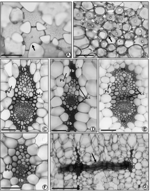
In all taxa we observed a single series of
collateral vascular bundles arranged in the central
portion of the mesophyll (Fig. 1A–F), along with
slender commissural bundles (Fig. 3G). The
larger vascular bundles are surrounded by slightly
thickened and lignified cells (also between the
xylem and phloem) and covered by a cap of fibers
on the xylem and phloem poles (Fig. 3C, E). This
cap of fibers can be totally lignified in
A. alba
,
A.
lamarchei
and
A. maasii
or incompletely lignified in
A. bromeliifolia
var.
bromeliifolia
,
A. bromeliifolia
var.
albobracteata
and
A. triangularis
(Table I). The
slightly thickened and lignified cells surrounding the
smaller vascular bundles do not appear between the
xylem and phloem and the cap of fibers can extend to
both poles (Fig. 3D) or not (Fig. 3F). In association
with the lignified cells and the cap of fibers, we
observe a sheath of parenchymatic cells contained
starch grains (Fig. 1A–F; 3C–F), smaller than the
remaining chlorenchymatic cells of the mesophyll.
DISCUSSION
The anatomical foliar features of
Aechmea
subgenus
Macrochordion
are in accordance with
the general patterns observed for Bromeliaceae
(Tomlinson 1969, Smith and Downs 1974,
Figure 2 - Details of epidermal cells, trichomes, stomata and mechanical hypodermis.A. Epidermal cell (Ep) with reduced lumen (arrow) and peltate trichome (Pt) on adaxial surface, with the sunk stalk (bracket). B. Stomata (St) sunk in epidermal depressions.
Benzing 2000b). The parenchymatic sheath and
fibers involving the vascular bundles are treated
as the endodermis and the pericycle, respectively,
following the nomenclature of Van Fleet (1961).
P.B. Pita (unpublished data), Sajo et al. (1998),
Arruda and Costa (2003) and Scatena and Segecin
(2005) have also described these structures in other
bromeliad species. Although Smith and Downs
(1974) suggested that a palisade chlorenchyma is not
a common character in the Bromeliaceae, Proença
and Sajo (2004) reported the presence of this tissue
in an exemplar of
A. bromeliifolia
from São Paulo
state, southeastern Brazil. The authors, however,
did not specify whether the specimen analyzed
Figure 3 - Detail of the air channels and vascular bundles. A.A. bromeliifolia var. bromeliifolia: stellatecells (arrow). B.A. lamarchei: isodiametric cells with short projections (arrow). C, D.A. lamarchei:
belonged to the typical variety or to the variety
albobracteata
. Both varieties of
A. bromeliifolia
occur in São Paulo state (Faria et al. 2010), and in
this study, we observed anticlinally extended cells
arranged like a palisade in var.
albobracteata
.
The species analyzed show several xeromorphic
characters commonly reported for Bromeliaceae
(e.g. epidermal cells with lignified thick walls and
silica body inclusions, mechanical hypodermis,
water parenchyma, sunken stomata and lignified
fibers in the mesophyll). Silica bodies have been
associated with resistance against herbivory due to
their inedibility (MacNaughton et al. 1985, Prychid
et al. 2004), but are also important for refracting
excess of light, helping individuals to establish in
sun-exposed environments (Krauss 1949). These
inclusions are also commonly found in other
monocots families within Poales, suggesting that
this is a plesiomorphic character for the order
(Pereira et al. 2011).
According to Tomlinson (1969), sunken stomata
below the remaining epidermal cells help to prevent
dehydration by transpiration. Several plant families
from arid zones, particularly succulents, have stomata
deeply sunken in depressions in the epidermis
(crypts), such as Agavaceae (Fahn and Cutler 1992)
and Cactaceae (Calvente et al. 2008). However, this
trait is not restricted among plants exposed to drought
TAXA
CHARACTERS 1 2 3 4 5 6
Hypodermic cells walls slightly thickened + - - + +
-Hypodermic cells walls thickened - + + - - +
Water parenchyma with 1-2 layers + - - + +
-Water parenchyma with 3-5 layers - + + - - +
Chlorenchymatic cells anticlinally extended - - + - - +
Chlorenchymatic cells slightly extended or
almost isodiametric + + - + +
-Air channels connected to the substomatal
chambers + - - + +
-Air channels not connected to the substomatal
chambers - + + - - +
Isodiametric cells with short projections
interrupting the air channels + - - + +
-Stellate cells interrupting the air channels - + + - - +
Vascular bundles recovered by fibers totally
lignified + - - + +
-Vascular bundles recovered by fibers
incompletely lignified - + + - - +
TABLE I
Leaf anatomy characters observed for Aechmea subgenus Macrochordion with
taxonomic relevance in the delimitation of groups of species.
1: A. alba; 2: A. bromeliifolia var. bromeliifolia; 3: A. bromeliifolia var. albobracteata; 4: A. lamarchei; 5: A. maasii; 6: A. triangularis.
(Cutler et al. 2007), and recent studies indicate that
it is unlikely that the primary function of crypts is
to reduce transpiration (Roth-Nebelsick et al. 2009).
Some species of
Aechmea
subgenus
Macrochordion
(e.g.
A. bromeliifolia
) can occupy both xeric and
mesic habitats (Faria et al. 2010). Previous studies
also indicate the occurrence of sunken stomata in
other
Aechmea
species from mesic environments
(Proença and Sajo 2004, Sousa et al. 2005) suggesting
that the presence of this character may be not a
significant adaptation to environmental conditions of
low water availability. As discussed by Proença and
Sajo (2004), although many xeromorphic characters
found in the Bromeliaceae allow the exploration of
extreme environments, they can represent ancestral
adaptations selected during the diversification of
the family and should not be interpreted only as
traits related to a particular environment where they
currently live.
Besides the position below the other
ordinary epidermal cells, the stomata of all
Macrochordion
taxa are restricted to the abaxial
surface. Hypostomatism is a common condition
for Bromeliaceae (Tomlinson 1969) and was
also reported for other
Aechmea
species from
subgenera
Lamprococcus
(Aoyama and Sajo 2003),
Platyaechmea
,
Pothuava
,
Ortgiesia
(Proença
and Sajo 2004) and
Chevaliera
(Sousa et al. 2005).
According to Fahn and Cutler (1992) stomata restricted
to the abaxial surface are more common in plants from
humid environments. Other authors have suggested
that hypostomatic leaves may be a strategy to minimize
water loss by convection currents or breezes that could
remove water vapor from the leaf surface (Nobel
1999). Scatena and Segecin (2005) investigated the
foliar anatomy of some
Tillandsia
(Tillandsioideae) in
which xeromorphic traits are predominant, and argued
that the presence of hypostomatic leaves in these
species, may be more of a plesiomorphic condition
retained than a response to environmental selection.
The hypodermis, along with the thick and
lignified epidermal cells and the vascular and
extravascular fiber bundles develop a mechanical
function of strengthing the foliar structure.
These tissues can also protect against hydric and
temperature stress by reducing water evaporation
from internal tissues and preventing mesophyll
collapse in unfavorable abiotic conditions (Krauss
1949, Brighigna et al. 1984, Fahn and Cutler 1992).
Besides avoiding water loss, the water parenchyma
also protects the chlorenchymatic cells against
excessive luminosity (Brighigna et al. 1984). The
thick-walled hypodermic cells and greater amount of
water parenchyma observed in
A. bromeliifolia
var.
bromeliifolia
,
A. bromeliifolia
var.
albobracteata
and
A. triangularis
may be adaptations to the particular
environmental conditions and habits of these species.
The main habitats occupied by
A. bromeliifolia
var.
bromeliifolia
and
A. bromeliifolia
var.
albobracteata
are dry and/or sun-exposed (Faria et al. 2010), such
as the rocky grasslands (
campos rupestres
),
caatingas
and savannas (
cerrados
). Although
A. triangularis
is restricted to humid habitats of the Atlantic Forest
(Faria et al. 2010), it grows only as epiphyte, and
mostly in the higher strata of trees. Besides the water
stressful condition created by the epiphytic life form,
this species receives intense levels of luminosity. The
remaining taxa (
A. alba
,
A. maasii
and
A. lamarchei
)
grow as terrestrials or epiphytes, preferentially in
humid and/or shaded habitats of dense ombrophile
and semideciduous forests, shrubby and wooded
sandy coastal plains (
restingas
) and tableland forests
in leaves of mesophytic bromeliads. In the subgenus
Macrochordion
, the occurrence of this character may
also suggest an adaptation of the species to their
respective habitats.
As observed by Versieux et al. (2010) for
closely related species of the genus
Alcantarea
(Tillandsioideae), most of the anatomical features
examined in this study are also very homogeneous
within the
Aechmea
subgenus
Macrochordion
.
However, some of the characters that were
investigated are useful to characterize particular
taxa, as well as circumscribing groups of species,
strengthening the relationships indicated by their
external morphology.
Aechmea triangularis
exhibits unique anatomical traits, such as a
thick cuticle layer on the epidermal surface,
anticlinally extended and sclerified hypodermic
cells, and a palisade chlorenchyma on both faces
of the leaf. Some aspects of the vegetative and
reproductive external morphology also distinguish
A. triangularis
from other
Macrochordion
species,
such as leaves with caudate and recurvate apices
and a blue corolla (Faria et al. 2010). The leaf
structure of
A. triangularis
is more similar to the
same observed for both varieties of
A. bromeliifolia
due to the presence of a well-developed water
parenchyma, hypodermic cells with thick walls,
air channels not connected to the substomatal
chambers and filled with stellate cells, and vascular
bundles covered by a cap of incompletely lignified
fibers. The close relationship among these taxa is
also reflected by some aspects of their external
morphology, such as the presence of leaf spines
longer than 3 cm, emarginate, symmetric to slightly
asymmetric and half connate sepals, and spatulate
petals with emarginated apices (Faria et al. 2010).
Similarly,
A. alba
,
A. lamarchei
and
A. maasii
have
leaf spines up to 3 mm long, obtuse and distinctly
asymmetric sepals and lingulate petals with
obtuse apices (Faria et al. 2010). These species
also share some anatomical features not observed
in
A. bromeliifolia
and
A. triangularis
, such as a
less developed water parenchyma, hypodermic
cells with thinner walls, air channels filled with
nearly isodiametric cells with short projections
and connected to the substomatal chambers, and
vascular bundles covered by a totally lignified
cap of fibers. Variations in the shape of the cells
interrupting the air channels were also reported by
Aoyama and Sajo (2003) and Sousa et al. (2005),
and showed to be useful in the delimitation of
species within
Aechmea
subgenera
Lamprococcus
and
Chevaliera
.
This study contributed to a better understanding
of some anatomical traits responsible for the
adaptation of the
Macrochordion
species to their
respective habitats and habits, as well as revealed
some useful taxonomical characters to characterize
particular species and to delimit groups of species.
We also contributed with potential data to be
explored in future phylogenetic studies within the
subgenus and between
Aechmea
and other related
Bromelioideae genera.
ACKNOWLEDGMENTS
We thank the Coordenação de Aperfeiçoamento
de Pessoal de Nível Superior (CAPES) for the
scholarship to Ana Paula G. de Faria and the
Conselho Nacional de Desenvolvimento Científico
e Tecnológico (CNPq) for a productivity grant to
Tânia Wendt. This paper is part of a PhD thesis
undertaken at the Graduate Program in Botany of
the Universidade Federal do Rio de Janeiro by
the first author.
RESUMO
A anatomia foliar de espécies de Aechmea subgênero Macrochordion foi analisada visando obter caracteres úteis para sua delimitação taxonômica e identificar adaptações
anatômicas aos seus respectivos habitats e hábitos. Todas as folhas são hipoestomáticas e apresentam: tricomas
hipoderme mecânica; parênquima aquífero; clorênquima com feixes de fibras e canais de aeração; feixes vasculares envolvidos por bainha parenquimática e calotas de fibras.
Os resultados são avaliados dentro de um contexto adaptativo e taxonômico. Variações no espessamento da
hipoderme, na quantidade de parênquima aquífero, na
posição dos canais de aeração e na forma das células que
preenchem os canais de aeração mostraram-se úteis para separar grupos de espécies, corroborando relações de
similaridade apontadas pela morfologia externa.
Palavras-chave: Aechmea, Bromeliaceae, anatomia foliar, Macrochordion.
REFERENCES
ALMEIDA VR,COSTA AF,MANTOVANI A,GONÇALVES-ESTEVES
V,ARRUDA RCO AND FORZZA RC.2009. Morphological phylogenetics of Quesnelia (Bromeliaceae, Bromelioideae). Syst Bot 34: 660-672.
ALVES MV,ESTELITA MEM,WANDERLEY MGL AND THOMAS WW.2002. Aplicações taxonômicas da anatomia foliar das
espécies brasileiras de Hypolytrum Rich. (Cyperaceae). Rev Bras Bot 25: 1-9.
AOYAMA EM AND SAJO MG.2003. Estrutura foliar de Aechmea
Ruiz & Pav. subgênero Lamprococcus (Beer) Baker e espécies relacionadas. Rev Bras Bot 26: 461-473.
APG - ANGIOSPERM PHYLOGENY GROUP. 2009. An update of
the Angiosperm Phylogeny Group classification for the orders and families of flowering plants: APG III. Bot J Lin
Soc 161: 105-121.
ARRUDA RC AND COSTA AF.2003. Foliar anatomy of five Vriesea sect. Xiphion (Bromeliaceae) species. Selbyana
24: 180-189.
BENZING DH.1976. Bromeliad trichomes: structure, function,
and ecological significance. Selbyana 1: 330-348.
BENZING DH. 2000a. Introduction. In: Benzing DH (Ed),
Bromeliaceae: profile of an adaptive radiation. Cambridge: Cambridge University Press, p. 3-15.
BENZING DH.2000b. Vegetative structure. In: Benzing DH (Ed),
Bromeliaceae: profile of an adaptive radiation. Cambridge: Cambridge University Press, p. 19-77.
BRAGA MMN. 1977. Anatomia foliar de Bromeliaceae da
Campina. Acta Amaz 7: 1-74.
BRIGHIGNA L,FIORDI AC AND PALANDRI MR.1984. Structural characteristics of mesophyll in some Tillandsia species.
Phytomorphology 34: 191-200.
BUKATSCH F.1972. Bemerkungen zur Doppelfärbung Astrablau-Safranin. Mikrokosmos 61: 255.
CALVENTE AM,ANDREATA RHP AND VIEIRA RC.2008. Stem anatomy of Rhipsalis (Cactaceae) and its relevance for
taxonomy. Pl Syst Evol 276: 1-7.
CARDOSO CMV, PROENçA SL AND SAJO MG.2009. Foliar
anatomy of the subfamily Myrtoideae (Myrtaceae). Aust J Bot 57: 148-161.
CUTLER DF, BOTHA T AND STEVENSON DW. 2007. Plant
Anatomy: an applied approach. Malden: Blackwell Publishing, 302 p.
FAHN A AND CUTLER DF.1992. Xerophytes. Encyclopedia of
Plant Anatomy. Band 13, Teil 3. Berlin: Gebrüder
Borntraeger, 175 p.
FARIA APG, WENDT T AND BROWN GK. 2004. Cladistic relationships of Aechmea (Bromeliaceae: Bromelioideae) and allied genera. Ann Missouri Bot Gard 91: 303-319.
FARIA APG,WENDT T AND BROWN GK.2010. A revision of Aechmea subgenus Macrochordion (Bromeliaceae) based on phenetic analyses of morphological variation. Bot J
Linn Soc 162: 1-27.
FLORES EM. 1975. Algunos aspectos de anatomia foliar comparada de dos especies de Bromeliaceae (Aechmea mexicana Baker y Hechtia glomerata Zucc.). Rev Biol Trop 23: 29-52.
GERLACH D.1984. Botanische Mikrotechnik: eine einführung.
Stuttgart: Georg Thieme Verlag, 298 p.
GILMARTIN AJ, BROWN GK, VARADARAJAN GS AND
NEIGHBOURS M.1989. Status of Glomeropitcairnia within
evolutionary history of Bromeliaceae. Syst Bot 14: 339-348.
GIVNISH TJ,MILLAM KC,BERRY PE AND SYTSMA KJ.2007.
Phylogeny, adaptive radiation, and historical biogeography of Bromeliaceae inferred from ndhF sequence data. Aliso
23: 3-26.
JOHANSEN D.1940. Plant microtechnique. New York:
McGraw-Hill, 523 p.
KRAUSS BH.1949. Anatomy of the vegetative organs of the pineapple, Ananas comosus (L.) Merr. II. The leaf. Bot
Gaz 110: 333-404.
LUTHER HE.2008. An alphabetical list of bromeliad binomials,
11th ed., Orlando: BSI, 114 p.
MACNAUGHTON SJ,TARRANTS JL,MACNAUGHTON MM AND
DAVIS RH.1985. Silica as a defense against herbivory and
a growth promoter in African grasses. Ecology 66: 528-535.
NOBEL PS.1999. Physicochemical and environmental plant physiology, 2nd ed., San Diego: Academic Press, 540 p. OLIVEIRA RJ,LONGHI-WAGNER HM AND LEITE KRB.2008.
A contribuição da anatomia foliar para a taxonomia de
Raddia Bertol. (Poaceae: Bambusoideae). Acta Bot Bras 22: 1-19.
PEREIRA TAR,OLIVEIRA TSO,SILVA LC AND AZEVEDO AA. 2011. Comparative leaf anatomy of four species of Bromelioideae (Bromeliaceae) occurring in the Atlantic
Forest, Brazil. Botany 89: 243-253.
PROENçA SL AND SAJO MG.2004. Estrutura foliar de espécies de Aechmea Ruiz & Pav. (Bromeliaceae) do Estado de
São Paulo. Acta Bot Bras 18: 319-331.
PRYCHID CJ,RUDALL PJ AND GREGORY M.2004. Systematics
and biology of silica bodies in monocotyledons. Bot Rev
ROBINSON H.1969. A monograph on foliar anatomy of the genera Connelia, Cottendorfia, and Navia (Bromeliaceae).
Smithsonian Contrib Bot 2: 1-41.
ROTH-NEBELSICK A,HASSIOTOU F AND VENEKLAAS EJ.2009. Stomatal crypts have small effects on transpiration: a
numerical model analysis. Plant Physiol 151: 2018-2027.
SAJO MG, MACHADO SR AND CARMELLO-GUERREIRO SM.
1998. Aspectos estruturais de folha de bromélia e suas implicações no agrupamento de espécies. In: PEREIRA
MV (Ed), Bromélias da Mata Atlântica: Canistropsis, Rio de Janeiro: Salamandra, p. 102-111.
SASS JE.1951. Botanical microtechnique, 2nd ed., Iowa: Iowa
State College Press, 228 p.
SASS C AND SPECHT CS.2010. Phylogenetic estimation of the
core Bromelioids with an emphasis on the genus Aechmea
(Bromeliaceae). Mol Phylogenet Evol 55: 550-571.
SCARANO FR,DUARTE HM,RÔçAS G,BARRETO SMB,AMADO
EF,REINERT F,WENDT T,MANTOVANI A,LIMA HRP AND
BARROS CFB.2002. Acclimation or stress symptom? An
integrated study of intraspecific variation in the clonal
plant Aechmea bromeliifolia, a widespread CAM
tank-bromeliad. Bot J Linn Soc 140: 391-401.
SCATENA VL AND SEGECIN S. 2005. Anatomia foliar de Tillandsia L. (Bromeliaceae) dos Campos Gerais, Paraná,
Brasil. Rev Bras Bot 28: 635-649.
SCHULTE K AND ZIZKA G.2008. Multi locus plastid phylogeny of Bromelioideae (Bromeliaceae) and the taxonomic utility of petal appendages and pollen characters. Candollea 63: 209-255.
SMITH LB.1934. Geographical evidence on the lines of evolution
in the Bromeliaceae. Bot Jahrb 66: 446-465.
SMITH LB AND DOWNS RJ.1974. Pitcairnioideae
(Brome-liaceae). In: WURDACK JJ (Ed), Fl Neotropica Mon 14, part 1, New York: Hafner Press, p. 1-658.
SMITH LB AND DOWNS RJ. 1977. Tillandsioideae
(Bromeliaceae). In: ROGERSON CT (Ed), Fl Neotropica Mon 14, part 2. New York: Hafner Press, p. 663-1492.
SMITH LB AND DOWNS RJ. 1979. Bromelioideae
(Bromeliaceae). In: ROGERSON CT (Ed), Fl Neotropica Mon 14, part 3, New York: Hafner Press, p. 1493-2141.
SOUSA GM,ESTELITA MEM AND WANDERLEY MGL.2005.
Anatomia foliar de espécies brasileiras de Aechmea subg. Chevaliera (Gaudich. ex Beer) Baker,
Bromelioideae-Bromeliaceae. Rev Bras Bot 28: 603-613.
STARR JR AND FORD BA.2001. The taxonomic and phylogenetic utility of vegetative anatomy and fruit epidermal silica
bodies in Carex section Phyllostachys (Cyperaceae). Can J Bot 79: 362-379.
STUESSY TF.1990. Plant taxonomy: the systematic evaluation
of comparative data. New York: Columbia University Press, 514 p.
TOMLINSON PB. 1969. Commelinales-Zingiberales. In:
METCALF CR (Ed), Anatomy of the Monocotyledons, vol. 3, Oxford: Oxford University Press, p. 192-294.
VAN FLEET DS. 1961. Histochemistry and function of the endodermis. Bot Rev 27: 165-220.
VARADARAJAN GS AND GILMARTIN AJ. 1988. Taxonomic
realignments within the subfamily Pitcairnioideae
(Bromeliaceae). Syst Bot 13: 294-299.
VERSIEUX LM,ELBL PM,WANDERLEY MGL AND MENEZES
NL. 2010. Alcantarea (Bromeliaceae) leaf anatomical
characterization and its systematic implications. Nord J Bot 28: 385-397.
WEINER G AND LIESE W.1993. Generic identification key to
rattan palms based on stem anatomy characters. IAWA