RESUMO. - [Estudo clonal de Escherichia coli aviário
por análise de seqüências de DNA conservadas do gene fliC.] A relação clonal entre linhagens de Escherichia
coli de origem aviária e sua proximidade genética com E. coli patogênica para humanos, Salmonella enterica, Yersinia enterocolitica e Proteus mirabilis foi determina-da através determina-da utilização determina-das seqüências conservadetermina-das 5’ e 3’ do gene fliC (responsável pela codificação da flagelina). Entre as 30 linhagens comensais de E. coli aviária e as 49 linhagens patogênicas de E. coli para aves (APEC), 24 linhagens comensais e 39 APEC apresentaram o gene fliC, que foi encontrado em tamanhos que variam de 670pb a 1900pb. Um dendrograma representando similaridade genética foi obtido a partir do seqüenciamento das
regi-ões 5’ e 3’ conservadas do gene fliC das linhagens de E. coli de origem aviária, das seqüências dos antígenos H de E. coli de origem humana, de S. enterica, Y. enteroco-litica e de P. mirabilis. A análise do dendrograma demons-trou que este apresenta dois grupos principais: um com-posto principalmente por isolados APEC e por antígenos H de E. coli de origem humana e outro formado por isola-dos comensais de E. coli aviária, S. enterica e por antíge-nos H de E. coli. No geral, o presente trabalho demons-trou que as regiões conservadas do gene fliC podem es-tar associadas à diferenciação clonal de linhagens de E. coli aviária, e que existe uma grande similaridade genéti-ca entre estas linhagens e antígenos H de E. coli huma-na. Estes dados podem adicionar evidências de que li-nhagens APEC podem apresentar riscos zoonóticos.
TERMOS DE INDEXAÇÃO: APEC, análise clonal, gene fliC.
INTRODUCTION
Avian pathogenic Escherichia coli (APEC) strainscause a variety of diseases in poultry, including respiratory tract
1 Received on October 26, 2007.
Accepted for publication on June 19, 2008.
2 Departamento de Microbiologia e Imunologia, Instituto de Biologia, Cx. Postal 6109, Universidade Estadual de Campinas (Unicamp), Cida-de Universitária Zeferino Vaz s/n, Barão Geraldo, Campinas, SP 3081-862, Brazil. *Corresponding author: wds@unicamp.br
Clonal study of avian
Escherichia coli
strains by
fli
C
conserved-DNA-sequence regions analysis
1Tatiana Amabile de Campos2, Gerson Nakazato2, Eliana Guedes Stehling2, Marcelo Brocchi2 e Wanderley Dias da Silveira2*
ABSTRACT.- Campos T.A., Nakazato G., Stehling E.G., Brocchi M. & Silveira W.D. 2008. Clonal study of avian Escherichia coli strains by fliC conserved-DNA-sequence
regions analysis. Pesquisa Veterinária Brasileira 28(10):508-514. Departamento de Microbiologia e Imunologia, Instituto de Biologia, Cx. Postal 6109, Universidade Estadual de Campinas, Cidade Universitária Zeferino Vaz s/n, Barão Geraldo, Campinas, SP 3081-862, Brazil. *Corresponding author: wds@unicamp.br
The clonal relationship among avian Escherichia coli strains and their genetic proximity with human pathogenic E. coli, Salmonela enterica, Yersinia enterocolitica and Proteus mirabilis, was determined by the DNA sequencing of the conserved 5’ and 3’regions fliC gene (flagellin encoded gene). Among 30 commensal avian E. coli strains and 49 pathogenic avian E. coli strains (APEC), 24 commensal and 39 APEC strains harbored fliC gene with fragments size varying from 670bp to 1,900bp. The comparative analysis of these regions allowed the construction of a dendrogram of similarity possessing two main clusters: one compounded mainly by APEC strains and by H-antigens from human E. coli, and another one compounded by commensal avian E. coli strains, S. enterica, and by other H-antigens from human E. coli. Overall, this work demonstrated that fliC conserved regions may be associated with pathogenic clones of APEC strains, and also shows a great similarity among APEC and H-antigens of E. coli strains isolated from humans. These data, can add evidence that APEC strains can exhibit a zoonotic risk.
infection, septicemia, omphalitis, swollen-head syndrome and enteritis, being responsible for significant economic losses in the poultry industry (Gross 1994). The patho-genesis and role of virulence factors present in these E. coli strains were not fully elucidated yet, although consi-derable efforts have been made in recent years to establish the mechanisms of pathogenesis (Dho-Moulin & Fairbrother 1999). Besides, clonal population studies based on REP and ERIC-PCR sequences, PFGE, and MLEE have demonstrated that APEC strains present a complex structure that could represent the reason for the absence of a clear definition of pathotypes in these strains (White et al. 1993, Ngeleka et al. 1996, Moura et al. 2001, Silveira et al. 2002, Brito et al. 2003, Ewers et al. 2004, Brochi et al. 2006).
The genes expressing proteins located on the surface of bacterial cells are candidates as potential biomarkers to assess intra-species genetic variation, because such proteins show a much greater rate of divergence in the amino acid sequence than those located internally. Among surface bacterial cell proteins, flagella proteins have been used as a target to population variation studies (Winstanley & Morgan 1997).
The flagellum is the cell structure responsible for motility in the majority of bacterial species. Flagellar activity is coupled to a sensory apparatus in a control system that allows movement of a cell towards attractive environments and away from repellent ones. The basic structure of bacterial flagella can be sub divided into three parts: (i) the basal body, which consists of a series of rings and a central rod, is anchored in the inner and outer membranes of the cell; (ii) the hook is located external to the cell and provides the link between the basal body and filament; and (iii) the flagellar filament, the largest portion of the flagella, consists of repeating sub-units of the protein flagellin in a helical arrangement and that often extends many times the length of the cell (Macnab 1992, Winstanley & Morgan 1997).
Flagellin proteins have a distinctive domain structure, comprising conserved N- and C-terminal regions, and a central domain that may vary considerably in both amino acid sequence and size (Macnab 1992, Winstanley & Morgan 1997). In Escherichia coli, flagellin proteins are encoded by the fliC gene that has conserved 5’ and 3’ regions and a high polymorphic central region (Kuwajima et al.1986, Winstanley & Morgan 1997, Fields et al. 1997, Wang et al. 2003, Botelho et al. 2003, Amhaz et al. 2004, Tominaga 2004, Beutin et al. 2005, Moreno et al. 2006). Joys (1985) suggested that the accumulation of random mutations maintained by functional pressure absence is responsible by the polymorphism presented in the central region of fliC gene. However, Honda et al. (1999) demonstrated a primordial function for the central region of flagellin. They proposed the hairpin model, suggesting that flagellin monomers fold into a hairpin-like conformation, with the conserved N- and C- domains located prevalently on the inside and being responsible for defining the basic filament structure, whilst the central, variable domain is exposed on the surface. Reid et al.(1999) and Strauch &
Beutin (2006) suggested that host immune selective pressure, recombination and lateral genetic transfer would be responsible for the polymorphism of the fliC central region and, consequently, by the flagellin polymorphism. The genetic variation of the fliC central region reflects the restriction fragment length polymorphism (RFLP) variability presented by E. coli strains belonging to the same H antigen. Such variability renders difficult the development of primers and molecular test to detect H antigens associated with pathogenic clones of E. coli (Fields et al. 1997, Wang et al. 2003, Tominaga 2004, Beutin et al. 2005, Moreno et al. 2006). The fliC gene has been used in bacterial systematic and population genetic studies because its variability reflects the selective procedures involved on the bacterial flagella antigen (H antigen) diversity. For phylogenetic trees and intra-specific population variation analysis the fliC sequences are divided in three regions: two corresponding to the 5’ and 3’ conserved regions and one corresponding to the central region (Winstanley & Morgan 1997).
The purpose of the present study was to establish the clonal relationship of commensal and pathogenic avian E. coli strains (APEC) by the DNA sequencing and comparison of the fliC conserved 5’ and 3’ regions. The fliC of the E. coli H-serotypes, of nine Salmonella enterica serovars and of the flagellin gene sequences of Proteus mirabilis and Yersinia enterocolitica were also used to assess the clonal population analysis of the commensal strains and APEC strains herein studied and to verify the genetic relationship of these avian E. coli strains with human E. coli and enterobacteria.
MATERIALS AND METHODS
Bacterial strains
Twenty-four septicemic (S), 14 swollen head syndrome (SHS), and 11 omphalitis (O) Escherichia coli strains isolated from different outbreaks, and 30 commensal strains (C) isolated from foals showing no signs of any of the above mentioned diseases and belonging to the Laboratory of Microbial Molecular Biology, DMI, UNICAMP, were studied in the present work. Strains from septicemic cases were isolated from liver, air sac and lung; swollen head syndrome strains were isolated from infra-orbitary sinuses and omphalitis strains were isolated from the yolk sacs of embryos chickens; commensal strains were collected from the cloacae region. All strains were kept at -80ºC in LB medium containing 15% glycerol final concentration.
Motility assay
All avian E. coli strains had their motility tested. Each strain was cultivated on MacConkey agar plates for 18 hours at 37ºC. One colony forming unit (CFU) of each strain was inoculated in LB medium added of 0.3% agar with a sterile needle and incubated for 18 hours at 37ºC. Motility was determined as the cloudy growth of each strain in LB (Sambrook & Russel 2001) medium containing 0.5% agar.
Genomic DNA extraction and detection of the fliC gene by
PCR
harvested in sterilized deionized water and its integrity was determined by using 0.7% agarose gels in TE buffer as described by Sambrook & Russell (2001).
The detection of the fliC gene on each strain was accomplished with polymerase chain reactions (PCR) by using the primers fliC1 (5’- ATGGCACAAGTCATTAATACCCAAC-3’) and fliC2 (5’- CTAACCCTGCAGCAGAGACA-3’) described by Fields et al. (1997). E. coli K12 HB101 strain was used as a positive control for the detection of fliC. The PCR reactions were prepared to contain 20ng of DNA, 10pmol of each primer, 10mM of the four deoxynucleoside triphosphates (Invitrogen), PCR buffer (Invitrogen), and 1 unit of Taq-polymerase high fidelity (Invitrogen). All amplification reactions were performed at a “Mastercyle” thermocycle (Ependorff). PCR were performed in 35 cycles of amplification as follow: 1min at 95°C, 1min at 61ºC, and 2min at 72ºC. PCR products were analyzed by submersed gel agarose (1.0%) electrophoresis as described by Sambrook et al. (1989).
fliC gene sequencing
All fliC genes fragments amplified by PCR were purified by using the GFX purification kit (GE - Healthcare Amershan). After purification, 600ng of PCR products were submitted to the DNA sequencing reaction after adding 4μL of ET (Dye Terminator, GE, Healthcare Amershan), 10pmol of primer, and sterile deionized water to a 10μL final volume. The PCR-sequencing reactions, performed at the “Mastercyle” thermocycle (Ependorff) in 96 wells sequencing-plates, consisted of 30 cycles as follow: 20sec at 90ºC, 15sec at 50ºC, and 1.20min at 60ºC. Each fliC fragment was sequenced forward (primer: 5’-ATGGCACAAGTCATTAATACCCAAC-3’) and reverse (primer: 5’- CTAACCCTGCAGCAGAGACA-3’) at least three times.
Precipitation of the PCR-sequencing products
After PCR-sequencing, each product was precipitated as follow: 200μL of Ammonium acetate 7.5M was added on each well of the sequencing plate. The sequencing plate was mixed in vortex and spun at 900rpm. 55μL of ethanol 100% (room temperature) were added in each well, and the plate was mixed and maintained at room temperature with absence of light for 30 minutes. The plate was centrifuged for 60 minutes (4,000 rpm at 20°C), and the supernatant discarded. 100μL of ethanol 70% (4ºC) were added in each well and the plate was again submitted to centrifugation (4,000 rpm at 4ºC) during 10 minutes. The supernatant was discarded, and the plate maintained at 4ºC during 2 hours in the absence of light. 10μL of loading solution were added in each well, the plate was carefully mixed (900 rpm) and maintained at 4ºC light protected, during 3 hours. The sequencing samples were then processed on the MegaBace apparatus (GE Healthcare Bio-Sciences, Little Chalfont, UK) to sequence reading.
DNA-sequence analysis
The fliC sequences obtained were analyzed by the Bioedit software (Hall 1999). For each strain, it was obtained one consensus sequence resulting from the three sequencing reactions realized. All concensus sequences from avian E. coli strains, fliC sequences described for the E. coli H-antigen (Wang et al. 2003), fliC sequence described for Salmonella enterica serovars (Popoff et al. 1997), and flagellin DNA sequences described for Yersinia enterocolitica (Kapatral & Minnichi 1995) and Proteus mirabilis (Belas & Flaherty 1994) were aligned to obtaining the final dendrogram of similarity.
DNA-similarity analysis and dendrogram obtaining
The dendrogram was generated with the Mega 3.1 software (Kumar et al. 2004) by the UPGMA algorithm.
RESULTS
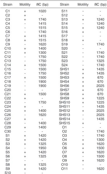
Forty-two avian Escherichia coli strains presented positive motility after 18 hours of growth into LB 0.3% agar at 37ºC. Among these strains, 15 were commensal, 11 were isolated from septicemic cases, 11 were isolated from swollen head syndrome cases (SHS), and 6 were omphalitis strains (Table 1).
The fliC gene was detected in 62 strains by the PCR assays (Table 1). Fragments varying from 670 bp to 1,900 bp were detected (Table 1 and Fig.1). Among fliC+ strains, 24 were commensal, 16 were septicemic, 14 were SHS, and 9 were omphalitis strains.
Thirty-six mobile strains harbored the fliC gene, 26 fliC+ strains showed to have negative motility and seven mobi-le strains were negative for fliC amplification.
Table 1. Motility and fliC detection by PCR among avian Escherichia coli strains
Strain Motility fliC (bp) Strain Motility fliC (bp)
C1 + 1020 S11 -
-C2 + - S12 -
-C3 + 1740 S13 + 1240
C4 - 1415 S14 - 1240
C5 - 1515 S15 + 1240
C6 - 1740 S16 +
-C7 + 1415 S17 -
-C8 + 1515 S18 +
-C9 - 1620 S19 - 1740
C10 + 1400 S20 -
-C11 + 1300 S21 - 1740
C12 - 1750 S22 - 1740
C13 - 1750 S23 - 1325
C14 + 1500 S24 - 1740
C15 - 1500 SHS1 + 1435
C16 - 1750 SHS2 + 1435
C17 + 1500 SHS3 + 670
C18 - 1900 SHS4 + 670
C19 - 1900 SHS6 + 2025
C20 - - SHS7 + 670
C21 + 1500 SHS8 + 670
C22 + - SHS9 + 1225
C23 - 1750 SHS10 + 1225
C24 + - SHS11 - 1435
C25 + 1400 SHS12 + 2025
C26 - 1620 SHS13 - 2025
C27 + - SHS14 - 1435
C28 - 1400 SHS15 +
-C29 + 1400 O1 -
-C30 - - O2 - 1740
S1 + 1420 O3 - 1740
S2 + 1420 O4 + 1300
S3 + 1325 O5 + 1620
S4 - 1950 O6 + 1500
S5 + 1420 O7 + 1620
S6 + 1325 O8 - 1500
S7 - - O9 + 1620
S8 + 1325 O10 -
-S9 + 1420 O11 - 1620
-About 400 bp of the fliC 5’region and 500bp of the fliC 3’ region were sequenced by the described primers. All obtained sequences were analyzed with the Bioedith (Hall 1999) and BLAST (http://www.ncbi.nlm.nih.gov/blast) to verify the homology of the fliC gene. After the homology confirmation, all sequences were aligned together with the fliC sequence of H-antigen from human E. coli, from 9 serovars of S. enterica, and with the flagellin sequences from Y. enterocolitica and P. mirabilis. The conserved sequences were used to obtain the dendrogram showed in Figure 2.
The dendrogram obtained presented four main clusters, named 1, 1A, 1A.1 and 1A.2 (Fig.2). Cluster 1 was compounded by four strains, two SHS (SHS1, SHS2), one commensal (C1), and one omphalitis (O6). Cluster 1.A possessed three commensal strains (C12, C21, C9), one SHS strain (SHS3), and the flagellin genes from P. mirabilis and Y. enterocolitica. Cluster 1.A.1 was compounded by 12 commensal (C14, C7, C15, C5, C28, C29, C25, C8, C17, C4, C10, C22) strains, two septicemic (S1, S5), one omphalitis strain (O8), by six H-antigens from human E. coli (H21, H11, H27, H16, H8, H2) and by the fliC from the nine S. enterica serovars. Finally, cluster 1.A.3 was compounded by 38 H-antigens from human E. coli and by 38 avian E. coli strains. Among these, 8 strains were commensal (C11, C18, C23, C13, C6, C26, C19, C3), 14 were originated from septicemic cases (S24, S19, S15, S23, S9, S8, S3, S22, S21, S13, S6, S14, S2, S4), 9 were from SHS cases (SHS6, SHS8, SHS14, SHS13, SHS7, SHS11, SHS12, SHS10 e SHS4), and 7 were from omphalitis cases (O5, O4, O11, O3, O9, O7, e O2) (Fig.2).
DISCUSSION
In recent years, several works demonstrated the presen-ce of genetic similarities among APEC strains, human Escherichia coli and other Enterobacteriaceae species, which suggested that APEC strains present a zoonotic
risk (Stocki et al. 2002, Johnson et al. 2003, Mokady et al. 2005, Rodriguez-Siek et al. 2005). The aim of the present study was to verify if there is a genetic similarity, as determined by the DNA sequencing and comparison of the fliC gene among avian pathogenic E. coli and other strains of human bacterial pathogens such as E. coli, Salmonella enterica, Yersinia enterocolitica, and Proteus mirabilis. At the same time, the DNA sequencing and comparison of the fliC gene allowed us to insert the different APEC strains into clusters of similarity.
The PCR-amplified DNA-fragments of the different avian E. coli strains presented variable molecular weights (Table 1 and Fig.1). These data are in agreement with previous studies that demonstrated the high genetic polymorphism of fliC gene from E. coli. This polymorphism was attributed to the duplication of DNA sequences, genetic recombination and the presence of insertion elements in the central region of the fliC gene. All together, these genetic events would be responsible by the H-antigen variability (Fields et al. 1997, Reid et al. 1999, Tominaga 2004, Beutin et al. 2005, Strauch & Beutin 2006). However, the fragment variability observed in our study was different from that observed by Moreno et al. (2006), where fliC fragments varying from 1,300 bp to 1,980 bp, among immobile E. coli strains, and from 1,190 to 4,170 among mobile E. coli strains were detected. Among the avian E. coli strains analyzed in the present work, fliC presented fragments varying from 670 bp to 1,900 bp (Table 1).
Although bacterial motility is considered as an indicative for the fliC gene presence, several fliC+ bacterial strains did not present positive motility, and seven mobile strains did not have the fliC fragment amplified (Table 1). The absence of motility among fliC+ strains may be attributed to the fliC non expression or to the non expression of other genes needed to bacterial motility or even the inexistence of these genes. Bacterial motility is a result from the expression of about 40 genes organized as a regulon (Macnab 1992). By the other hand, mobile strains with fliC- genotype would suggest that fliC gene, at least for these strains, is not responsible by the expression of the flagellar filament or is the result of sequence variations in the primers annealing regions. We believe that the former hypothesis is more reliable since the sequences choose for the primers annealing are very much conserved. In addition, studies realized by Raitner (1998) and by Tominaga (2004) demonstrated that genes flmA and flkA are responsible by the flagellin expression in E. coli strains belonging to serogroups H53 and H54.
As it was previously observed by other research groups (Kuwajima et al. 1986, Fields et al. 1997, Reid et al. 1999, Botelho et al. 2003, Wang et al. 2003, Amhaz et al. 2004, Beutin et al. 2005, Moreno et al. 2006, Strauch & Beutin 2006), our results also demonstrate, as determined by DNA-sequencing, that the fliC gene from avian E. coli strains presented conserved extremities and central regions with high polymorphism and as proposed by Fig.1. Agarose gel (1%) with the fliC PCR products from avian
Winstanley & Morgan (1997) and Wang et al. (2003), we used the conserved fliC regions (5’ and 3’) to establish possible phylogenetic proximity among avian E. coli strains, E. coli H-antigen, S. enterica, Y. enterocolitica, and P. mirabilis.
The dendrogram obtained by the comparison of these regions (Fig.2) demonstrated two distinct main groups: one (1.A.2) compounded, in its majority, by APEC strains (82%) and other (1.A.1) compounded mainly by commensal avian E. coli strains (Fig.2). Thirty-eight (38) human H- E. coli (Wang et al., 2003) were also grouped in cluster 1.A.2. The genetic proximity of the fliC genes among APEC and human E. coli strains may suggests that the flagellar filament of these bacterial have a similar ancestor origin with the occurrence of divergence in the central region of fliC gene probably collaborating to differentiate the flagellar filament of these groups of bacteria. In this way, the genetic proximity of fliC conserved regions among APEC and human E. coli strains could indicated genetic similarity among these strains and may suggest a possible zoonotic risk to be present on APEC strains.
Clusters 1.A and 1.A.1 were compounded by 35 different strains. From these 14 were commensal avian E. coli strains, 6 were human E. coli H-antigen, 9 were different S. enterica serovars, and three were APEC strains (one omphalitis, one septicemia and one swollen head syndrome) (Fig.2). All strains from cluster 1.A.1 presented 90% similarity. These data suggest that flagellar antigen from commensal avian E coli presents similarity with S. enterica flagellar antigen, which, again, may indicate a si-milar genetic origin of commensal avian E. coli and S. enterica. These data are also corroborated by those published by Wang et al. (2003) who proposed, based on the same approach, to exist a common ancestor fliC gene for E. coli of human origin and S. enterica. Inside the same line of thought, genetics events probably occurred to differentiate among the APEC strains and E. coli H-antigen strains that were grouped in the cluster 1.A.2 (Fig.2). In this late case, the proximity among these strains supports the status of zoonotic bacteria to APEC, a hypothesis that is reinforced by the sharing of pathogenic traits among avian and human E. coli.
In conclusion, this work demonstrated that there is a genetic proximity among APEC and human E. coli strains, as assessed by the DNA sequencing and comparison of the fliC conserved regions of these strains. These data add evidences that APEC strains exhibits a zoonotic risk.
Acknowledgments.- This study was supported by a fellowship granted to Tatiana Amabile de Campos by Campinas State University (Unicamp), by Grant no303301/2002-3 from the Brazilian Research Council (CNPq), and by Grant no 03/08407-0 from Fundação de Amparo à Pesquisa do Estado de São Paulo (FAPESP).
REFERENCES
Amhaz J.M.K., Andrade A., Bando S.Y., Tanaka T.L., Moreira-Filho C.A. & Martinez M.B. 2004. Molecular typing and phylogenetic analysis of enteroinvasive Escherichia coli using the fliC gene sequence. FEMS Microb. Lett. 235:259-264.
Ausubel F.M., Brente R., Kingstone R.E., Moore D.D., Smith J.A., Seidman J.G. & Struhl K. 1988. Curr. Protoc. Mol. Biol., Green Publishing Associates, Brooklyn, N.Y.
Belas R. & Flaherty D. 1994. Sequence and genetic analysis of multiple flagellin – encoding genes from Proteus mirabilis. Gene 148:33-41. Beutin L., Struch E., Zimmermann S., Kaulfuss S., Schaudinn C., Männel
A. & Gelderblom H.R. 2005. Genetical and functional investigation of fliC genes encoding flagellar serotype H4 in wild type strains of
Escherichia coli and in a laboratory E. coli K-12 strain expressing flagellar antigen type H48. BioMedCentral., http:// www.biomedcentral.com/1471-2180/5/4.
Botelho B.A., Bando S.Y., Trabulsi L.R. & Moreira-Filho C.A. 2003. Identification of EPEC and non- EPEC serotypes in the EPEC O serogroups by PCR-RFLP analysis of the fliC gene. J. Microb. Meth. 54:83-93.
Brito B.G., Gaziri L. & Vidotto M.C. 2003. Virulence factor and clonal relationships among Escherichia coli strains isolated from broiler chickens with cellulites. Infect. Immun. 71:4175-4177.
Brocchi M., Ferreira A., Lancellotti M., Stehling E.G., Campos T.A., Nakazato G., Pestana de Castro A.F. & Silveira W.D. 2006. Typing of avian pathogenic Escherichia coli by REP-PCR. Pesq. Vet. Bras. 26:69-73.
Dho-Moulin M. & Fairbrother J.M. 1999. Avian pathogenic Escherichia coli (APEC). Vet. Res. 30:299-241.
Ewers C., JanBen T., KieBling S., Philpp H.C. & Wieler L.H. 2004. Molecular epidemiology of avian pathogenic Escherichia coli (APEC) isolated from colisepticemia in poultry. Vet. Microb. 104:91-101. Fields P.I., Bom H., Hughes L.O., Feng P. & Swaminathan B. 1997.
Molecular characterization of the gene encoding H antigen in
Escherichia coli and development of a PCR-restriction of length polymorphism test of identification of E. coli O157:H7 and O157: NM. J. Clin. Microb. 35:1066-1070.
Gross W.B. 1984. Effect of a range of social stress severity on Escherichia coli challenge infection. Am. J. Vet. Res. 45:2074-2076.
Hall T.A. 1999. BioEdit a user friendly biological sequence alignment editor and analysis program for Windows 95/98/NT. Nucl., Ac. Symp. Ser. 41:95-98.
Honda S., Uedaira H., Vondervistzt F., Kidokoro S. & Namba K. 1999. Folding energetics of a multidomains protein, flagellin. J. Mol. Biol. 293:719-732.
Johnson J.R., Murray A.C., Gajewski A., Sullivan M., Snippes P., Kuskowisk M.A. & Smith K.E. 2003. Isolation and molecular characterization of nalidixic-acid resistant extraintestinal pathogenic
Escherichia coli from retail chickens products. Antimicrob. Ag. Chemother. 47:2161-2168.
Joys T.M. 1985. The covalent structure of the phase-1 flagellar filament protein of Salmonella typhimurium and its comparison with other flagellin. J. Biol. Chem. 260:15758-15761
Kapatral V. & Minnich S.A. 1995. Co-ordinate, temperature sensitive regulation of the three Yersinia enterocolitica flagellin genes. Mol. Microb. 17:49-56.
Kumar S., Tamura K. & Nei M. 2004. MEGA3: Integrated software for Molecular Evolutionary Genetics Analysis and sequence alignment. Brief. Bioinf. 5:150-163.
Kuwajima G., Asaka J.I., Fujiwara T., Fujiwara T., Node K. & Kondo E. 1986. Nucleotide sequence of the hag gene encoding flagellin of
Escherichia coli. J. Bacteriol. 168:1479-1483.
Macnab R. 1992. Genetics and biogenesis of bacterial flagella. Annu. Rev. Genet. 26:131-158.
Mokady D., Gophna U. & Ron E.Z. 2005. Extensive gene diversity in septicemic Escherichia coli strains. J. Clin. Microb. 43:66-73. Moreno A.C., Guth B.E.C. & Martinez M.B. 2006. Can the fliC
PCR-restriction fragment length polymorphism technique replace classic serotyping methods for characterizing the H antigen of enterotoxigenic
Moura A.C., Irino K. & Vidotto M. 2001. Genetic variability of avian
Escherichia coli strains evaluated by enterobacterial repetitive intergenic consensus and repetitive extragenic palindromic polymerase chain reaction. Avian Dis. 45:173-181.
Ngeleka M., Kwaga J.K.P., White D.G., Whittam T.S., Riddell C., Goodhope R., Potter A.A. & Allan B. 1996. Escherichia coli cellulitis in broiler chickens: clonal relationships among strains and analysis of virulence-associated factor of isolates from diseased birds. Infect. Immun. 64:3118-3126.
Popoff M.Y., Bockemuhl J. & Brenner F.W. 1997. Supplement 1997 (no.41) to the Kauffmann-White scheme. Res. Microb. 149:601-604. Ratiner Y.A. 1998. New flagellin-specifying genes in some Escherichia
coli strains. J. Bacteriol. 180:979-984.
Reid S.D., Selander R.K. & Whittam T.S. 1999. Sequence diversity of flagellin (fliC) alleles in pathogenic Escherichia coli. Infect. Immun. 181:153-160.
Rodriguez-Siek K.E., Giddings C.W., Doetkott C., Johnson T.J., Fakhr M.K. & Nolan L.K. 2005. Comparison of Escherichia coli isolates implicated in humans urinary tract infection and avian colibacillosis. Microb. 151:2097-2110.
Sambrook J. & Russel D.W. 2001. Molecular Cloning:A laboratory manual. 3rd ed. Cold Spring Harbor Laboratory Press, Cold Spring Harbor, New York, NY, p.999.
Silveira W.D., Ferreira A., Lancellotti M., Barbosa I.A.G.C.D., Leite D.S., Castro A.F.P. & Brocchi M. 2002. Clonal relationships among avian
Escherichia coli isolates determined by enterobaterial repetitive intergenic consensus. Vet. Microb. 89:323-328.
Stocki S.L., Babiuk L.A., Rawlyk N.A., Potter A.A. & Allan B.J. 2002. Identification of genomic differences between Escherichia coli strains pathogenic for poultry and E. coli K-12 MG1655 using suppression subtractive hybridization analysis. Microb. Pathog. 33:289-298. Strauch E. & Beutin L. 2006. Imprecise excision of insertion elements
IS5 from the fliC gene contributes to flagellar diversity in Escherichia coli. FEMS Microb. Lett. 256:195-202.
Tominaga A. 2004. Characterization of six flagellin genes in the H3, H53 and H54 standard strains of Escherichia coli. Gen. Genet. Syst. 79:1-8.
Wang L., Rothemund D., Curd H. & Reeves P.R. 2003. Species-wide variation in the Escherichia coli flagellin (H-antigen) gene. J. Bacteriol. 185:2936-2943.
White D.G., Wilson R.A., Emery D.A., Kakambi V.N. & Whittam T.S. 1993. Clonal diversity among strains of Escherichia coli incriminated in turkey colisepticemia. Vet. Microb. 34:19-34.