O
r
IG
IN
A
l
A
r
T
IC
lE
Authors
ElzaFTBelo1 CalilKFarhat1 ElizabethNDeGaspari1
1ImmunologySection, InstitutoAdolfoLutz, SãoPaulo,SP,Brazil.
Submittedon:06/07/2009 Approvedon:11/14/2009
Correspondence to:
Dr.ElizabethNDeGaspari InstitutoAdolfo Lutz.355,11andar CEP:01246-902,São Paulo,Brasil
Tel.:(55)(11)3068-28-98. Fax:(55)(11)3085-35-05 E-mail:
Wedeclarenoconflict ofinterest.
ABSTRACT
Dot-ELISAusingtheoutermembranecomplexantigensofNeisseria meningitidisasatargetwas standardizedforrapiddetectionofmeningococcal-specificantibodiesinhumanserum.Weinves-tigatedthelevelofmeningococcal-specificIgG,IgA,andIgMinserumusingdot-ELISAwithouter membraneantigenspreparedfromNeisseria meningitidis serotypeB:4.19:P1.15,3,7,9(astrainiso-latedfromaBrazilianepidemic).Thedot-ELISAisbasedonthesameprinciplesasthestandard ELISA and is useful for detection of anti-N. meningitidis B antibodies in serum of patients with meningococcal infections. For the assay, outer membrane complexes (OMCs) were absorbed by nitrocellulosemembraneandblockedwitha5%skimmilksolution.Serumsamplesweredrawn uponhospitaladmissionandduringconvalescencefrompatientswithmeningococcalsepticemia, and single samples were drawn from uninfected controls.We retrospectively examined a total of 57serumsamples:35frompatientsinfectedwithN. meningitidisB,12frompatientsinfectedwith
Haemophilus influenzaeb,and10fromhealthindividuals.Whenperformedatroomtemperature, dot-ELISAtookapproximatelyfourhourstoperform,andtheoptimumantigenconcentrationwas 0.42μgperdot.ThespecificityofIgG,IgM,andIgAdemonstratesthatdot-ELISAusingOMCsfrom
N. meningitidis Basatargetissuitableforserologicverificationofclinicallysuspectedmeningococ-caldiseaseinpatientsandfortiterdeterminationofantibodiesproducedduringdifferentphases ofnaturalinfection.Furthermore,thesensitivityofdot-ELISAwascomparabletothatofstandard ELISA.Overall,dot-ELISAissimpletoperform,rapid,andlowcost.Furthervalidationofthetestas ascreeningtoolisrequired.
Keywords: Neisseria meningitidis,dot-ELISA,ELISA,antibodies,outermembranecomplex.
[Braz J Infect Dis 2010;14(1):35-40]©ElsevierEditoraLtda.
Comparison of dot-ELISA and standard ELISA for detection of
Neisseria meningitidis
outer membrane complex–specific antibodies
INTRODUCTION
Bacterialmeningitisremainsaseriousthreat toglobalhealthandannuallyaccountsforan estimated 170,000 deaths worldwide. Even withtheavailabilityofantimicrobialtherapy andsophisticatedintensivecare,thecasefa-tality rate remains 5-10% in industrialized countriesandisevenhigherinthedevelop- ingworld.Between10-20%ofsurvivorsde-velop permanent sequelae, such as epilepsy, mental retardation, or sense-neural deaf-ness. Three species (Haemophilus influen-zae,Streptococcus pneumoniae,andNeisseria meningitidis) are responsible for most cases of bacterial meningitis that occur after the neonatal period. Since the introduction of
H. influenzaetypeb(Hib)conjugatevaccine,
N. meningitidis andS. pneumoniae have be-come the most common cause of bacterial
meningitisintheworld.Moreover,N. menin-gitidis istheonlybacteriumcapableofgener-atingepidemicsofmeningitis.1
Between2000and2006,therewere74,449 reported cases of meningitis in the state of SãoPaulo;however,themicrobiologicaleti-ologywasdeterminedinonly24.7%ofthese cases. Among these, 8,710 cases (11.7%) were diagnosed with meningococcal men-ingitis, 3,497 (4.7%) with pneumococcal meningitis, and 489 (0.6%) with menin-gitis caused by Hib. An additional 14,990 cases(20%)wereconsideredtobepossibly causedbyabacterialinfection;howeverthe etiologicagentwasnotidentified.2
Dot-ELISAhasbeenusedtodetectava-riety of bacterial and protozoal antigens.3-6
serogroup B meningococcal antigens in cerebrospinal fluidthathasbecomeavaluabletoolinthedetectionof meningococci.Furthermore,in1989Oprandy&Sippel7
usedamonoclonalantibodythatwasimmobilizedona nitrocellulose membrane for an antigen capture assay. ThedetectionofaN. meningitidis serogroupApolysac- charideusingthisassaywasmoresensitivethanenzyme-basedimmunoassays.7Dot-ELISAusingmeningococcal
outermembranecomplexes(OMCs)astheantigenhas been developed and used to investigate the isotypes of antibodiesproducedinpatientswithmeningococcaldis-easecausedbyserogroupB.However,dot-ELISAusingthe outermembranecomplexhasnotbeenevaluatedforanti-bodiesdetectionofinhumanserumfrompatientsinfected withN. meningitidis .Therefore,thepresentstudywascon-ducted to standardize and evaluate dot-ELISA in order to establishtheoptimalconditionsforthedetectionofN. men-ingitidis-specific IgG, IgM, and IgA in sera during natural infection.Dot-ELISAwascomparedwithstandardELISA.
MATERIAL AND METHODS
Clinical samples
Atotalof35serumsamplesfrom15individualswitha clinicaldiagnosisofbacterialmeningitiswereanalyzed. The clinical diagnosis was made on the basis of symp-toms including fever, headache, vomiting, neck muscle rigidity,cerebraldysfunction,andtoxemia.8,9
Thecere- brospinalfluid(CSF)sampleswereassayedusingcoun-terimmunoelectrophoresis (CIE) in the Immunology SectionoftheAdolfoLutzInstitute.CSFwascollected forimmunodiagnosisandbacterialcultureaccordingto thestandardtechnique,aspreviouslydescribed.10,11
Counterimmunoelectrophoresis
CIE was performed on cellulose strips (Cellogel; Chemet-ron, Italy) that had been stored in a 40% methanol so-lution.11 Before use, the strips were washed in 0.05 M sodium veronal buffer, pH 8.6 (Merck, Rio de Ja-neiro) and dried between filter papers. For each test, 7μLofantiserumwasappliedattheanodeand10μLofthe treatedsamplewasappliedatthecathode,withthesample wells0.6cmapart.CIEwascarriedoutfor10minwitha 15-20mAelectriccurrentusingsodiumveronalbuffer.Af-terelectrophoresis,thestripswerewashedsixtimesfor10 minin0.15MNaCl,stainedin50%methanolcontaining 0.5% (w/v) amido black, and destained in 50% methanol containing5%(v/v)aceticacid.
Bacterial culture
AllCSFsampleswereculturedforbacterialgrowthand forserotypingattheBacteriologySectionofAdolfoLutz
Institute.12Serumsampleswerecollectedfrompatients
intheacutephaseofthedisease(uponadmissiontothe hospital;day0),duringtheconvalescentphase(day10), andduringthelateconvalescentphase(4weeks).8,9All
of the serum samples were collected in 1998 and were stored at -70° C until use. To determine the specificity and cross-reactivity of the tests, 12 samples obtained frompatientsintheacutephaseofHaemophilus influen-zaebinfectionand10samplesfromhealthyindividuals werealsoexamined.AN. meningitidisoutermembrane complexfromstrainB:4.19:P1.15,3,7,9,whichwasiso-latedfromthecerebrospinalfluidofapatientinfectedin the1998Brazilianepidemic,wasusedasantigensource. Since 1986, serogroup BN meningitidis has been re-sponsibleforapproximately80%ofthemeningococcal diseaseinBrazil.13Theserotypewasdeterminedinthe
Bacteriological Section ofAdolfo Lutz Institute. Bacte-riaweregrownonbrainheartinfusionagar,and10mL oftheculturewastransferredto500mLoftrypticsoy broth (Difco Laboratories, Detroit, MI) and incubated at37°Cinarotaryshaker(120rpm)for18hours.
Isolation of outer membrane complex
Thebacteriawereharvestedbycentrifugationat10,000´g for 15 min. The outer membrane complexes were ex-tractedin5mLofbuffersolution(0.1Msodiumacetate and0.2MLiCl,pH5.8)pergramofcells(wetweight) byshakingwith2mmdiameterglassbeadsinagyratory water bath at 45° C for 2 hours.14 The purified OMCs was obtained by removing cells by centrifugation at 12,000gfor20min.Thesupernatantwasdialyzedover-night in 0.15 M NaCl, and protein concentration was determinedaspreviouslydescribed.15
SDS-PAGE and by immunoblot
The antigen used in dot-ELISA was analyzed by SDS-PAGEina13%acrylamidegel,asdescribed.16
Approxi-mately 20 μg of the OMC antigen was stained with Coomassiebrilliantblue,and2μgwasusedforlipopol-ysaccharidedetermination(detectedbysilverstaining), asdescribed.17
The murine monoclonal antibodies against LPS deter-minants (WBE12-C10-C10, L3,7,9-specific; 6E7-10, L8-specific;and3G3-1-8C,L1-specific;allgenerouslysupplied by Dr. Zollinger, Walter Reed Army Institute of Research, Washington, DC) were used to analyze the immunotypes present in the strains isolated from the 15 patients. Based ontheantigenicityoftheLPS,N. meningitidis can be di-vided into 12 immunotypes. Immunotypes L1 to L8 were found among the Group B and C strains18,19 and
immunotypesL9toL12wereintheGroupAstrains.20,21
Theapparentmolecularweightoftheseparatecellcom-ponentswasdeterminedbycomparingtheirmigration with those of molecular weight markers (Pharmacia, Uppsala,Sweden).
Dot-ELISA
The membrane antigen–specific antibody titer was quantified using an enzymatic dot-ELISA. All opera- tionswerecarriedoutatroomtemperatureandallin- cubationswereperformedonashakingplatformtopro-vide gentleagitation.Anitrocellulosemembrane(BioRad, Richmond,CA)wascuttofitthetopofa96-wellplateand 1μLoftheOMCssample(0.21–0.84μg)wascarefullyplaced as a dot on the membrane in a position corresponding to the center of each well. The nitrocellulose membrane was blockedwith5%nonfatdrymilkinTBS(0.01MTris-HCl, 0.14MNaCl).Thissolutionwaspre-heatedina100°Cwater bathfor5mintoinactivatetheendogenousmilkproteases andthenfiltered.Inpreviousexperiments,weoptimizedthe incubation time with blocking solution: no differences in backgroundreactivitywereseenwhentheblockingsolution wasincubatedfor1hour,2hours,orovernight.Afterblock-ingthemembranes,100μLofserumsamplesseriallydiluted in2.5%nonfatmilkinTBSwereaddedtoeachwellandin- cubatedfor2hours.Thenitrocellulosemembranewasblot-ted in order to remove excess moisture and placed on the microtiter plate. The nitrocellulose membrane was sealed with a layer of parafilm by rolling a pipet across the sur-face.SheetsofWhatman3MMpaperwereadded,andthe lidwasclampeddownwithspring-typeclamps.Attheend ofthefirstincubation,thenitrocellulosemembranewasre-movedandrapidlywashedinPBSwith0.05%NP-40(Shell QuímicadoBrasil,SãoPaulo,Brazil)for1minwithshaking andthenwashedinseveralchangesofthesamebufferfor30 min.Thewashingsolutionwasremovedand50μLofdiluted horseradish peroxidase–conjugated anti-human IgG, IgM, orIgAantibody(Sigma,StLouis,MO)wereaddedtoeach well and incubated for 2 hours. The optimum dilution of thesecondaryantibodywasfoundtobe1:500foranti-IgG, 1:1000foranti-IgM,and1:500foranti-IgA.Theconditions ofthesecondaryantibodypreparationandevaluationhave beendescribedpreviously.22
Thesecondaryantibodywasre-movedandthemembranewaswashedasdescribedabove. Thechromogen4-chloro-1-naphthol(Sigma)wasdissolved ina(stocksolution,3mg/mL;Merck,Darmstadt,Germany) andstoredinadarkbottleatroomtemperatureforupto 10 days. Immediately before use, 10 mL of PBS, 10 μL of 30%H2O2,and2mLofthechromogenstocksolutionwere mixedtogetherandincubatedfor20min.Thechromogen solutionwasremovedandthemembranewaswashed,asde-scribedabove.Samples,inwhichbluedotsdeveloped,when comparedwiththenegativecontrolseraandtheantigenand secondaryantibodycontrols,wereconsideredtobepositive.
Theoptimalantigenconcentrationfordot-ELISAwasde-terminedusingthreeconcentrationsofantigens(0.21,0.42, and0.84μg/dot)andtestedwithpositiveseraandnegative controlsera.
ELISA
A standard ELISA was performed for meningo-coccal-specific antibodies. The OMCs from strain B:4.19:P1.15,3,7,9wasplatedintriplicateinmicrodilu-tionplates(Nunc),asdescribedbyHarthuget al.23with
somemodification.Apolystyreneimmunoplate(Nunc) with96wellswascoatedwith100uLofOMCsperwell, correspondingto2ugofproteinpermLin0.1MTris buffer,pH8.5,andincubatedfor18hours.Allincuba-tions were performed at 37° C. The plates were stored at4°Cforupto2weeksandwerewashedfourtimesin phosphate-bufferedsaline,pH7.4,with0.05%Tween20 and 0.02% sodium azide immediately before use. Two serumsamplesthatshowedstrongimmuneresponsesto OMCsantigenswereusedasinternalstandards.Allse-rum samples were diluted 1:200 in phosphate-buffered saline,pH7.4,with0.05%Tween20,0.1%bovineserum albumin,and0.02%sodiumazideandanalyzedintrip-licate. Diluted serum (100 μL) was added to each well, incubated for 3 hours, and then washed as described above.A horseradish peroxidase–conjugated goat anti-humanIgA,IgM,orIgG(SigmaChemicalCo.,StLouis, MO) detection system was used to detect the bound antibodies.Incubationswerecarriedoutat37ºCfor1 hourinallsteps,exceptforthatwiththesubstrate(15 min). The reaction was interrupted with 4 N H2SO4. Theabsorbancewasobtainedat450nminaplatereader (SLT-Spectra,US).
RESULTS
TheelectrophoreticprofileoftheN. meningitidis anti-gens(bothproteinandlipopolysaccharide)usedinthe assayisshowninFigure1A.Theproteinsaremainly20 to120kDainweightandtheexpressionofL3,7determi-nants could be seen when monoclonal antibodies were used.24Wetestedtheserumfrompatientswithclinically
Figure 1:
(A) The SDS-PAGE of the native outer membrane complex of representative N. meningitidis B strains isolated from patients are shown in lanes 1, 4, 7, and 10 after Coomassie blue staining. Lanes 2, 5, 8, and 11 demonstrate the analysis of LPS variants by PAGE and silver staining, and lanes 3,6,9, and 12 are immunoblots. The immunoblots were sequentially reacted with three different monoclonal antibodies. The band at position (a), (b), and (c) reacted with the WBE12-C10 (L3,7,9-specific; 5.9 kDa), the 3G3-1-8C (L1-specific; 4.8 kDa), and the 6E7-10 (L8-specific; 3.6 kDa) monoclonal antibody, respectively. The outer membrane complex of the strain) B:4.19:P1.15,3,7,9 shown in lanes 1 was used in the dot-ELISA and the ELISA assay.
(B) Dot-ELISA for the detection of N. meningitis B outer membrane complex–specific IgG in patient serum at a 1:100 (A) to 1:12,800 (H) dilution demonstrating the sensitivity of the assay. For each spot, 0.42 µg of the OMC was spotted onto a nitrocellulose membrane. Lanes 1 through 10 demonstrate the immunoreactivity of patient serum in different phases of the disease. Serum samples were collected from patients in the acute phase (upon admission to the hospital; day 0), during the convalescent phase (day 10), and during the late convalescent phase (4 weeks). Lanes 11 and 12 are serum samples from normal controls. The color intensity was judged visually, and the intensities were assigned values on an arbitrary scale (0, +, ++, +++, or ++++) in reference to the negative control (assigned a value of 0).The dots are blue in color.
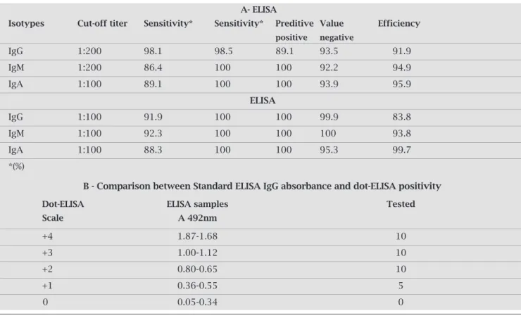
Table 1. Performance of assays for detecting antibodies in this sample studies
A- ElISA
Isotypes Cut-off titer Sensitivity* Sensitivity* Preditive Value Efficiency
positive negative
IgG 1:200 98.1 98.5 89.1 93.5 91.9
IgM 1:200 86.4 100 100 92.2 94.9
IgA 1:100 89.1 100 100 93.9 95.9
ElISA
IgG 1:100 91.9 100 100 99.9 83.8
IgM 1:100 92.3 100 100 100 93.8
IgA 1:100 88.3 100 100 95.3 99.7
*(%)
B - Comparison between Standard ElISA IgG absorbance and dot-ElISA positivity
Dot-ElISA ElISA samples Tested
Scale A 492nm
+4 1.87-1.68 10
+3 1.00-1.12 10
+2 0.80-0.65 10
+1 0.36-0.55 5
0 0.05-0.34 0
activemeningitisthaninnormalsubjects.Thereactiv- ityofseveralstandardpositiveandnegativecontrolse- rumsamplesformeningococcalantigens(0.42μganti-gens) suggested the possibility of using serum samples diluted more than 1:100. Importantly, while the serum
samples from patients withH. influenzae b presented cross-reactivity,thiscross-reactivitydecreasedwiththe dilutionoftheserumsamples.AsseeninTable1A,the IgMandIgApresentedalowerdegreeofcross-reactivity whencomparedwiththeIgGinthesameserumsample.
A
B
1 2 3 4 5 6 7 8 9 10 11 12
12 11 10 9 8 7 6 5 4 3 2 1
A
B
C
D
E
F
G
H a b c 94
64
43
Toobtainahigherefficiencyfordot-ELISA,cut-offval-ues were selected to better discriminate between men-ingitis patients and the normal controls (Table 1A). Furthermore,thelongitudinaltiterofthedifferentan- tibodyisotypesduringthecourseofmeningococcalin- fectionwasdetermined.TheIgGtiterwaslowatadmis-sion (p < 0.05) and increased progressively thereafter. IgMtiterwasgreaterthanIgGtiterandalsoincreasedin laterstagesofinfection.IgAtiterwashigherthanaver-ageduringearlyconvalescence(p<0.05)anddecreased during the convalescent stage. The color intensity was judgedvisually,andtheintensitieswereassignedvalues onanarbitraryscale(0,+,++,+++,or++++)inrefer-encetothenegativecontrol(assignedavalueof0).For determinationofthesensitivity,specificity,andpositiveand negativepredictivevaluesofdot-ELISA,astandardELISA was carried out and analyzed. Our results demonstrated a goodcorrelationbetweenthevisualgradingofdot-ELISA andELISAabsorbance(Table1B).
Theantibodiesmeasuredinthisstudyweremostlikely directedagainstthe20–120kDaproteinsandtheL3,7LPS datenotshown.
DISCUSSION
LPSisanimportantcomponentoftheoutermembraneof meningococcalbacteria.Neisseria producesonetosixdiffer- entLPSmolecules(3,200to7,200Da)thatcanbecharacter-izedbytheirelectrophoreticmobilityinSDS-PAGE.22The
differenceinmobilityappearstobeduetotheheterogeneity ofthechemicalcompositionofoligosaccharides.Antibodies directedagainststrain-specificepitopesanddifferentouter membraneproteinsofN. meningitidisBhavebeendetected inserumfromconvalescentpatients.5,6
Inthisstudy,adot-ELISAusingtheOMCsderivedfromN. meningitidis sero-typeBwasfoundtobeaspecifictechniqueforestablishing the level of antigen-specific IgG, IgM, and IgA present in serumofpatientsduringthedifferentstagesofinfection.It alsohastechnicaladvantages,suchasattheuseofminute amountsofantigens,antibodyconjugates,andachromog-enicsolution.
The procedure of dot-ELISA has a number of advan- tagesoverthestandardELISAcurrentlyperformedinlabo-ratory.Thenitrocellulosemembraneiscapableofbinding moreantigensthanthemicrotiterplates.25Therefore,only
asmallamountoftheOMCsisrequiredasanantigenfor the test.Additionally, the reaction on nitrocellulose mem-brane is viewed against the memmem-brane white background, makingitmucheasiertoidentifyapositiveornegativere-action.Anotheradvantageofdot-ELISAisthatitdoesnot requirespectrophotometricreadings,whicharerequiredin classicplate-orcuvette-basedELISAs,animportantconsid-eration for laboratories in developing countries. Most im-munologicassays,includingthedot-ELISA,areinfluenced
byboththeantibodyconcentrationandaffinity.However, dot-ELISAappearstobeoneofthemoresensitiveandleast affinity-dependent procedures.3 In contrast with antibody
responseagainstserotypeBpolysaccharide,whichhasbeen showntobemediatedmostlybyIgM,theresponseagainst theOMCsantigensusingdot-ELISAseemstobemediated byotherisotypes,suchasIgGandIgA.Thisassaywasalso useful in the longitudinal analysis of the antigen-specific immunoglobulintitersforuseinimmunologicalstudiesof this severe disease. OMC-specific IgM was detected in all
N. meningitidispatients,withoutanyH. influenzae bcross-reactivity.Thissuggeststhatthisassaywillbesuitableforuse inhospitallaboratories,especiallywhenthebacterialculture ofCSFsamplesproduceanegativeresult.Applicationofthis techniqueinpatientdiagnosisandscreeningisvalidbecause ofthetesteasyexecutionandthecharacteristicsdescribed above.Thesensitivityofdot-ELISAandstandardELISAwas similar,aspreviouslyreported.26Dot-ELISAcouldbeused
for screening meningococcal disease in less well-equipped laboratories.N. meningitidis-specificIgG,IgM,andIgAwere detectedintheserumfromall35serainvestigatedusingdot-ELISAwithgoodsensitivity.
AmultianalyteDot-enzyme-linkedimmunosorbentas-say(Dot-ELISA-Multi)withTrypanosoma cruzi epimastig-otealkalineextract(EAE),trypomastigoteexcreted–secreted antigen(TESA),recombinantproteinderivedfrom19-kDa C-terminalregionofthePlasmodium vivax merozoitesur-face protein 1 (PvMSP119),Plasmodium falciparum Zwit-tergentR extract (Pf-Zw), andTreponema pallidum Zwit-tergentRextract(Tp-Zw)wasstandardizeandevaluatedas a method for surveying IgG-specific antibodies in Chagas disease,malaria,andsyphilisinasingletest.27
In conclusion, dot-ELISA presented here is very useful andeasytohandle;itusesminuteamountsofantigen,can measurethelevelofantibodiesIgG,IgMandIgAin Neisse-ria meningitidisinfectionwithfacility,andtheresultscanbe seendirectlybythenakedeye.
ACKNOWLEDGEMENTS
We gratefully acknowledge Dr Wendell Zollinger for LPS monoclonal antibodies. The study was sponsored by IAL through number Mem. C.T.A.Nro 09/89 and CNPq/ Nro 20275691/92.
Theserumsampleswereusedtoguaranteeandpreserve strict anonymity of the individuals originally involved as well as the conservation and ethically correct. Use of the materialandinformationobtainedfromit.Resolution196 ofOctober10,1996TheNationalBoardofHealth.
REFERENCES
1. WorldHealthOrganization:http://www.who.int/vaccine_re-search/diseases/soa_bacterial/en/index4.htm
3. Pappas MG, Hajkowski R, Hockmeyer WT. Standardization ofthedotenzyme-linkedimmunosorbentassay(Dot-ELISA) forhumanvisceralleishmaniasis.AmJTropMedHyg1984; 33:1105-11.
4.
BeutinL,BodeL,RichterT,PeltreG,andStephanR.Rapidvis-ualdetectionofEscherichiacoliandVibrio choleraeheat-labile
enterotoxinsbynitrocelluloseenzyme-linkedimmunosorbent assay.J.Clin.Microbiol1984;19:371-5.
5. GaspariEN,StolfAMS,AbrahamsohnIA.Enhancedsensitiv-
ityofdot-immunobindingassaysforthedetectionofantibod-iesinmicebytheuseofmilddetergentextractedTrypanosoma
cruziantigens.RevInstAdolfoLutz1990;50:231-3.
6. Coll P, Borche L, Ausina V, Mirelis B, Prats G. Dot-immu-nobindingassaywithamonoclonalantibodyfordetectionof group B meningococcal antigen. Eur J Clin Microbiol 1998; 65:44-6.
7. OprandyJJ,SippelJE.Evaluationofperformanceparameters of a membrane-based dot immunoassay for meningococcal polysaccharide.JClinMicrobiol1989;27:74-7.
8. DeGaspariEN,Tavares,AV,Ribeiro,Cl,FarhatCR.IgGclass
antibodiesagainstgroupBNeisseria meningitidis
outermem-braneantigensofdifferentserotypesprevalentinBrasil.AmJ TropMedHyg1991;45:43.
9. deGaspariEN,RibeiroCL;FarhatCK.Studyoftheantigenic stabilityofthe50kDapeptideusingnativeoutermembrane
vesiclesofNeisseria meningitidis
Bandserafrominfectedpa-tients.ImmunolLet1997;56:3.
10. AlkminMG,ShimizuSH,LandgrafI,GaspariEN,MellesCE.
ProductionandImmunochemicalcharacterizationof
Neisse-ria meningitidis
groupBantiserumforthediagnosisofpuru-lentmeningitis.BrazJMedBiolRes1994;27:1627-34
11. AlkminMG,LandgrafIM,ShimizuSH.DetectionofN.
men-ingitidisgroupBantigensbyMB-Dot-ELISAinpatientswith
meningitis.BullPanAmHealthOrgan1996;30:212-7.
12. deLemosAP,YaraTY,GorlaMCIet al.Clonaldistribution
ofinvasiveNeisseria meningitidis
serogroupCstrainscircu-latingfrom1976to2005ingreaterSãoPaulo,Brazil.JClin Microbiol2007;45:1266-73.
13. SacchiCT,PessoaLL,RamosSRet al.Clin.Microbiol1992;
30:1734-8.
14. Frasch CE, ZollingerWD, Poolman JT. Serotype antigens of
Neisseria meningitidisandaproposedschemefordesignation
ofserotypes.RevInfectDis1985;7:504-10.
15. Lowry OH, Rosebrough NJ, Farr AL, Randall, RJ. Protein measurement with the Folin phenol reagent. J Biol Chem 1951;193:265-75.
16. LaemmliUK.Cleavageofstructuralproteinsduringassembly oftheheadofbacteriophageT4,Nature1970;227:680-5. 17. Tsai CM, Frasch CE. A sensitive silver stain for detecting
li-popolysaccharidesinpolyacrylamidegels.AnalBiochem1982; 119:115-9.
18. MandrellRE,ZollingerWD.Lipopolysaccharideserotypingof
Neisseria meningitidis by hemagglutination inhibition. Infect
Immun1977;16:471-5.
19. Zollinger WD, Mandrell RE. Outer-membrane protein and
lipopolysaccharideserotypingofNeisseria meningitidis
byin-hibition of a solid-phase radioimmunoassay. Infect Immun 1977;18:424-33.
20. ZollingerWD,MandrellRE.Type-specificantigensofgroupA
Neisseria meningitidis:lipopolysaccharideandheat-modifiable
outermembraneproteins.InfectImmun1980;28:451-8. 21. Kim JJ, Phillips NJ, Gibson BW, Griffiss JM, Yamasaki R.
MeningococcalgroupAlipooligosaccharides(LOS):prelimi- narystructuralstudiesandcharacterizationofserotype-asso-ciated and conserved LOS epitopes. Infect Immun 1994; 62; 1566-75.
22. Guimarães MC, Celeste BJ, Franco EL. Evaluation of dot enzyme-linked immunosorbent assay for mucocutaneous leishmaniasis and comparison with microplate enzyme im-munoassay.JClinMicrobiol1986;24:364-7.
23. HarthugS,RosenqvistE,HøibyEA,Gedde-DahlTW,Frøholm LO.AntibodyresponseingroupBmeningococcaldiseasede-termined by enzyme-linked immunosorbent assay with se-rotype 15 outer membrane antigen. J. Clin Microbiol 1986; 24:947-53.
24.
MoranEE,BrandtBL,ZollingerWD.ExpressionoftheL8li-popolysaccharidedeterminantincreasesthesensitivityof
Neis-seria meningitidis to serum bactericidal. Infect Immun1994;
62:5290-5.
25. Burt SM, Carter TJ, Kricka LJ. Thermal characteristics of microtitre plates used in immunological assays. J Immunol Methods1979;31:231-6.
26. CamargoED,NakamuraPM,VazAJ,daSilvaMV,ChieffiPP, de Melo EO. Standardization of dot-ELISA for the serologi-caldiagnosisofToxocariasisandcomparisonoftheassaywith ELISA.RevInstMedTropSaoPaulo1992;34:55-60.
27. Santos CJ, Soares IS, Antunes LEet al. A multianalyte