Effe cts o f e xe rcise training o n
auto no m ic and m yo cardial dysfunctio n
in stre pto zo to cin-diabe tic rats
1Laboratório de Fisiologia Cardiovascular, Departamento de Fisiologia,
Instituto de Ciências Básicas da Saúde, and 2Laboratório de Pesquisa do Exercício,
Faculdade de Educação Física, Universidade Federal do Rio Grande do Sul, RS, Brasil
3Unidade de Hipertensão e Divisão de Experimentação, Instituto do Coração (Incor),
Hospital das Clínicas, Faculdade de Medicina, Universidade de São Paulo, São Paulo, SP, Brasil
K.L.D. De Angelis1,
A.R. O liveira1,2, P. Dall’Ago1,
L.R.A. Peixoto1,
G. Gadonski1, S. Lacchini1,
T.G. Fernandes1 and
M.C. Irigoyen1,3
Abstract
Several investigators have demonstrated that diabetes is associated with autonomic and myocardial dysfunction. Exercise training is an efficient non-pharmacological treatment for cardiac and metabolic diseases. The aim of the present study was to investigate the effects of exercise training on hemodynamic and autonomic diabetic dysfunc-tion. After 1 week of diabetes induction (streptozotocin, 50 mg/kg, iv), male Wistar rats (222 ± 5 g, N = 18) were submitted to exercise training for 10 weeks on a treadmill. Arterial pressure signals were obtained and processed with a data acquisition system. Autonomic function and intrinsic heart rate were studied by injecting methylatropine and propranolol. Left ventricular function was assessed in hearts perfused in vitro by the Langendorff technique. Diabetes (D) brady-cardia and hypotension (D: 279 ± 9 bpm and 91 ± 4 mmHg vs 315 ± 11 bpm and 111 ± 4 mmHg in controls, C) were attenuated by training (TD: 305 ± 7 bpm and 100 ± 4 mmHg). Vagal tonus was decreased in the diabetic groups and sympathetic tonus was similar in all animals. Intrinsic heart rate was lower in D (284 ± 11 bpm) compared to C and TD (390 ± 8 and 342 ± 14 bpm, respectively). Peak systolic pressure developed at different pressures was similar for all groups, but +dP/dt max was decreased and -dP/dt max was increased in D. In conclusion, exercise training reversed hypotension and bradycardia and improved myocardial function in diabetic rats. These changes represent an adaptive response to the demands of training, supporting a positive role of physical activity in the management of diabetes.
Co rre spo nde nce
M.C. Irigoyen Unidade de Hipertensão Instituto do Coração
Av. Enéas de Carvalho Aguiar, 44 05403-000 São Paulo, SP Brasil
Fax: + 55-11-3069-5048
E-mail:
hipirigoyen@ incor4.incor.usp.br
Presented at the III International Symposium on Vasoactive Peptides, Belo Horizonte, MG, Brasil,
O ctober 8-10, 1999.
Research supported by CNPq, CAPES and FAPERGS. Publication supported by FAPESP.
Received November 26, 1999 Accepted February 21, 2000
Ke y wo rds
·Experimental diabetes
·Exercise training
·Arterial pressure
·Autonomic control
·Myocardial contractility
Intro ductio n
Diabetes is a chronic metabolic disorder associated with secondary complications in the cardiovascular system and autonomic control in humans and animals (1-4). Higher morbidity and mortality have been observed
presented lower arterial pressure (AP) and heart rate (HR), as well as a reduction in intrinsic HR (IHR) and in vagal tonus, sug-gesting early autonomic dysfunction (2,8). Those changes in AP, HR and autonomic function, evaluated by AP variability, were reversible by chronic insulin treatment (3). Studies on animals and humans have shown that diabetic subjects exhibit depressed myo-cardial contractile performance, particularly under stress conditions such as enhanced workload (1,4).
In our laboratory, exercise training ap-plied to young and aged normotensive and young hypertensive rats improved cardio-vascular and autonomic control (9,10). More-over, we demonstrated that physical activity enhanced the peripheral action of insulin in aged normotensive and hypertensive rats (9,11). Exercise training has also been shown to limit the depression in myocardial pump function in diabetic subjects (12-14). How-ever, there are few data in the literature about the effects of exercise on autonomic and hemodynamic function in experimental diabetes. In the present study, we evaluated changes induced by exercise training in AP, HR and autonomic control, as well as in myocardial function in STZ-diabetic rats.
Mate rial and Me tho ds
Experiments were performed on 18 male Wistar rats (222 ± 5 g) from the Animal House of Universidade Federal do Rio Gran-de do Sul, Porto Alegre, RS, Brazil,
receiv-ing standard laboratory chow and water ad
libitum. The animals were housed in
indi-vidual cages in a temperature-controlled room
(22o
C) with a 12-h dark-light cycle. All sur-gical procedures and protocols used were in accordance with the Guidelines for Ethical Care of Experimental Animals and were ap-proved by the International Animal Care and Use Committee.
The rats were randomly assigned to one of three groups: sedentary control (C, N = 5),
sedentary diabetic (D, N = 6) or trained diabetic (TD, N = 7). Animals were made diabetic by a single injection of STZ (50 mg/
kg, iv;Sigma Chemical Co., St. Louis, MO,
USA) dissolved in citrate buffer, pH 4.5. Rats were fasted overnight before STZ injec-tion.
The animals were submitted to
low-in-tensity exercise training (50% VO2 max) one
week after diabetes induction on a treadmill twice a day (one hour each time), 5 days a week for 10 weeks, with gradually progres-sion to a speed of 31 m/min, as described in detail elsewhere (15).
After the last training session, two cath-eters filled with 0.06 ml saline were im-planted under ether anesthesia into the femo-ral artery and vein (PE-10) for direct meas-urements of AP and drug administration, respectively. Rats receiving food and water ad libitum were studied 1 day after catheter
placement; the rats were conscious and al-lowed to move freely during experiments. The arterial cannula was connected to a strain-gauge transducer (P23Db, Gould-Statham, Oxnard, CA, USA), and blood pressure sig-nals were recorded over a 20-min period by a microcomputer equipped with an analog-to-digital converter board (CODAS, 1-kHz sampling frequency, Dataq Instruments, Inc., Akron, OH, USA). The recorded data were analyzed on a beat-to-beat basis to quantify changes in mean AP and HR.
Vagal and sympathetic tonus and IHR were studied (2) by injecting methylatropine
(3 mg/kg, iv; Sigma) and propranolol (4 mg/
kg, iv; Sigma) in a maximal volume of 0.2 ml
propranolol and methylatropine. On the sub-sequent day, the sequence of injections was inverted, first propranolol and then methylat-ropine. IHR was evaluated after simulta-neous blockade with propranolol and meth-ylatropine. Sympathetic tonus was deter-mined as the difference between maximum HR after methylatropine injection and IHR. Vagal tonus was obtained by the difference between the lowest HR after propranolol injection and IHR.
After the hemodynamic measurements the rats were killed by a blow to the neck. The chest was opened and the heart excised and perfused using a Langendorff system. The aorta was retroperfused with a modified Krebs solution (120 mM NaCl, 5.4 mM KCl,
1.8 mM MgCl2, 1.25 mM CaCl2, 27 mM
NaHCO3, 2 mM NaH2CO3, 1.8 mM Na2SO4,
and 11 mM glucose), pH 7.4. The solution
was maintained at 37o
C, gassed with 95% O2
and 5% CO2 and used for perfusion at a
constant flow (11 ml/min). A latex balloon filled with water and connected to a pressure transducer (Isotec P23 XL - Hugo Sachs Elektroniks, March, Freiburg, Germany) was placed inside the left ventricle. The signals were processed using an analogic-digital con-version system including a microcomputer (IBM - PC 486), an analogic-digital con-verter board and an Isoheart software (Hugo Sachs Elektroniks). A stabilization period of 30 min was allowed to elapse for standardi-zation of cardiac function. After stabiliza-tion, the balloon volume was adjusted to generate a diastolic pressure of 0 mmHg. The pressure was increased in steps of 10 mmHg at 5-min intervals, reaching 10, 20, 30, 40 and 50 mmHg. The variables ana-lyzed for each diastolic pressure used were left ventricular systolic pressure, maximum rate of rise (+dP/dt max) and maximum rate of fall (-dP/dt max).
Body weight was monitored each week during the period of physical activity. Blood samples were collected at rest in the fasting state one week after diabetes induction.
Plasma glucose was measured by a colori-metric enzymatic test (Enz Color, Bio Diagnostica, Piraquara, PR, Brazil).
Data are reported as means ± SEM, and two-way ANOVA was used to compare groups, followed by the Student-Newman-Keuls test. Correlation was determined by linear regression analysis.
Re sults
All rats given STZ developed severe hy-perglycemia (479 ± 8 for the D group and 467 ± 17 mg/dl for the TD group) compared with controls (125 ± 7 mg/dl). Body weight was reduced in both diabetic groups as com-pared with sedentary control (329 ± 16 g) (P<0.01). Trained diabetic rats (241 ± 11 g) showed an increase in body weight com-pared with sedentary diabetic rats (195 ± 15 g) (P<0.04).
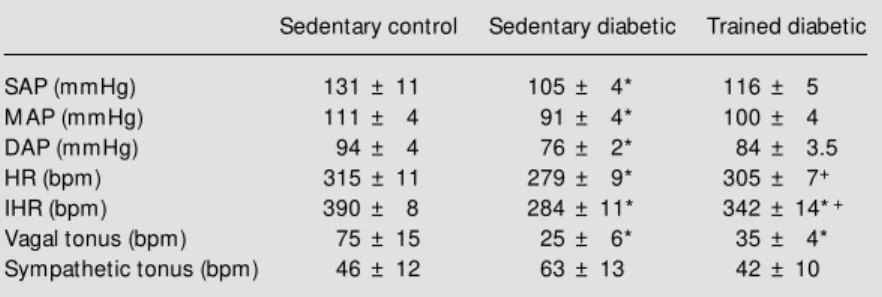
As can be seen in Table 1, exercise train-ing induced attenuation in diabetic hypoten-sion and bradycardia. Figure 1A shows the control HR, IHR and HR responses to drug blockade. Sedentary diabetic rats presented lower basal AP values than sedentary con-trol animals. No differences were observed between trained diabetic rats and sedentary groups. Basal evaluations of HR showed bradycardia in the STZ sedentary group (279 ± 9 bpm) compared with the STZ trained and sedentary control groups (305 ± 7, P<0.04
Table 1 - Cardiovascular characterization of sedentary and trained diabetic rats. Data are reported as means ± SEM . * P<0.05 compared to sedentary control rats; +P<0.05 compared to sedentary diabetic rats (Student t-test). SAP = Systolic arterial pressure; M AP = mean arterial pressure; DAP = diastolic arterial pressure; HR = heart rate; IHR = intrinsic heart rate.
Sedentary control Sedentary diabetic Trained diabetic
SAP (mmHg) 131 ± 11 105 ± 4* 116 ± 5
M AP (mmHg) 111 ± 4 91 ± 4* 100 ± 4
DAP (mmHg) 94 ± 4 76 ± 2* 84 ± 3.5
HR (bpm) 315 ± 11 279 ± 9* 305 ± 7+
IHR (bpm) 390 ± 8 284 ± 11* 342 ± 14*+
Vagal tonus (bpm) 75 ± 15 25 ± 6* 35 ± 4*
and 315 ± 11 bpm, P<0.03). The IHR ob-tained after methylatropine and propranolol blockade was significantly lower in seden-tary diabetic (284 ± 11 bpm) than in trained diabetic and control rats (342 ± 14, P<0.01 and 390 ± 8 bpm, P<0.00). A significant reduction in IHR was observed in trained diabetic rats compared to control animals (P<0.02). A positive correlation determined by linear regression was found between rest-ing HR and IHR, showrest-ing higher HR values at higher IHR (r = 0.74, P<0.04) (Figure 1B). Vagal tonus was decreased in diabetic rats compared to sedentary control rats and sym-pathetic tonus was similar for all groups (Table 1).
The left ventricular function obtained for the various groups is shown in Figure 2, where pooled results of total left ventricular isovolumetric systolic pressure are plotted against left ventricular diastolic pressure. It can be seen that the increase in isovolumet-ric systolic pressure as a function of diastolic pressure was similar in control, diabetic and trained diabetic rats. Isolated hearts from sedentary control and trained diabetic rats developed a reduced maximum rate of rise (+dP/dt max) and an increased maximum rate of fall (-dP/dt max) compared to isolated
hearts from sedentary diabetic rats (P<0.05). The overall spontaneous HR was not signif-icantly altered by diabetes or training. HR for the three groups (bmp) was as follows: controls, 201 ± 22; diabetic rats, 170 ± 34; trained diabetic rats, 204 ± 25.
D iscussio n
The present study confirms our prelimi-nary findings that STZ diabetes induces hy-potension, bradycardia and autonomic dys-function. Also, we observed decreased myo-cardial function in sedentary diabetic rats (2,3). However, the major finding of this investigation is that exercise training im-proves diabetes-induced dysfunction. Jack-son and Carrier (16) suggested that the de-crease in AP may be the result of a dede-creased cardiac output in diabetic sedentary rats due to hypovolemia caused by hyperglycemic osmotic diuresis. However, Cohen et al. (17) observed that these animals were polyuric with a high urine flow, reflecting the osmotic diuretic effects of glucose. The hypotension observed in the sedentary diabetic group may also have been due to an increase in parasympathetic outflow, although Maeda et al. (2) and the present data demonstrated a decrease in vagal function, suggesting that changes in AP are not related to an increase in parasympathetic outflow. Previous stud-ies have shown that exercise training im-proves cardiac output in diabetic rats (12,13). Exercise also improves glucose homeosta-sis, reducing the glucose/insulin ratio and increasing insulin sensitivity (9,11). More-over, exercise not only attenuates the reduc-tion in myocardial GLUT-4 transporters (18) but also increases sarcolemmal GLUT-4 pro-tein in diabetic rats (14). Insulin plays a critical role in this process since GLUT-4 depression and hemodynamic changes were reversed by insulin treatment (3,19). These metabolic effects may have contributed to AP normalization in trained diabetic rats.
Reduction in HR in sedentary diabetic
H
R
(
b
p
m
)
450
VT 375
425 400
350 325 300 275
R
R
R
436 ± 12
390 ± 8IHR
315 ± 15 279 ± 9 315 ± 11
346 ± 13
305 ± 7+
284 ± 11*
259 ± 6*
383 ± 10
342 ± 14*+
307 ± 4*
ST VT
VT ST
ST
IHR
IHR
Sedentary control
Sedentary diabetic
Trained diabetic
IH
R
(
b
p
m
)
450
400
350
300
250
200
200 220 240 260 280 300 320 340 360 HR (bpm)
r = 0.74 Figure 1 - A, Graphs show ing
in-trinsic heart rate (IHR), sympa-thetic (ST) and vagal tonus (VT) in sedentary control, sedentary diabetic and trained diabetic rats. Arrow s indicate basal heart rate (HR). Dat a are report ed as means ± SEM . * P<0.05 com-pared to sedentary control rats; +P<0.05 compared to sedentary
diabetic rats (ANOVA). B, Posi-tive relationship betw een rest-ing HR and IHR expressed by linear regression line (r = 0.74) in the diabetic groups (P<0.04).
A
animals has been attributed to changes in the sinoatrial node (2,20), although functional alterations in the cholinergic mechanism can-not be excluded as a causal factor. In the present study we observed an increase in resting HR in trained diabetic rats that was correlated with changes in IHR (r = 0.74), confirming the important role of the sinoatrial node in changes in HR in experimental dia-betes. In contrast, previous studies have dem-onstrated that exercise training decreases resting HR in normotensive rats (21) and humans. The decreased IHR previously ob-served in our laboratory in trained control rats (11,21) as well as a decreased sympa-thetic tonus in spontaneously hypertensive rats after training may be the mechanisms involved in exercise bradycardia (10). In the present experiments we did not observe changes in sympathetic tonus between groups, suggesting that the increase in rest-ing HR in trained diabetic rats may be related to the improvement of intrinsic pacemaker regulation. The reduction in vagal tonus in-dicated a reduction of vagal function in rats with STZ-induced diabetes, probably related to vagal cardiac neuropathy, since a decrease in acetylcholine concentration (22) and a defect in cardiac cholinergic nerves (23) were described. Exercise training did not modify the changes in parasympathetic func-tion of diabetic rats, although a slight in-crease in vagal tonus was observed in trained rats. Impairment of vagal function evaluated by the reduced bradycardia elicited by in-creasing AP or electrical vagal stimulation was observed in normal rats after exercise training (21).
The hemodynamic changes induced by diabetes are usually accompanied by myo-cardial abnormalities in patients (1) and ex-perimental animals (4,13). Although in the present study we did not find differences in left ventricular isovolumetric systolic pres-sure between groups, cardiac contractility and relaxation, respectively represented by +dP/dt and -dP/dt, were all reduced in hearts
isolated from STZ-treated rats, particularly at left ventricular diastolic pressures higher than 10 mmHg. These findings are in agree-ment with the results reported by DeBlieux et al. (13) showing a slight reduction in cardiac output and no changes in other indi-ces of cardiac work in diabetic rats. How-ever, reduction in myocardial performance has been previously reported (12) and sev-eral authors (24,25) have demonstrated that these changes may be related not only to depressed myosin ATPase but also to de-creased calcium uptake by the sarcoplasmic reticulum (13). However, the most impres-sive finding in the present study is that exer-cise training reverses the changes in contrac-tile properties of the heart induced by STZ-diabetes in rats. Paulson et al. (26) reported that exercise training improved cardiac func-tion in diabetic animals by decreasing the severity of the diabetic state. In the present study the increase in body weight in trained rats seems to indicate metabolic changes.
S
y
s
to
lic
p
re
s
s
u
re
(
m
m
H
g
)
140
80 120
100
60 40
20
10 20 30 40 50
0
Diastolic pressure (mmHg)
+
d
P
/d
t
m
a
x
(
m
m
H
g
/s
)
-d
P
/d
T
m
a
x
(
m
m
H
g
/s
)
1600
1200
800
400
-300
-450
-750
-900 -600
0 10 20 30 40 50
0 10 20 30 40 50
Diastolic pressure (mmHg)
Sedentary control Sedentary diabetic Trained diabetic
Figure 2 - Influence of diastolic pressure on systolic pressure (A), maximum rate of rise (+dP/ dt max) (B), and maximum rate of fall (-dP/dt max) (C) developed by the left ventricle of hearts iso-lated from sedentary control, sedentary diabetic and trained diabetic rats. Data are reported as means ± SEM . * P<0.05 com-pared to sedentary control rats; +P<0.05 compared to sedentary
diabetic rats (ANOVA).
A
B
C
Moreover, it is well known that training is able to increase sensitivity to insulin (9,11). Since STZ-treated rats did not receive insu-lin treatment in the present study, the im-provement in metabolic state was probably due to maintenance of increased insulin sen-sitivity during the post-exercise period (27). Indeed, exercise training increases whole body insulin sensitivity and glucose oxida-tion by skeletal and cardiac muscle (28). This improvement may be due to the in-crease in myocardial sarcolemmal GLUT-4 in the diabetic hearts (14). Changes in myo-cardial metabolism involving a shift from glucose to fat metabolism (28) have been reported in diabetes mellitus. Hence, rats treated with STZ have increased plasma lev-els of triglycerides and cholesterol (26) that are lowered by exercise training, leading to
improved myocardial sarcoplasmic reticu-lum function.
Finally, while low-intensity exercise train-ing seems to improve cardiovascular func-tion some types of endurance training can exacerbate the diabetes-induced decrease in
myocardial Ca2+
-activated ATPase and B-adrenergic receptor number (29). The results of the present study provide evidence for the effectiveness of exercise training in reduc-ing some of the cardiovascular complica-tions associated with diabetes mellitus.
Ackno wle dgm e nts
The authors are grateful to Imbramed Ltda. for its technical support in physical training equipment.
Re fe re nce s
1. Kannel WB (1978). Role of diabetes in cardiac disease: conclusions from popula-tion studies. In: Zonaraich S (Editor), Dia-betes and Heart. Thomas, Springfield, IL, 97-112.
2. M aeda CY, Fernandes TG, Timm HB & Irigoyen M C (1995). Autonomic dysfunc-tion in short-term experimental diabetes. Hypertension, 26: 1100-1104.
3. Schaan BD, M aeda CY, Timm HB, M edei-ros S, M oraes R, Ferlin E, Fernandes TG, Ribeiro JP, Schmid H & Irigoyen M C (1997). Time-course of changes in heart rate and blood pressure in rats w ith strep-tozotocin-induced diabetes treated w ith insulin. Brazilian Journal of M edical and Biological Research, 30: 1081-1086. 4. Fein FS, Kornst ein LB, St robeck JE,
Capasso JM & Sonnenblick EH (1980). Altered myocardial mechanics in diabetic rats. Circulation Research, 47: 922-933. 5. Sampson M J, Wilson S, Karaggianis P,
Edmonds M E & Watkins PJ (1990). Pro-gression of diabetic autonomic neuropa-thy over a decade in insulin dependent diabetics. Quarterly Journal of M edicine, 75: 635-646.
6. M onckton G & Pehow ich E (1980). Auto-nomic neuropathy in the streptozotocin diabetic rat. Journal Canadien des Sci-ences Neurologiques, 7: 135-142.
7. Yagihashi S & Sima AAS (1986). Diabetic autonomic neuropathy in the BB rat: Ul-trastructural and morphometric changes in parasympathetic nerves. Diabetes, 34: 558-564.
8. M aeda CY, Fernandes TG, Lulhier F & Irigoyen M C (1995). Streptozotocin diabe-tes modifies arterial pressure and barore-flex sensitivity in rats. Brazilian Journal of M edical and Biological Research, 28: 497-501.
9. De Angelis KLD, Oliveira AR, Werner A, Bock P, Belló-Klein A & Irigoyen M C (1997). Exercise training in aging: hemo-dynamic, metabolic, and oxidative stress evaluations. Hypertension, 30: 767-771. 10. Gava NS, Véras-Silva AS, Negrão NE &
Krieger EM (1995). Low -intensity exercise training attenuates cardiac beta-adrener-gic tone during exercise in spontaneously hypert ensive rat s. Hypert ension, 26: 1129-1133.
11. De Angelis KLD, Gadonski G, Fang J, Dall’Ago P, Albuquerque VL, Peixoto LRA, Fernandes TG & Irigoyen M C (1999). Ex-ercise reverses peripheral insulin resis-tance in trained L-NAM E-hypertensive rats. Hypertension, 34: 768-772. 12. Paulson DJ, Stephen JK, Peace DG & Tow
JP (1988). Improved post-ischemic recov-ery of cardiac pump function in exercised
trained diabetic rats. Journal of Applied Physiology, 65: 187-193.
13. De Blieux PM , Barbee RW, M cDonough KH & Shepherd RE (1993). Exercise train-ing improves cardiac performance in dia-betic rats. Proceedings of the Society for Experimental Biology and M edicine, 203: 209-213.
14. Osborn BA, Darr JT, Laddaga RA, Romano FD & Paulson DJ (1997). Exercise training increases sarcolemmal GLUT-4 protein and mRNA content in diabetic heart. Jour-nal of Applied Physiology, 82: 828-834. 15. Tancrede G, Rousseau-M igneron S &
Nadeau A (1982). Beneficial effects of physical training in rats w ith a mild strep-tozotocin-induced diabetes mellitus. Dia-betes, 31: 406-409.
16. Jackson CV & Carrier GO (1983). Influ-ence of short-term experimental diabetes on blood pressure and heart rate in re-sponse to norepinephrine and angiotensin II in the conscious rat. Journal of Cardio-vascular Pharmacology, 5: 260-265. 17. Cohen AJ, M cCarthy DM & Rosseti RR
19. Garvey WT, Hardin D, Juhaszova M & Dominguez JH (1993). Effects of diabetes on myocardial glucose transport system in rats: implications for diabetic cardiomy-opathy. American Journal of Physiology, 264: H837-H844.
20. Dall‘Ago P, Fernandes TG, M achado UF, Belló AA & Irigoyen M C (1997). Barore-flex and chemoreBarore-flex dysfunction in strep-tozotocin-diabetic rats. Brazilian Journal of M edical and Biological Research, 30: 119-124.
21. Negrão CE, M oreira ED, Santos M CLM , Farah VM A & Krieger EM (1992). Vagal function impairment after exercise train-ing. Journal of Applied Physiology, 72: 1749-1753.
22. Kubtscherova J & Vlk J (1970). Influence of alloxan diabetes on acetylcholine syn-thesis in tissues of the albino rat.
Physi-ologia Bohemoslovaca, 19: 431-434. 23. Tomlison DR & Yusof APM (1983).
Auto-nomic neuropathy in the alloxan-diabetic rats. Journal of Autonomic Pharmacology, 3: 257-263.
24. M akino N, Dhalla KS, Elimban V & Dhalla NS (1987). Sarcolemmal Ca2+ transport in streptozotocin-induced diabetic cardiomy-opathy in rats. American Journal of Physi-ology, 253: E202- E207.
25. Rodrigues B, Ross JR, Farahbakshian S & M cNeill JH (1990). Effects of in vivo and in vitro treatment w ith L-carnitine on iso-lated hearts from chronically diabetic rats. Canadian Journal of Physiology and Phar-macology, 68: 1085-1092.
26. Paulson DJ, M athew s R, Bow man J & Zhao J (1992). M etabolic effects of tread-mill exercise training on the diabetic heart. Journal of Applied Physiology, 73:
265-271.
27. King DS, Baldus PJ, Sharp RL, Kesl LD, Feltmeyer TL & Riddle M S (1995). Time course for exercise-induced alterations in insulin action and glucose tolerance in middle-aged people. Journal of Applied Physiology, 78: 17-22.
28. Tomlinson KG, Gardiner SM , Hebden RA & Bennett T (1992). Functional conse-quences of streptozotocin-induced diabe-tes mellitus, w ith particular reference to the cardiovascular system. Pharmacologi-cal Review s, 44: 103-150.