Radio auto grapho lo gy: the pro po sal
o f a ne w co nce pt
Department of Anatomy and Cell Biology, Shinshu University School of Medicine, Matsumoto, Japan
T. Nagata
Abstract
A new concept termed radioautographology is advocated. This term was synthesized from radioautography and ology, expressing a new science derived from radioautography. The concept of radioauto-graphology (RAGology) is that of a science whose objective is to localize radioactive substances in the biological structure of objects and to analyze and study the significance of these substances in the biological structure. On the other hand, the old term radioautography (RAG) is the technique used to demonstrate the pattern of localization of various radiolabeled compounds in specimens. The specimens used in biology and medicine are cells and tissues. They are fixed, sectioned and placed in contact with the radioautographic emulsions, which are exposed and developed to produce metallic silver grains. Such speci-mens are designated as radioautographs and the patterns of pictures made of silver grains are named radioautograms. The technicians who produce radioautographs are named radioautographers, while those who study RAGology are scientists and should be called radioautog-raphologists. The science of RAGology can be divided into two parts, general RAGology and special RAGology, as most natural sciences usually can. General RAGology is the technology of RAG which consists of three fields of science, i.e., physics concerning radioactiv-ity, histochemistry for the treatment of cells and tissues, and photo-chemistry dealing with the photographic emulsions. Special RAGology, on the other hand, consists of applications of general RAGology. The applications can be classified into several scientific fields, i.e., cellular and molecular biology, anatomy, histology, embryology, pathology and pharmacology. Studies carried out in our laboratory are summa-rized and reviewed. All the results obtained from such applications should be systematized as a new field of science in the future.
Co rre spo nde nce
T. Nagata
Department of Anatomy and Cell Biology
Shinshu University School of Medicine Matsumoto 390 Japan
Fax: + 81-263-33-6458
Research supported in part by Grants-in-Aid for Scientific Research from the Ministry of Education, Science and Culture of Japan, JEO L Co. Ltd. and FAPESP.
Presented at the 5th International Symposium on Radioautography, São Paulo, SP, Brasil, August 24-26, 1997.
Received July 11, 1997 Accepted May 21, 1998
Ke y words
•Light microscopy •Electron microscopy •Radioautography •Technique •Application
1. Introduction
The term radioautography (RAG) means the technique used to demonstrate the pat-tern of localization of various compounds labeled with radioactive isotopes in speci-mens (1). The specispeci-mens used in biology and medicine are usually cells and tissues which
autora-diography, autoradiographs and autoradio-grams are sometimes used alternatively (2). Historically, the principle of radioautog-raphy was first reported by Becquerel (1896) one year after the famous discovery of X-rays by Roentgen (1895) who produced the first radiogram. This was a negative picture (white images on a black background) pro-duced on photographic plates after the pen-etration of X-rays through the tissues. Becquerel found that photographic plates were activated by radiations from uranium which produced positive images (black im-ages on a white background) after develop-ment of the plates. He first gave a talk at the annual meeting of the Paris Academy in 1896. This picture was regarded as the first radioautogram produced by radiation from the minerals. Later, London (3) reported that he could produce the picture of a frog which incorporated radium and which was placed on a photographic plate. This report appears to be the first radioautograph obtained from animal tissues. However, London (3) did not use any term for this picture. The first radio-autograph from the human body was ob-tained by Kotzareff (4) who, when treating a patient suffering from metastatic cancer of the iliac bone with radium, placed a photo-graphic plate on the bone and obtained a picture of the bone after photographic devel-opment. He named the picture radiumgraphie or curiegraphie after Curie. This nomencla-ture, however, was not used by other scien-tists. Lacassagne and Lattés (5) reported that the radiation emitted by radium or polonium given to rabbits could be detected on photo-graphic plates applied to the sectioned smooth surface of paraffin blocks of embedded rab-bit organs. These pictures surprised the sci-entist by demonstrating the distribution of radioactive substances in the organs but they did not provide good resolution. It was Lacassagne who first named these pictures autohisto-radiographie. This term was later abbreviated to autoradiography. One of his students, Leblond (6,7), moved to North
America (USA and Canada) and developed a new technique to paint the tissue sections which contained radioactive compounds with warmed liquid photographic emulsion using a camels hair brush, thus obtaining a better resolution. Pelc (8) used the stripping film method, while Joftes and Warren (9) devel-oped the dipping method and used the term autoradiography originating from the word autohisto-radioautography used by Lacassagne. Leblond first used the term ra-dioactive autography according to Lacassagne in the 1940s, but he later pro-posed to use the term radioautography by condensing the words radioactive autogra-phy into radioautograautogra-phy which has been used since the 1950s (10). The above his-torical facts tell us how the two kinds of terms were developed.
As was described above from the histori-cal viewpoint, the two terms, radioautogra-phy and autoradiograradioautogra-phy, are originally due to the two words radioactive autography or autohisto-radiography. The two terms are now generally regarded as synonyms. How-ever, the author prefers the term radioautog-raphy because of etymological reasons (2). In order to select terminology in scientific description, the preference for a term should depend on the etymological meaning of this term. The etymological difference between the two terms, radioautography and autora-diography, is as follows.
There-fore, etymologically the term radioautogram means the positive picture produced by ra-diation which is emitted from the object itself resulting in the autogram. In contrast, in the word autoradiogram the suffix auto means automatic, while the term radiogram means the picture of the object which is penetrated by rays resulting in negative im-ages such as chest X-ray films. Thus, autora-diogram etymologically means a negative picture of the specimen produced automati-cally with radiation emitted from another radiation source away from the specimens. Anyhow, it is now accepted that the terms radioautography and autoradiography are synonyms (2).
On the other hand, the term radioautogra-phy or autoradiograradioautogra-phy means only the tech-nique used to demonstrate the patterns of silver grains in specimens. Radioautography is only the technique used to produce the specimens (radioautographs) in order to dem-onstrate the pictures (radioautograms). In contrast, the present author now proposes a new concept, named radioautographology. This new term was coined from radioautog-raphy and ology, expressing a new sci-ence derived from radioautography. The con-cept of radioautographology (RAGology) is that of a science whose objective is the local-ization of radioactive substances in the struc-ture of the objects and to analyze and study the significance of these substances in the structure (11).
RAGology can be divided into two parts, general RAGology and special RAGology, as most natural sciences usually can. Gen-eral RAGology is the technology of RAG which consists of three fields of sciences, i.e., physics concerning radioactivity, his-tochemistry for the treatment of cells and tissues, and photochemistry dealing with the photographic emulsions.
Since experimentally produced radioau-tograms include a variety of specimens, i.e., human biopsy material, experimental ani-mal tissues and cells or even whole bodies of
small animals, the size of the specimens varies according to the samples. Larger speci-mens such as whole bodies or large organs are designated as macro-radioautograms which are observed with the unaided eye. In contrast, small samples such as small pieces of tissues or cells are observed under the light or electron microscope and are called microscopic radioautography. Macroscopic radioautography can be classified into two levels, whole body radioautography and or-gan radioautography, while the microscopic radioautography can be classified into light microscopic radioautography (LMRAG) and electron microscopic radioautography (EMRAG). These two types of radioautog-raphy are divided into two categories, soluble compound radioautography and insoluble compound radioautography, on the basis of the solubility of the radioactive compounds (4). The classifications of these two combi-nations are listed in Table 1. Thus, all radio-autographs can be classified into 8 catego-ries. These technologies should be desig-nated as general RAGology.
Special RAGology, on the other hand, consists of applications of general RAGology to biology and medicine (12). From the view-point of radioactive compounds, it can be classified into insoluble and soluble RAG. The former concerns macromolecules such as DNA, RNA, proteins, sugars or lipids, while the latter concerns small molecules such as hormones, neurotransmitters, vita-mins, inorganic substances, drugs and tox-ins. The applications of RAG to biology and medicine can also be classified into several
Table 1 - Classification of radioautography.
M acroradioautography M icroradioautography
w hole body organ LM EM
Insoluble RAG insoluble insoluble insoluble insoluble w hole body RAG organ RAG LM RAG EM RAG
Soluble RAG soluble soluble soluble soluble
scientific fields, i.e., cellular and molecular biology, anatomy, histology, embryology, pathology and pharmacology. All the results obtained from such applications should be systematized as a new field of science desig-nated as special RAGology.
The present paper is a review of general RAGology including the techniques of light and electron microscopic radioautography which were developed in our laboratory as well as special RAGology including the re-sults applied to histology, histopathology and pharmacology of organ systems in ani-mals and humans in our laboratory over the past 40 years.
2. Ge ne ral radioautographology
The science named radioautographology deals with the technology of radioautogra-phy and the applications of these techniques to various objects. It demonstrates the sites of incorporation, syntheses and discharge of various substances in living organisms by macroscopic and microscopic radioautogra-phy to localize intracellular sites of metabo-lism at the cellular and organelle levels in various objects. This technology should be designated as general radioautographology and includes all the natural sciences involved in the preparation of the specimens which contain radioactive compounds, the proce-dures for tissue preparations and the
meth-ods used to place tissues in contact with the photographic emulsions, followed by expo-sure for a certain period of time to produce the latent images of the radioactive sub-stances in the specimens. The emulsion is then developed to produce the silver metal grains, thus permitting a comparison of the specimens and radioautograms in order to determine the localization of radioactive sub-stances in the specimens. In order to carry out these procedures, three fields of knowl-edge, i.e., physics concerning radioactivity, histology and histochemistry dealing with the fixation, embedding, sectioning and stain-ing of tissues and cells, and photographic chemistry treating photographic emulsions are necessary.
2.1. Physics of radioactive substance s
2.1.1. Radioactive isotope s
The radioactive compounds used in ra-dioautography are mainly composed of inor-ganic or orinor-ganic compounds which are arti-ficially labeled with radioisotopes (RI) and can be incorporated into human or animal bodies. The radioactivity emitted from the radioactive isotopes is divided into three kinds of rays, i.e., alpha, beta and gamma rays. The alpha ray is the helium nucleus in nature and has low energy, weak penetrabil-ity, strongest ionization and the shortest range (several µm). The beta ray is the electron and has medium energy, medium penetrability, medium ionization and a shorter range (µm-mm), while the gamma ray is the electromag-netic wave and has high energy, strong pen-etrability, low ionization and the longest range (m). Among these 3 rays, the beta ray is the best for radioautography because of its shorter range and strong ionization. The various kinds of RI used for radioautography are listed in Table 1. Among them, 3
H, 14
C, 35
S and 125
I are very often utilized for both macro- and microradioautography because they can be used to label various inorganic compounds
Table 2 - Radioisotopes used in microscopic radio-autography.
Nuclide Half-life Rays emitted Particle energy (KeV)
3H 12.5 years beta 18.5
14C 5760 years beta 155
32P 14.2 days beta 1710
35S 87 days beta 167
45Ca 165 days beta 250
56Fe 3 years Auger electron 14 125I 60 days Auger electron 35
habitually used in biological and medical research. Since the radioactivity of the re-spective RI decreases time-dependently but never reaches zero, the life of an RI is de-fined as half-life which is the time required for the RI to lose 50% of its initial radioactiv-ity. The half lives of 3
H or 14
C are longer (years) than those of 35S or 125I (days) as
listed in Table 2, so that the experiments using shorter half-life compounds should be completed within a shorter period.
2.1.2. RI-labeled compounds
In histology, histopathology and pharma-cology the RI-labeled compounds used for radioautography can be classified into 2 cat-egories, i.e., the precursors which are incor-porated into macromolecules such as nucleic acids (DNA and RNA), proteins, glucides and lipids, and the other target tracers which are small molecular compounds such as hor-mones, neurotransmitters, vitamins, inor-ganic substances, drugs and others (Table 3). All RI should be treated only in the RI laboratories according to the regulations con-cerning radiation damage enforced in each country.
2.1.3. Administration of radioactive
co m po unds
In order to study the localization of radio-active compounds in animal bodies, com-pounds labeled with a specific RI are usually administered orally to small animals such as rats and mice or by injection given subcuta-neously, intramuscularly, intravenously or intraperitoneally. For the purpose of demon-strating macromolecular synthesis, animals were injected intraperitoneally with radioac-tive precursors for macromolecular synthe-sis at concentrations ranging from 1 to 50 µCi (37-1850 kBq)/gram body weight for macro- and for LMRAG or 10-100 µCi (370-3700 kBq)/g body weight depending on the
characteristics of the compounds and RI used. The RI-labeled precursors used in our ex-periments were [3
H]-4-thymidine (Amersham, Buckinghamshire, England; specific activity 877 GBq/mM) for DNA synthesis, [3
H]-uridine (Amersham; 1.11 TBq/mM) for RNA, [3
H]-4,5-leucine (Amersham; 1.04 TBq/mM) and [3H]-taurine (New England Nuclear,
Boston, MA, USA; specific activity 74 GBq/ mM) for proteins, [3
H]-1-glucosamine (Amersham; 105 GBq/mM) for polysaccha-rides, [35
S]-sulfuric acid (Amersham; 1.11 TBq/mM) for mucosubstances, and [3
H]-1,2,3-glycerol (Amersham; 7.4 GBq/mM) for lipids.
Sometimes in vitro labeling can be car-ried out in cultured cells, both established cell lines or primary culture and tissue blocks obtained from human biopsy or necropsy material, in media containing radioactive compounds at concentrations of 1-100 µCi (37-3700 kBq)/ml medium, using a CO2
in-cubator at 37oC with 5% CO
2 in air under
normal or pathological conditions, for time intervals ranging from several minutes up to several hours.
Table 3 - Radio-labeled compounds used in microscopic radioautography.
M acromolecular precursor Radio-labeled compounds
Nucleic acids
DNA [3H]-thymidine
RNA [3H]-uridine
Proteins
secretory granules [3H]-glycine, [3H]-leucine, etc.
collagen [3H]-proline, [3H]-hydroxyproline
Glucides
simple polysaccharides [3H]-glucose, [3H]-glucosamine, etc.
mucosubstances 35SO4
Lipids [3H]-glycerol, [3H]-fatty acids, etc.
Target tracers (low molecular w eight compounds)
Hormones [3H]-steroids, [3H]-insulin, etc.
Neurotransmitters [3H]-GABA, [3H]-dopamine, etc.
Vitamins [3H]-vitamin A, B, C, D, etc.
Inorganic substances 22NaCl, 45CaCl2, 203HgCl2, etc.
Drugs [3H]-antibiotics, [3H]-anti-allergics, etc.
2.2. Histology and histoche mistry
for radioautography
2.2.1. Animal tre atme nt and
tissue proce ssing
Small animals such as mice and rats are anesthetized by intraperitoneal injections with pentobarbital sodium (Nembutal, Abbott Laboratories, Chicago, IL, USA), after the administration of radioactive compounds by either oral administration or injection, and are sacrificed at a given time, usually 1 h after RI administration, either by decapita-tion or by perfusion via the left ventricles of the heart with 2.5% glutaraldehyde in 0.1 M cacodylate buffer, pH 7.2, depending on whether insoluble or soluble radioautogra-phy will be carried out. For conventional insoluble radioautography perfusion fixation can be used and the tissues from various organs are dissected out, cut into small pieces (1 x 1 x 1 mm), soaked in the same glutaral-dehyde fixative at 4o
C for 1 h and postfixed in 1% osmium tetroxide in the same buffer for 1 h, dehydrated with graded ethanol and embedded in epoxy resin (Epon 812 or Epok 812, Oken Co., Tokyo, Japan). In contrast, in vitro labeling of cultured cells and tissue blocks obtained from either animal or hu-man biopsy material are incubated in media containing radioactive compounds, using a CO2 incubator under normal conditions at
37o
C for a given time, usually 1 h or up to a few hours. They are then rinsed in Hanks solution, fixed in the same buffered glutaral-dehyde and osmium tetroxide solutions, de-hydrated and embedded in epoxy resin as described above.
For soluble radioautography, however, perfusion fixation cannot be used. The whole bodies of the small animals or organs and tissues obtained after decapitation without using any perfusion methods should be im-mediately cryo-fixed by the metal contact method or by the method of immersion in
liquid nitrogen and processed by cryo-sec-tioning or freeze-drying or freeze-substitu-tion (13). Macroscopic RAG of whole bod-ies of small animals usually employs cryo-fixation by immersion in a dry ice and ac-etone mixture and cryo-sectioning with a large cryostat microtome (14).
Tissues embedded in epoxy resin can be used for either LMRAG or EMRAG. For LMRAG, 2-µm thick sections are cut with an ultramicrotome, picked up onto clean glass slides and warmed for extension and drying. For electron microscopy, ultrathin sections of 100-nm thickness are cut and usually examined under a conventional trans-mission electron microscope with the accel-erating voltage at 100 kV. It is generally accepted that the thinner the section the bet-ter the resolution, but the less the radioactiv-ity it contains and the longer the exposure time for radioautography. If an intermediate high voltage electron microscope is avail-able with accelerating voltages of 200, 300 or 400 kV, thicker sections of 200 or 300 nm can be used. We prefer to use semithin 200-nm thick sections at 400 kV in order to shorten the exposure time (15). Semithin sections are cut with a Porter-Blum MT-2B ultramicrotome (Dupont-Sorvall, Newtown, CN, USA). Ultramicrotomes of the mechan-ical feeding type are preferable to the ther-mal feeding type because of the accuracy of the section thickness, which affects the num-ber of silver grains by radioautography. Ul-trathin or semithin sections are picked up onto either platinum or gold grid meshes in order to prevent the copper meshes from rusting through the histologic and radioauto-graphic treatment especially during devel-opment. Alternatively, collodion-coated cop-per grid meshes can be used. For collodion coating, copper grid meshes (100-200 meshes) are soaked in 2% collodion solution for a few minutes, spread on filter paper in a Petri dish and dried at 37o
2.2.2. Fixation of tissue s and ce lls
We have developed simple routine tech-niques to demonstrate insoluble compounds in various cells and tissues of experimental animals and to quantify the contents of newly synthesized macromolecules in each cell and cell organelle by both light and electron microscopy. The localization of silver grains developed by means of ordinary radioautog-raphy, however, demonstrates only the in-soluble radioactive substances bound to the macromolecules fixed in the cell with the chemical fixatives used (1). In contrast, ra-dioisotopes bound to the small molecules which are not fixed with ordinary chemical fixatives are washed away by conventional routine procedures such as fixation, dehy-dration, embedding, sectioning, and radio-autographic procedures, so that these compounds cannot be demonstrated. Ordi-nary radioautographic procedures can be designated as wet-mounting radioautogra-phy, since the tissues are processed by both conventional wet treatments and by apply-ing wet radioautographic emulsions to the specimens. In order to demonstrate any soluble radioactive compound, special tech-niques are required in accordance with the
characteristics of the radioisotopes used for radioautography (Table 4). The techniques for microscopic radioautography developed in our laboratory can be divided into 2 cat-egories, i.e., wet-mounting radioautography for insoluble compounds resulting from mac-romolecular synthesis and dry-mounting ra-dioautography for soluble compounds such as small molecular compounds, and each can be subdivided into two methods, i.e., light microscopy and electron microscopy (Table 4).
2.2.2.1. Che mical fixation for
we t-mounting radioautography
Small animals such as mice and rats are anesthetized and sacrificed either by decapi-tation or by perfusion via the left ventricle of the heart with 2.5% glutaraldehyde in 0.1 M cacodylate buffer, pH 7.2. The tissues from various organs are removed, cut into small pieces, soaked in the same glutaraldehyde fixative at 4oC for 1 h and postfixed in 1%
osmium tetroxide in the same buffer for 1 h, dehydrated with graded ethanol and embed-ded in epoxy resin. On the other hand, cul-tured cells and tissue blocks, incubated in media containing radioactive compounds in
Table 4 - Principle techniques for microscopic radioautography demonstrating soluble and insoluble compounds.
vitro, using a CO2 incubator under normal
conditions at 37oC for given times, are rinsed
in Hanks solution, fixed in the same buff-ered glutaraldehyde and osmium tetroxide solutions, dehydrated and embedded in ep-oxy resin as described above. The tissue blocks are cut with an ultratome for either LM (2 µm thick) or EM (0.1-2 µm) and picked up onto either glass slides or grid meshes using water. In some cases, the whole mount cells on glass coverslips or coated meshes are fixed, dried and used for wet-mounting radioautography to observe the whole cells without sectioning (15). In order to observe isolated cells obtained from the tissues in vivo, small tissue blocks are oscil-lated in Ranviers alcohol and isooscil-lated, smeared on glass slides, fixed in Carnoys fluid, and wet-mounted for LMRAG (16).
2.2.2.2. Che mical fixation for soluble
co m po unds
The techniques of radioautography of soluble compounds can be theoretically clas-sified into two categories according to the fixation used, as shown in Table 4 (17,18). In the precipitation method, the labeled soluble compound is fixed in a mixture con-taining a substance which reacts with the soluble radioactive compounds forming a precipitate, so that the fixed tissues can be processed by routine histological procedures followed by a routine wet-mounting radio-autography to demonstrate the labeled pre-cipitation. This principle was first used to detect soluble 45
Ca by Nagata and Shimamura (19-21) in light microscopic radioautogra-phy of several tissues fixed in formaldehyde solution containing ammonium oxalate to form a calcium oxalate precipitate. At the electron microscopic level, Mizuhira et al. (22) later used this method by fixing the tissues labeled with [3H]-thiamine with a
glutaraldehyde solution containing platinum chloride to form thiamine-PtCl6
precipita-tion, or fixing labeled [3
H]-cholesterol with
a mixture containing digitonin. This proce-dure, however, is limited to the radioactive compounds which can be precipitated with any other specific compounds. Moreover, there are many possibilities of diffusion of the labeled compounds when they are pre-cipitated with the fixatives.
2.2.2.3. Cryo-fixation for soluble compounds
By the freezing method, on the other hand, the labeled tissues are quickly frozen in a liquid such as isopentane or propane cooled to its melting point with liquid nitro-gen. The tissues can then be cut by cryo-microtomy. At the light microscopy level, the frozen tissues can be cut with a cryostat and the frozen sections are placed in contact with radioautographic emulsions by various methods. At the light microscopy level, the frozen tissues can be cut with a cryostat at a thickness around 20-30 µm and the frozen sections are placed in contact with radioau-tographic emulsions by various techniques. Many papers have been published on this problem. We first used a large-wire loop to produce dry films which were air-dried and applied to cryostat sections placed on glass slides (23). This procedure is very conve-nient and will be described in detail. At the electron microscopy level, however, only a few papers have been published on the prin-ciple of cryo-ultramicrotomy. Appleton (24) and Christensen (25) discussed the possibili-ties of this method but they did not demon-strate the results, while we (17) reported the results for the first time. In contrast, Mizuhira et al. (26), and Futaesaku and Mizuhira (27) reported a new technique for ultramicro-tomy utilizing interposed films, which, how-ever, is not an ideal procedure because it employs cryosectioning after the tissues are immersed in sucrose solution. The cryosec-tions are then picked up with sucrose drop-lets and placed in contact with wet radioau-tographic emulsion.
freeze-substituted tissues can be embedded in par-affin or resin for light microscopy and dry sections can be mounted on precoated slides. Edwards and Udupa (28) and Smitherman et al. (29) used freeze-dried and paraffin-em-bedded sections. Miller Jr. et al. (30) applied the wire loop method as part of the dry-mounting procedure at the light microscopy level. We improved this technique first at the light microscopy level (23) and then applied it at the electron microscopy level (17). Stirling and Kinter (31) embedded freeze-dried tissues in silicon-araldite and the plas-tic sections were wet-mounted with a drop-ping procedure but not by dry-mounting.
To demonstrate soluble low molecular weight compounds, cryo-fixation and dry-mounting radioautography should be rou-tinely employed for both LMRAG and EMRAG.
2.2.2.3.1. Pre paration of tissue blocks
After the tissues are removed without using any solution from the experimental animals previously injected with radioactive compounds, they are trimmed to a size as small as 1 x 0.5 x 0.5 mm with two pieces of razor blades on a plate cooled to 0o
C with ice and water. The tissue blocks are attached to small pieces of aluminum foil, 5 x 5 mm in size (13), or directly to the surface of a metal block. Cultured cells are centrifuged at 500 rpm for 10 min after incubation with media containing radioactive compounds, and the pellet is placed on a piece of aluminum foil or on metal blocks.
2.2.2.3.2. Fre e zing proce dure
For quick fixing by cryo-fixation, we soak the tissues in a quenching fluid which is cooled with a cooling agent. The quenching fluids we usually employ are propane (melt-ing point, -169o
C), isopentane (-161o
C), or hexane (-94oC). These reagents are liquids at
low temperatures and conduct heat very well.
They also bubble when they contact tissues. Among them, propane is explosive, while hexane has a rather higher melting point. Therefore, isopentane is very often used (13). Instead of quenching fluids, a pure copper block is sometimes used in direct contact with the tissues.
The following substances or mixtures of two substances are generally used as cooling agents: liquid helium (boiling point, -269o
C), liquid nitrogen (-196oC), liquid air (-190oC),
dry ice and ether (-60o
C), and dry ice and nonane (-53o
C). Among these, liquid nitro-gen is most frequently used. We usually employ the combination of isopentane or propane as the quenching fluid and liquid nitrogen as the cooling agent, or a copper block as metal contact and liquid nitrogen as the cooling agent. Cryo-instruments such as RF-2 (Eiko, Tokyo, Japan), JFD-RFA (JEOL, Tokyo, Japan), cryoblock or cryovacublock (Reichert-Jung, Jena, Germany) have recently become commercially available. Liquid ni-trogen (200-300 ml) is carefully poured into a Dewer flask. A 50-ml beaker is placed in liquid nitrogen, and 20-30 ml of isopentane or propane is poured into the beaker. We prefer isopentane to propane. Within a few minutes the liquid isopentane begins to so-lidify at its melting point of -161o
C as it is cooled by the liquid nitrogen at -196oC. The
cryo-instruments, frozen tissues are removed from the copper blocks and also stored in liquid nitrogen.
2.2.2.3.3. Cryo-se ctioning
The frozen tissues can be cut directly by cryo-sectioning without any embedding. The procedures for cryo-sectioning can be di-vided into light microscopy and electron microscopy.
2.2.2.3.3.1. Light microscopic cryo-se ctioning
The frozen tissues can be cut with a cryostat at -80o
C, and the frozen sections are then placed on glass slides in contact with radioautographic emulsions for light micros-copy. Various techniques are employed. Kinter et al. (32) placed sections directly on the photographic plates and clamped them for exposure. Fitzgerald (33) applied dry films to sections mounted on slides. Appleton (24) placed sections on precoated slides at low temperature. Stumpf and Roth (34-36) transferred freeze-dried sections on either siliconized slides or Teflon slides. Hammar-strom et al. (37) used cellophane tapes to attach sections to plates. Since Appleton (24) first applied cryostat sections to precoated slides at very low temperature, many investigators such as Rogers et al. (38) have used this technique at the light micro-scope level. All of these techniques require handling of the sections, glass slides and emulsions in the dark room. When cryostat sections picked up onto precoated slides are air-dried at room temperature, the sections melt and cause diffusion of soluble com-pounds. This procedure is called thaw-mount radioautography and is applicable when the diffusion is negligible.
In contrast, we use the cryostat in a con-ventional bright room and then coat the cryo-sections with the emulsion in a darkroom using the large wire loop method (23). We use a conventional rotary type cryostat. The
frozen tissues are transferred to a cryostat kept at around -30oC and dry sections of
20-30 µm are cut. They are transferred to glass slides and either air-dried at room tempera-ture or freeze-dried at -30oC for a few hours.
The procedure for coating the slides with dry emulsion film by the large wire loop method will be described later.
2.2.2.3.3.2. Ele ctron microscopic
cryo -se ctio ning
The technique of ultrathin cryo-section-ing, or simply cryo-ultramicrotomy, was first reported by Bernhard and Leduc (39) and was later improved by Tokuyasu (40) em-ploying preincubation in sucrose solution and picking up sections with sucrose drop-lets. For radioautography, however, his tech-nique is not applicable because of the diffu-sion of radiolabeled compounds. We use an LKB ultratome 4800 equipped with an LKB cryokit 14800 or an LKB-NOVA (LKB, Bromma, Sweden). Other types of ultrami-crotomes such as DuPont-Sorvall or Reichert-Jung with cryokits can also be used. The temperature of the specimens is usually set at -100o
C to -120o
C and that of the glass knives at -80o
C to -100o
C. The optimal tem-perature depends on the kind of tissue used. Dry sections are picked up with dry eyelash probes onto grids, covered with another grid as a sandwich and pressed with copper rods according to Christensen (25) or Sakai et al. (41). The authors use a modified apparatus of the Sakai type. Grids used for this purpose are coated with collodion applied by soaking in 1% collodion solution and dried at 37o
C for 1 h. As controls, wet sections are picked up with sucrose droplets (42). Grids carrying dry sections are dried by freeze-drying at -50o
both cell structure and radioisotopes (13). In order to freeze-dry cryosections, we use a rotary cryotransfer apparatus. The rotary disc, which has spaces for 5 grids with cryosec-tions, is changed every 5 grid and transferred to a carrier, which consists of a tube and a cylinder containing up to 5 discs on its shelves, for a total of 25 grids (13). The carrier is transferred to the desiccator of the freeze-drying apparatus, which is operated for 3 h at -80oC. After drying the
cryosec-tions are coated with carbon and processed by the dry-mounting radioautographic pro-cedure. Cryotransfer apparatuses have re-cently become commercially available as attachments of cryo-kit-equipped ultrami-crotomes such as LKB, Sorvall or Reichert instruments.
2.2.2.3.4. Fre e ze -drying proce dure
The freeze-drying technique was first used by Altmann (43), and was then applied to light and electron microscopy by Gersh (44,45). The freeze-drying apparatus we use was designed and constructed in our labora-tory (13,16). It consists of a cold trap, a desiccator, which is set in the cold trap, three Geisler discharge and ionization vacuum gauges, and two rotary and oil diffusion vacuum pumps. The cold trap consists of a stainless steel cylinder containing liquid ni-trogen. The desiccator, which is set in the cold trap, has 96 sets of thermoelements which can be controlled with electric current at temperatures between -80 and +60o
C. The whole apparatus is capable of maintaining a pressure of less than 10-6 Torr. A similar
apparatus is now commercially available from several manufacturers. The frozen tis-sues are transferred to the freeze-drying ap-paratus in a small aluminum cup. The freeze-drying should be carried out at first with the operation of the rotary pump for about an hour until the pressure reaches 10-3
Torr. The two pumps (RP and DP) are then oper-ated for about 24 h to complete drying, while
the desiccator is kept at -80o
C. After the completion of freeze-drying, the tempera-ture is slowly increased over several hours by adjusting the electric current of thermo-element, so that the whole drying procedure takes about 30 h for tissue blocks. When cryo-sections on grids are used, however, freeze-drying for only 12 h is sufficient. Before completion of the procedure, the ep-oxy embedding mixture is placed in the drip-ping unit which should be evacuated for 10 min by means of another rotary pump at a pressure of 10-3 Torr. After completion of
drying, the embedding medium, epoxy resin mixture, is dripped down into the specimen chamber, the tissues are infiltrated and the two pumps are stopped. When the drying procedure is completed, the tissues adhering to the aluminum foil will sink in the embed-ding medium. Fixation is not required before embedding, although Pearse (46) maintains that the unfixed freeze-dried tissues are eas-ily damaged. When fixation is preferable before embedding in order to enhance con-trast, freeze-dried tissues can be exposed to osmium vapor for 30 min in a tight jar con-taining a small piece of osmium tetroxide crystal. Freeze-dried tissues are taken out and infiltrated with fresh embedding medi-um overnight at room temperature, and poly-merized at 35o
C, 45o
C and 60o
C for 12 h each by the method of Luft (47). When cultured cells or cryo-sections on grids are freeze-dried, no embedding is necessary. The cultured cells freeze-dried on grids are pro-cessed directly by radioautography and can be observed by high voltage electron micros-copy (15,48,49).
2.2.2.3.5. Fre e ze -substitution
have been published (52,53). The principle of freeze-substitution is to dehydrate the fro-zen tissues in a solvent at very low tempera-ture without thawing the tissues, and to sub-stitute the ice with the solvent. The usual solvents are acetone, ethanol or ether. The coolants used are a dry ice-acetone mixture (-78oC) or dry ice-ethanol (-78oC). We
ap-plied this principle to radioautography for the first time (54). The routine procedure is as follows.
1) Dry ice and acetone are mixed in a Dewer flask. 2) A small test tube or sample tube containing 20-30 ml of absolute ac-etone is placed in the Dewer flask and cooled to -78o
C. 3) Tissues are frozen according to the procedure described above in liquid ni-trogen, and transferred with aluminum foil cups to the test tube containing absolute acetone. 4) The transferred tissues are kept in the substituting fluid for 72 h to exchange the ice with the solvent. The present authors use a deep freezer (Tabai, Tokyo, Japan) in which the Dewer flask is stored and the temperature is kept at -80oC. 5) After the
substitution of ice with solvent is completed, the temperature is gradually raised to 20o
C over several hours. The tissues are trans-ferred to an Epon/acetone mixture, then pro-cessed through Epon mixtures and polymer-ized. In general, it is useful to use cryopro-tective agents such as glycerin, DMSO, or sucrose in order to reduce ice crystal forma-tion artifacts. However, for the purpose of demonstrating soluble radioactive com-pounds by means of radioautography, it is preferable not to employ such techniques. Recently, freeze-substitution instruments such as CS-auto (Reichert-Jung) which can be controlled automatically have become commercially available.
2.2.2.3.6. Dry se ctioning of fre e ze -drie d or
fre e ze -substitute d mate rials
After freeze-drying or freeze-substitution the embedded tissues should be cut dry
with-out using any water. We usually cut dry sections from epoxy resin-embedded tissues for both light and electron microscopy with-out water. To section the Epon or Epok blocks of freeze-dried or freeze-substituted tissues, it is necessary to cut sections without using water in the knife trough in order to prevent diffusion artifact of labeled soluble compounds. Complete dry sectioning with-out any liquid is very difficult in terms of expanding dry sections. It is also difficult to pick them up onto glass slides or grid meshes. Among many knife trough liquids which we tested, ethylene glycol was the best for flota-tion and expansion of the dry secflota-tions (17). It wets the glass knife to the very edge but does not wet the plastic sections. It does not dis-solve soluble labeled compounds. Sections are not so easily expanded on ethylene gly-col as on water, but can be expanded when they are warmed with a tungsten lamp for a few minutes. They are picked up onto collo-dion-coated glass slides or 150 mesh grids. We use glass slides or grids which are previ-ously coated with collodion by dipping them in 1% collodion solution and drying them at 37o
C for 1 h.
2.2.3. O bse rvation of radioautograms by
m icro sco py
After the specimens are coated with ra-dioautographic emulsions according to the various procedures described in the follow-ing sections, the developed radioautograms are stained and observed by light or electron microscopy.
2.2.3.1. Light microscopy
confus-ing due to densely stained structures such as secretory granules. Epon thick sections are usually stained with toluidine blue, while paraffin sections can be stained with con-ventional hematoxylin and eosin or any other routine stain after the radioautographic emul-sions are developed. If the sections were stained before the application of emulsion, the staining can sometimes cause chemical background fogs. On the other hand, by the latter method silver grains appear as bright specks over the dark sections like stars in the dark sky, so that we can recognize the silver grains but cannot observe the sections well. In this case, staining is not necessary. Either method has its advantages and disadvan-tages. It depends on the preference of each investigator.
2.2.3.2. Ele ctron microscopy
Electron microscopy can usually be car-ried out using conventional transmission elec-tron microscopes with standard voltage around 100 kV. However, we prefer to use interme-diate high voltage electron microscopes with accelerating voltages at 200, 300 or 400 kV when available in order to obtain better con-trast between the silver grains and the cell structures as well as to shorten the exposure time using semithin sections which contain more RI than ultrathin sections. We use ei-ther a Hitachi H-700 electron microscope at 200 kV or a JEOL JEM-4000EX electron microscope at 300 or 400 kV (15).
2.2.3.3. Image analysis of radioautographs
To analyze these radioautograms quanti-tatively, we can use various kinds of image analyzers now commercially available (55,56). The number of labeled nuclei per total cell population labeled with [3
H]-thy-midine is determined to calculate the label-ing index, or the number of silver grains per cell body or per unit area labeled with other macromolecular precursors is determined to
calculate the relative incorporation rates. On the other hand, direct quantification of silver grains on EMRAG is possible by using en-ergy dispersive X-ray microanalyzers equipped with intermediate high voltage elec-tron microscopes with either STEM or TEM modes (57).
2.3. Photoche mistry and radioautographic
e m ulsio ns
2.3.1. The radioautographic e mulsions
The photographic emulsions for normal photography generally consist of both matri-ces of gelatin and photosensitive silver ha-lide crystals which are embedded in gelatin. The nuclear emulsions used for radioautog-raphy are sensitive to radiation and consist of a gelatin matrix and silver bromide crys-tals. The silver bromide crystals are uniform in size ranging from 70 to 400 nm in diam-eter depending on their brands and are pro-duced by several photo-industry manufac-turers such as Konica (Japan), Eastman-Kodak (USA), and Ilford (UK). Several kinds of emulsions are commercially available. They are classified into four types, i.e., gel form or bulk liquid emulsions for light and electron microscopy, stripping films for LMRAG, coated plates or films for macro-RAG and LMmacro-RAG, and coated films for macro-RAG. We prefer to use bulk liquid emulsions produced by Konica Co. (Tokyo, Japan), Konica NR-M2 for LMRAG and Konica NR-H2 for EMRAG, because of their fine grains and high sensitivity. Other types of emulsions such as Kodak NTB-2, 3, 5, or Ilford K-2 and L-4 can be used alternatively.
2.3.2. The application of radioautographic
emulsions to specimens
be examined. The procedures are described in detail according to the methodologies employed in the following sections. After the specimens are placed in contact with the emulsions, they are kept in a cold (4oC) dark
room, usually in a light-tight slide box stored in a refrigerator, for exposure for several weeks and finally developed. The develop-ment is a chemical reaction that reduces the silver bromide crystals in a developer and converts them to metallic silver grains. When the emulsion is soaked in a developer, the developer reduces the silver bromide crys-tals which contain specks of latent images, building up more and more metallic silver around the latent image. The size of silver grains depends on the constituents of the developer as well as the time and tempera-ture of development. The standard develop-ers such as Kodak D-19 or Konica SD-X1 consist of some reducing reagents such as methol and hydroquinone, which are abbre-viated as MQ developers. When MQ devel-opers are used for both LM and EMRAG, large spiral silver grains as long as a few µm are grown, which can be observed by light microscopy without problems but are too large for electron microscopy. On the con-trary, when a fine grain developer such as gold latensification and phenidon developer at a low temperature and shorter time is used, small dot-like silver grains less than 1 µm in
diameter are produced, which are preferable for electron microscopy (1,58).
2.3.2.1. Light microscopic
we t-mounting radioautography
For conventional light microscopic wet-mounting radioautography, the same tissue blocks used for electron microscopy, which were fixed in buffered glutaraldehyde and osmium tetroxide solutions and then embed-ded in epoxy resin, are cut with a Porter-Blum MT-2B ultramicrotome at 2-µm thick-ness, picked up onto clean glass slides and warmed for extension and drying. Other-wise, conventional formalin fixed, paraffin-embedded tissues can be used. In order to produce many radioautograms simulta-neously and also to compare them quantita-tively, the following procedures, developed in our laboratory (59,60), are carried out.
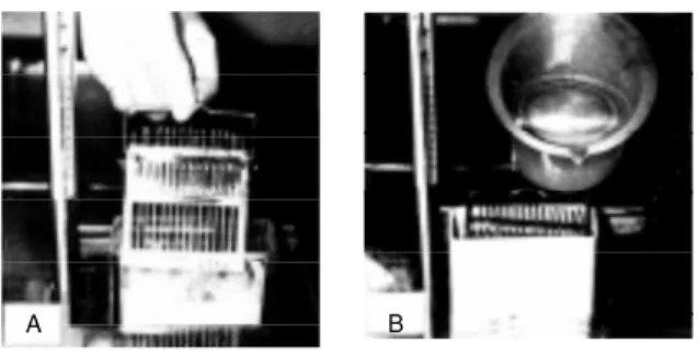
1) A bottle of bulk emulsion (we use Konica NR-M2 emulsion, Konica Ltd., To-kyo, Japan), is melted in a water bath at 45o
C for about 10 min, and an equal amount of distilled water is added to it and mixed for 5-10 min with a glass slide to remove all air bubbles. Then, a slide holder made of stain-less steel, holding 15 glass slides which carry several thick sections, is dipped in the melted emulsion for several seconds, and then pulled up vertically for about 3 s to assure equal thickness coating (3-4 µm) over the sections (Figure 1A). The faster the speed, the thinner becomes the emulsion coating. 2) The bot-tom of the slide holder is wiped with a paper towel to remove excess emulsion and the slide holder is placed in an electric incubator at 28oC with a humidity of about 80%,
con-taining a wet sponge at the bottom, and dried for 1 h. When the slides are dry, they are stored in a light-tight slide box containing a desiccant (silica gel). The edge of the box is sealed with black tape and the box is kept in a refrigerator at 4o
C for exposure. 3) After an appropriate exposure time, all the slides are developed at once by pouring the developer
Figure 1 - The standard procedure for preparing LM RAG by w et-mounting radioautography. A, Bulk emulsion is melted and diluted w ith distilled w ater in a staining jar, in w hich a slide holder holding 15 slides is dipped and pulled up vertically. B, The slide holder is stored in a light-tight slide box for exposure, into w hich the developer is finally poured after exposure for developing all the slides at once.
into the slide box (Figure 1B). We use Konica KD-X1 (formerly SD-X1) developer for Konica NR-M2 emulsion. Kodak D-19 may be used for any type of emulsion. After development, the slides are rinsed in a stop bath (2% aqueous acetic acid solution), fixed twice in a fixer (30% aqueous sodium thio-sulfate solution) for 5 min, washed gently in running tap water for 10 min, and finally stained in 1% toluidine blue solution for light microscopy.
2.3.2.2. Ele ctron microscopic
we t-mounting radioautography
The same embedded tissues in epoxy resin can be used for both LMRAG and EMRAG. For EMRAG, ultrathin sections of 0.1 µm (100 nm) or semithin sections of 0.2 µm (200 nm) are cut with an ultramicrotome and picked up on collodion-coated grid meshes. The semithin sections should be observed by high voltage electron micros-copy (49).
As for the radioautographic emulsions, several types of emulsions are commercially available. We use Konica NR-H2 emulsion (Konica Ltd.) because of the small sized silver bromide crystals and better sensitivity. To obtain a thin monolayer of silver bromide crystals, two techniques, dipping and wire loop methods, are now in general use. The choice lies between mounting the sections on a flat microscopic glass slide or on a grid mesh during exposure. By the former method, glass slides are covered with thin collodion films on which sections are placed and are coated with radioautographic emulsion by dipping as done in the light microscopy pro-cedure. After exposure and photographic processing, the sections and collodion films together with the emulsion are floated off the glass slide and picked up on a grid for exam-ining by electron microscopy. This proce-dure is called the flat substrate method and is highly complicated and cumbersome. On the other hand, by the latter method, sections
are placed on grid meshes coated with collo-dion films according to the normal section-ing method and are covered with a pre-formed monolayer emulsion by picking up thin bubbles of molten emulsion with a wire loop and allowing them to gel before touch-ing the grids. This is called the wire loop method. We prefer this procedure, which was developed in our laboratory, using small or larger wire loops, and appears to be rather easier. Our procedure is described in detail as follows (1,58,61).
1) A regular 1.25-cm square glass block is made from No. 4890-40 glass strips for the LKB knife maker (LKB-Produkter AB, Bromma, Sweden). A square piece of double-coated Scotch tape, 4 mm in length, is stuck on the surface of each glass block. Six grids are placed around the tape like a rosette, arranged clockwise to identify each grid (Fig-ure 2A). The grids are vacuum coated with carbon of 10-nm thickness. 2) The radioau-tographic emulsion is diluted with an equal part of distilled water at 45o
C. We use Konica NR-H2 emulsion from Konica (formerly Sakura) Ltd., Tokyo, Japan, but any other emulsion such as Kodak can be used. Ten ml of diluted emulsion is added to 0.2 ml of a 2% aqueous solution of dioctyl sodium sulfosuccinate (a surfactant) in order to pre-vent the emulsion film from bursting (14). A thin film of emulsion is obtained by dipping
Figure 2 - The standard procedure for preparing EM RAG by w et-mounting radioautog-raphy. A, A large w ire loop is dipped into melted emulsion and a thin film of emulsion is obtained, w hich is applied horizontally to a glass block on w hich 6 meshes, each carrying several sections, are placed. B, Ten glass blocks carrying 6 meshes each are attached to a glass slide w ith Scotch tape. Thus, several glass slides, each carrying 10 glass blocks, are stored in a slide box for exposure and finally all the meshes on glass blocks are developed, fixed and stained.
a wire loop, 2.5 cm in diameter, made of platinum wire or vinyl-coated iron wire into the solution. 3) After air-drying horizontally for 1 min, when the emulsion film is gelled but still wet, the film is applied to the grids on the glass block horizontally (Figure 2A). The glass block is warmed at 28o
C for 1 h to dry the emulsion. 4) For exposure, several glass blocks are attached to one side of a microscope slide with double-coated Scotch tape (Figure 2B). 5) Control emulsion films should be checked by electron microscopy before exposure. 6) Several glass slides car-rying several glass blocks are placed in a black light-tight plastic slide box containing a desiccant (silica gel), and the top is sealed with black tape. The slide box is kept in a refrigerator at 4o
C for exposure. 7) After an appropriate exposure time, the glass slides carrying glass blocks with grid meshes are processed for development, then stopped in a stop bath, fixed in a fixer and stained with lead citrate solution for electron staining.
Concerning the development of the emul-sions, when a conventional MQ-developer such as D-19 is used, long spiral silver grains are formed. In order to obtain smaller silver grains, phenidon developer after gold laten-sification (G-L) is recommended (Figures 3-8). We prefer the following procedure (1,62). 1) The glass slides carrying the grid meshes are first soaked in distilled water in a staining jar for 10 s, and then 2) soaked in G-L solution (0.2 ml 2% aqueous gold chloride solution, 0.05 g potassium thiocyanate, 0.06 g potassium bromide, 100 ml distilled water) for latensification at 16oC for 30 s. 3) The
slides are then rinsed in distilled water for 10 s, and 4) soaked in phenidon developer (1.5 g ascorbic acid, 0.25 g phenidon, 0.4 g potas-sium bromide, 1.3 g potaspotas-sium carbonate, 20 g sodium sulfite anhydrous, 4.0 g potassium thiocyanate, 100 ml distilled water) in a water bath at 16o
C for 60 s for development. 5) They are then soaked in a stopper solution (2% aqueous acetic acid solution) for 10 s, 6) fixed in a fixer (30% aqueous sodium
thio-sulfate solution) for 5 min, with 2 changes, and 7) rinsed in distilled water for 5 min, with 3 changes. 8) The sections are stained in lead citrate solution (63) for 3 min, and 9) rinsed in distilled water for a few minutes. 10) Finally the grids are carefully removed from the glass blocks with a forceps, placed on filter paper in a Petri dish and dried in an incubator at 37o
C for 1 h. 11) The specimens are coated with a 10-nm thick carbon layer in a vacuum coater before electron microscopy.
2.3.2.3. Dry-mounting proce dure
Both cryo-sectioned and freeze-dried specimens and freeze-dried or freeze-substi-tuted and embedded specimens should be coated with dry radioautographic emulsions without using any water. The procedure is designated as dry-mounting radioautogra-phy. Two types of procedures are used for dry-mounting radioautography, i.e., light microscopic and electron microscopic pro-cedures.
2.3.2.3.1. Light microscopic
dry-mounting procedure
Historically, various procedures have been employed for light microscopic dry-mounting radioautography as described pre-viously (24,32-37). Since the first applica-tion of cryostat secapplica-tions to precoated slides at very low temperature by Appleton (24), many authors have recommended this tech-nique for light microscopy. However, these procedures are highly complicated in terms of treatment of both specimens and emul-sions. We first used dry-films produced with a large wire loop which were air-dried and applied to cryostat sections placed on glass slides (23). We believe that this method is the most convenient one. The procedure is as follows.
drop of ethylene glycol without using any water. 2) Radioautographic emulsion is di-luted with an equal part of distilled water at 45o
C. We use Konica NR-M2 emulsion from Konica but any other emulsion such as Kodak can be used. Ten ml of diluted emulsion is added to 0.2 ml of a 2% aqueous solution of dioctyl sodium sulfosuccinate (a surfactant) in order to prevent the emulsion film from bursting (14). 3) A thin film of emulsion is obtained by dipping a wire loop, 2.5 cm in diameter, made of platinum wire or vinyl-coated iron wire and attached with Scotch tape to a glass slide as a handle. 4) The handle is set horizontally on a flat surface for air-drying. 5) After air-drying for 1-2 min, when the center of the emulsion film is gelled and dried and transparent but the peripheral zone is still wet and opaque, the film is applied to the slide horizontally. 6) The glass slide is kept in a Petri dish and warmed at 28oC in an incubator for 1 h for drying the
emulsion. 7) Several glass slides are placed in a black light-tight plastic slide box con-taining desiccant (silica gel), and the top is sealed with black tape. The slide box is kept in a refrigerator at 4o
C for exposure. 8) After an appropriate exposure time, the glass slides are processed for development, then stopped in a stop bath, fixed in a fixer and stained with toluidine blue solution. 9) Control tis-sues should be fixed with chemical fixative, dehydrated, embedded, wet-sectioned and wet-mounted by conventional dipping pro-cedures.
2.3.2.3.2. Ele ctron microscope
dry-mounting procedure
For tissues fixed by precipitation fixa-tion, ultrathin Epon sectioning and radioau-tography can be carried out according to the routine wet-mounting procedures for dem-onstrating soluble compounds. However, when the tissues are fixed by rapid freezing and freeze-dried or freeze-substituted, they are dry-sectioned and have to be
radioauto-graphed by the dry-mounting procedure (17,18). The grids carrying dry sections (ei-ther freeze-dried or freeze-substituted Epon-embedded sections or freeze-sectioned and freeze-dried) are coated with a 5-10-nm thick carbon layer before emulsion application. They are then placed on a grid holder made of a glass slide (25 mm x 75 mm) and 3 glass rods (3 mm in diameter and 10 mm in length). The routine procedures for dry-mounting are as follows.
1) The radioautographic emulsion is di-luted 1:10 with distilled water at 45o
C in a dark room. 2) Ten ml of the diluted emulsion is added to 0.2 ml of 2% aqueous solution of dioctyl sodium sulfosuccinate and maintained at 45oC in a thermobath for several minutes
to complete mixing. Dioctyl sodium sulfo-succinate, a surface activating agent, is used to prevent the emulsion films from bursting while they are being air dried (17,23). We use Konica NR-H2 emulsion. Other emul-sions for electron microscopic radioautogra-phy such as Kodak NTB or Ilford L4 could also be used. The procedures are as follows. 3) A thin film of the emulsion thus prepared is obtained by dipping a platinum wire loop, about 1 cm in diameter, into the emulsion (17). 4) Instead of a small platinum wire loop, a large vinyl-coated iron wire loop, 2.5 cm in diameter, can also be used (1,61). 5) The handle of the wire loop is set on a flat surface for air drying (for 1-2 min). 6) The best condition for applying the film to the grid is in such a way that the peripheral zone of the film appears gelled but wet (opaque) while the central zone is gelled and almost dry, but still transparent. The films are al-most 100% air-dried without breaking by using the dioctyl sodium sulfosuccinate. Without this agent, the films will burst most of the time (23). 7) The dried films are then applied to the grids on the holders like quoits and 8) the grids are transferred to Petri dishes and warmed at 37o
with black vinyl tape and kept in a refrigera-tor at 4oC for exposure.
On the other hand, when several grids are attached to a square glass block, 12.5 mm in length, a large wire loop, 2.5 cm in diameter, can be used as done in the wet-mounting procedure, and several blocks are placed on a slide. They are exposed, developed, fixed and stained simultaneously.
As a control, radioautography should be carried out by conventional procedures for insoluble compounds. The tissues are fixed in 2.5% glutaraldehyde and 1% osmium tetroxide in 0.1 M cacodylate buffer, dehy-drated in a graded ethanol series, embedded in Epon, sectioned with water in the knife trough, and wet-mounted with radioauto-graphic emulsion. After an appropriate ex-posure time, the meshes are transferred to the developer. The author uses gold-latensi-fication and phenidon developer (62). Other types of developer such as Kodak D-19 can also be used. Following processing with the stopper, fixer and several rinses in distilled water, the grids are stained in a lead citrate solution for 3 min for the purpose of both staining and removing the gelatin of the emul-sion. We use a lead citrate solution consist-ing of 10 ml distilled water and 30 mg lead citrate, adjusted to pH 12 with a few drops of 10 N NaOH. A lead citrate solution could also be used according to the method of Reynolds (63). Uranyl staining is usually not necessary.
3. Spe cial radioautographology
Special RAGology consists of applica-tions of general RAGology to various fields of biology and medicine. The applications of RAG to biology and medicine can also be classified into several scientific fields, i.e., cellular and molecular biology, anatomy, histology, embryology, pathology and phar-macology, etc. All the results obtained from such applications should be systematized as a new field of science to be designated as
special RAGology in the future.
In this section, the author would like to review briefly the results obtained in cellular and molecular biology, anatomy, histology, embryology, pathology and pharmacology from organ systems of animals and humans in our laboratory.
3.1. Ce llular and mole cular biology
In cellular and molecular biology, spe-cial radioautographology deals with the mac-romolecular synthesis and intracellular lo-calization of small molecular compounds in living organisms at the cellular and molecu-lar levels.
3.1.1. Macromole cular synthe sis
Macromolecular compounds such as nucleic acids (DNA and RNA), proteins, glucides and lipids which are synthesized in cells can be demonstrated at the subcellular level by conventional wet-mounting radio-autography in cells and tissues submitted to chemical fixation (64-66).
3.1.1.1. DNA synthe sis
DNA is the main component of nucle-oproteins and consists of deoxyribose, phos-phate, and bases which contain adenine (A), thymine (T), guanine (G) and cytosine (C). [3
H]-Thymidine is incorporated into the bases. Light and electron microscopic radio-autograms of the gastrointestinal tract of mice and rats labeled with [3
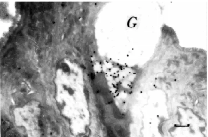
carried out by Leblond and coworkers in the 1960s (67). We have mainly studied the labeling sites and labeling indices as well as cell organelle changes in cell components of parenchymatous digestive organs such as the liver and pancreas, and in esophageal and intestinal epithelial cells of aging mice from the prenatal to the postnatal period by light and electron microscopic radioautogra-phy (64,66). We also demonstrated DNA synthesis in some other organ systems such as the respiratory, urogenital, endocrine (Fig-ure 3), circulatory, nervous and sensory sys-tems (12). Extranuclear DNA synthesis in mitochondria or peroxisomes of various cells was also studied under different experimen-tal conditions (68-70).
3.1.1.2. RNA synthe sis
RNA is composed of ribose, phosphate and the bases, A, U, G, and C. In order to demonstrate RNA synthesis, [3
H]-labeled cytidine was formerly used (71). However, [3
H]-cytidine can also be incorporated into DNA although at very low levels, so that demonstration of DNase digestion was nec-essary to confirm RNA synthesis. In con-trast, [3H]-5-uridine was shown to be
specif-ically incorporated into only RNA. There-fore, it has now come to be used routinely for the demonstration of RNA synthesis (1,12,72). When [3
H]-uridine is adminis-tered to animals, or cultured cells are incu-bated in a medium containing [3H]-uridine
in vitro and radioautograms are prepared, silver grains first appear over the chromatin of the nucleus and nucleolus of all the cells within several minutes, then spreading over the cytoplasm within 30 min, showing mes-senger RNA and ribosomal RNA (Figure 4). We studied quantitative changes of RNA synthesis in the liver and pancreas of aging mice by means of light and electron micro-scopic radioautography after injection of [3
H]-uridine (66). We also demonstrated in-tramitochondrial RNA synthesis in some
cells independently of the nuclei (72,73).
3.1.1.3. Prote in synthe sis
Proteins consist of polypeptides and the intracellular localization of protein synthe-sis can be demonstrated by light and electron microscopic radioautography after adminis-tration of RI-labeled amino acids such as [3
H]-glycine or [3
H]-leucine which are in-corporated into the endoplasmic reticulum and Golgi apparatus and then transferred to secretory granules or cytoplasmic ground
Figure 3 - Light microscopic radioautograms (LM RAG) of the adrenal gland of a 2-w eek-old mouse injected w ith [3H]-thymidine, demonstrating DNA
synthesis. Tw o nuclei in the zona fasciculata at the center are labeled w ith many silver grains. M agnification bar: 5 µm.
Figure 4 - Electron microscopic radioautogram (EM RAG) of HeLa cells cultured in vitro in a medium containing [3H]-uridine, demonstrating RNA
substance (Figure 5) and finally discharged outside the cells (64-66). On the other hand, [3
H]-proline and [3
H]-hydroxyproline are incorporated into endoplasmic reticulum and Golgi apparatus of fibroblasts and accumu-late into collagen in the extracellular matrix (74,75).
3.1.1.4. Glucide synthe sis
The glucides are classified biochemically into monosaccharides, disaccharides and polysaccharides. Monosaccharides and di-saccharides are small water-soluble mol-ecules which cannot be fixed by chemical fixation, but only by cryo-fixation for soluble compounds. Therefore, only polysaccharide synthesis is usually demonstrated by con-ventional light and electron microscopic ra-dioautography with chemical fixation. Poly-saccharides can be classified into simple polysaccharides and complex polysaccha-rides. Among complex polysaccharides, gly-coproteins are composed of proteins and sugar chains, which incorporate [3
H]-labeled glucose, galactose, fucose, N-acetyl-glucos-amine (Figure 6), etc. (1). On the other hand, mucosubstances or glycosaminoglycans are composed of mucous and proteins, which mainly contain chondroitin sulfate or hepa-rin, and incorporate the radiosulfate 35
SO4.
Therefore, the intracellular localization of mucosubstance synthesis can be demon-strated by light and electron microscopy us-ing 35SO
4 incorporation (Figure 7). The
half-life of radiosulfate is very short, only 87 days, so that experiments should be com-pleted within a few months. We showed incorporation of radiosulfate into the Golgi apparatus and mucigen granules of colonic goblet cells (76).
3.1.1.5. Lipid synthe sis
Lipids are esters of high fatty acids and can be classified into simple lipids and com-pound lipids. The former are composed of
Figure 5 - EM RAG of the pancreas of an adult mouse, 1 month after birth, injected w ith [3H]-leucine, demonstrating protein synthesis in pancreatic
acinar cells. M any silver grains are localized over the endoplasmic reticu-lum, Golgi apparatus and secretory granules. M agnification bar: 1 µm.
Figure 6 - EM RAG of a 14-day-old mouse injected w ith [3
H]-glucosa-mine, demonstrating glucide synthesis in a pancreatic acinar cell. M any silver grains are observed over the nucleus, nucleoli, secretory granules and cytoplasmic matrix. M agnification bar: 1 µm.
Figure 7 - EM RAG of a goblet cell in the colon of an adult mouse injected w ith 35SO4, demonstrating mucus synthesis. Silver grains are observed
glycerol and fatty acids, while the latter are composed of both lipids and other compo-nents such as phosphates, glucides or pro-teins. In order to demonstrate the intracellu-lar localization of lipid synthesis by light and electron microscopic radioautography, the incorporation of [3
H]-glycerol or [3
H]-fatty acids was examined (77,78).
3.1.1.6. Ge ne e xpre ssion
Recently, gene expression in cells has been demonstrated by in situ hybridization.
In situ hybridization with RI-labeled probes to demonstrate mRNA by light and electron microscopic radioautography has been fre-quently used. We have demonstrated gene expression of peroxisomal enzymes such as acyl-CoA oxidase mRNA with RI-labeled probes by LM and EMRAG (79-81). Other scientists prefer to use immunostaining rather than radioautography because they believe that the former is easier. However, we prefer radioautography to immunostaining because it has more advantages. The sensitivity of RAG is better than that of immunostaining. The electron density of silver grains is much higher than that of immunochemical depos-its when observed by electron microscopy. The intensity of reaction by RAG is quantifi-able by grain counting (81) but not by immu-nostaining. Therefore, this method should be used more often than immunostaining (82).
3.1.2. Small molecular compounds
The small molecular compounds such as precursors of macromolecular compounds, i.e., thymidine, uridine, amino acids, glucos-amines, fatty acids or target tracers such as hormones, neurotransmitters, vitamins, in-organic compounds, drugs or toxins cannot be demonstrated by wet-mounting radioau-tography. They are only demonstrable by means of dry-mounting radioautography (13). We have demonstrated many small
molecu-lar compounds in various organs by dry-mounting radioautography (Figure 8), as de-scribed in the following sections.
3.2. Anatomy and histology
The results obtained with the applica-tions of various precursors or tracers to all the organ systems should be described ac-cording to the conventional order of anatomy and histology of the organs. These results should be included in the histochemistry of the organs (83). The outline of the results obtained from various organ systems in our laboratory will be described briefly.
3.2.1. The organs of move me nt
3.2.1.1. The bone s and joints
DNA synthesis was studied in sala-manders of various ages. When salasala-manders were divided into 8 age groups from larvae to 4, 6, 8, 9,10 weeks and 8, 12 months after hatching and injected with [3
H]-thymidine and their forelimbs and hindlimbs were fixed, embedded in Epon, sectioned and radioauto-graphed, the aging changes of DNA
synthe-Figure 8 - EM RAG of a mast cell from adult rat peritoneum, incubated in medium containing [3H]-tranilast, a synthetic anti-allergic agent,
sis demonstrated that the labeling indices of chondrocytes in digital bones showed peaks at 4 weeks, then decreased after 6 weeks, reaching 0 at 8 months (84). We also studied DNA, RNA and protein synthesis of normal and rheumatoid synovial membranes surgi-cally obtained from rheumatoid patients and radioautographed by in vitro labeling with [3
H]-thymidine, [3
H]-uridine and [3
H]-leu-cine. No significant differences between nor-mal and rheumatoid tissues were observed (85).
3.2.1.2. The muscular syste m
The aging changes of DNA synthesis in ddY mouse intercostal muscle from prenatal day 13 through 24 months of postnatal life were studied by [3
H]-thymidine radioautog-raphy. The labeling indices revealed chrono-logical changes, reaching a peak at embry-onic day 13 and decreasing gradually to 0% at 3 months after birth (86). We also studied DNA synthesis in rat thigh muscle when injured and labeled with [3
H]-thymidine, demonstrating satellite cell regeneration, as well as DNA synthesis of satellite cells in the muscle of chickens with dystrophy, which will be described in detail in the following pathology section.
As for the incorporation of [3
H]-taurine, no difference was found between normal and dystrophy mice by chemical fixation and wet-mounting radioautography. When [3
H]-taurine was administered to normal or dys-trophy mice and their skeletal muscles were observed in freeze-substituted or freeze-dried specimens followed by dry-mounting radio-autography, however, silver grains appeared on the sarcoplasmic reticulum, myofilaments, mitochondria and sarcoplasmic membranes (87,88). More silver grains were observed in normal mice than in dystrophic mice. Only few silver grains were observed in the speci-mens conventionally fixed with glutaralde-hyde and osmium tetroxide followed by wet-mounting radioautography. Based on these
results, we conclude that [3
H]-taurine is dif-fusible and is associated with skeletal muscle cells. Taurine is an amino acid which regu-lates intracellular calcium transport and cell membrane excitability (88).
3.2.2. The dige stive syste m
We have published many papers from our laboratory dealing with the macromo-lecular synthesis of digestive organs from the oral cavity to the gastrointestinal tract and the digestive glands (66). The outline of the results will be described briefly in the order of systematic anatomy and special his-tology.
3.2.2.1. The oral cavity
DNA synthesis of mucosal epithelia of the lips and tongue as well as the subman-dibular glands of aging mice from fetal day 19 to 2 years of postnatal life were studied by LM and EMRAG with [3
H]-thymidine. The results showed that the labeling indices of stratified squamous epithelial cells in the oral mucosa increased from perinatal stages to postnatal day 14 and reached steady levels in adults (89), while the indices in the cell types of the submandibular glands increased from the fetal stage to postnatal day 1 and then steadily decreased up to 2 years. A-mong the cell types in the submandibular glands, the intercalated duct cells were the most frequently labeled and persisted for a longer period. The results suggested that intercalated duct cells are involved in the proliferation of other cell types (90).
3.2.2.2. The esophagus
DNA synthesis in the esophagus of aging mice labeled with [3


![Figure 3 - Light microscopic radioautograms (LM RAG) of the adrenal gland of a 2-w eek-old mouse injected w ith [ 3 H]-thymidine, demonstrating DNA synthesis](https://thumb-eu.123doks.com/thumbv2/123dok_br/15809552.650794/19.918.379.756.442.676/figure-microscopic-radioautograms-adrenal-injected-thymidine-demonstrating-synthesis.webp)
![Figure 8 - EM RAG of a mast cell from adult rat peritoneum, incubated in medium containing [ 3 H]-tranilast, a synthetic anti-allergic agent, demon-strating intracellular drug localization](https://thumb-eu.123doks.com/thumbv2/123dok_br/15809552.650794/21.918.382.746.747.1008/peritoneum-incubated-containing-tranilast-synthetic-allergic-intracellular-localization.webp)
