THIAGO ALVES MAGALHÃES
Respostas subcelulares a indução de galhas por
Baccharopelma dracunculifoliae (Psyllidae) em Baccharis dracunculifolia
(Asteraceae) e por Eriogallococcus isaias (Eriococcidae) em
Pseudobombax grandiflorum (Malvaceae)
Tese apresentada ao Programa de Pós-Graduação em Biologia Vegetal do Departamento de Botânica do Instituto de Ciências Biológicas da Universidade Federal de Minas Gerais, como requisito parcial à obtenção do título de Doutor em Biologia Vegetal. Área de Concentração: Morfologia, Sistemática e
Diversidade Vegetal
Orientadora: Profa. Dra. Rosy Mary dos Santos Isaias
Universidade Federal de Minas Gerais
Coorientador: Prof. Dr. Denis Coelho de Oliveira
Universidade Federal de Uberlândia
Magalhães, Thiago Alves.
Respostas subcelulares a indução de galhas por Baccharopelma
dracunculifoliae (Psyllidae) em Baccharis dracunculifolia (Asteraceae) e por Eriogallococcus isaias (Eriococcidae) em Pseudolombax grandiflorum (Malvaceae) [manuscrito] / Thiago Alves Magalhães. – 2014.
95 f. : il. ; 29,5 cm.
Orientadora: Rosy Mary dos Santos Isaias. Co-orientador: Denis Coelho de Oliveira.
Tese (doutorado) – Universidade Federal de Minas Gerais, Departamento de Botânica.
Sumário
Dedicatória...V Agradecimentos...VI Lista abreviaturas...VII
Resumo...01
Abstract...02
Introdução geral...03
CAPÍTULO I - Patterns of cell elongation in the determination of the final shape in galls of Baccharopelma dracunculifoliae (Psyllidae) on Baccharis dracunculifolia DC (Asteraceae)...14
Abstract...15
Introduction...15
Material and methods...16
Results……...16
Discussion...18
References...20
CAPÍTULO II - Are the sites of reactive oxygen species production and programmed cell death related during the development of Baccharopelma dracunculifoliae (Psyllidae) gall on Baccharis dracunculifolia (Asteraceae)?...22
Abstract...25
Introduction...26
Material and methods...27
Results...29
Discussion...31
Conclusion...34
References...35
Figures...42
CAPÍTULO III - Variação na estrutura péctica da parede celular durante o desenvolvimento da galha induzida por Eriogallococcus isaias (Eriococcidae) em Pseudobombax grandiflorum (Malvaceae)...48
Resumo...49
Introdução...50
Material e métodos...52
Resultados...54
Figuras...57
Discussão...61
CAPÍTULO IV - Impacto citológico decorrente do alto estresse oxidativo causado pelo desenvolvimento da galha de Eriogallococcus isaias (Eriococcidae) em
Pseudobombax grandiflorum (Malvaceae) ...71
Resumo...72
Introdução...73
Material e métodos...75
Resultados...77
Figuras...79
Discussão...85
Referências bibliográficas...88
vi AGRADECIMENTOS
A Deus que pela força e saúde para realizar este trabalho.
Em especial a Profa. Dra. Rosy Mary dos Santos Isaias pela orientação e confiança nestes quase sete anos. Obrigado pela sinceridade, disponibilidade, puxões de orelha e por todo conhecimento que me foi passado. É muito bom ter uma pessoa como você pra se espelhar.
Ao Prof. Dr. Denis Coelho de Oliveira pela coorientação, amizade e dedicação. Pelas acolhidas em sua casa, muito obrigado por tudo.
Ao Prof. Dr. Fenando Vale pela tutoria no REUNI. Pelos ensinamentos na sala de aula e pelos bons papos compartilhados.
À Capes pela bolsa, ao departamento de Botânica e a Pós-graduação em Biologia Vegetal da UFMG pela oportunidade. A Fapemig e ao CNPq que tem financiado os projetos do nosso grupo de pesquisa. Ao IEF e aos funcionários da Gruta da Lapinha pelo auxilio nas coletas de campo.
Ao Prof. Dr. Eduardo Alves, a técnica Eloisa e a Claudia Labory do Laboratório de Microscopia Eletrônica da UFLA pela ajuda durante com as análises de microscopia eletrônica. Aos funcionários do Centro de Microscopia Eletrônica da UFMG, em especial a Roberta por toda ajuda com a preparação de amostras. Ao CAPI UFMG pelo uso do Confocal. Aos professores Geraldo Wilson Fernandes e Maria de Fátima Horta pelas contribuições para desenvolvimento da tese.
Aos professores Elder, Denise do laboratório de Anatomia Vegetal e aos professores e funcionários (Selminha, Terezinha, Sonia, Denise, Sara, Márcia) do Departamento de Botânica da UFMG por toda ajuda dispensada quando foi preciso.
Ao Wagner pelas inúmeras ajudas prestadas nos experimentos. Muito obrigado por sua disponibilidade e pelos cafés compartilhados.
A Marina Neiva pela ajuda com as análises de confocal. A Jarina da Mata pela ajuda com as análises de morte celular programada e dicas sobre os experimentos de pectinas.
Lu Seabra, Advânio, Lucimara, Matheus, Sofia, Carol Marques, Deborah, Marcia Bacelar, Camila, Poliana e Nara foi muito bom conviver com todos vocês.
Ao grupo galhas (Rosy, Denis, Anete, Renê, Ariane, Cibele, Bruno, Cris, Aline, Gracielle, André, Priscila, Grazi, Graci) pela parceria de trabalho, pela convivência. Aprendi muito com todos vocês. Agradeço a Ariane, Aline e Bruno por terem participado diretamente do meu trabalho.
Gostaria de agradecer em especial a cada um que nesta reta final esteve ao meu lado e que muito me ajudou a finalizar esta tese. Muito obrigado por toda ajuda, pelos incentivos e conselhos, por terem me ensinado a ter paciência e principalmente por terem tido paciência comigo. É muito importante pra mim ter pessoas assim, ESPECIAIS, ao meu lado. Isso faz com que eu tenha cada vez mais certeza das minhas escolhas.
Aos amigos de Bom Sucesso pela confiança e pelo incentivo nessa caminhada. Ao Hugo, Júlio, Matheus, Hebert, Thalles, Ivan e Waguinho pelas cervejas, prosas e convivência durante esses anos.
viii
Lista de Abreviaturas
AGP – arabinogalactanos DAB - 3,3'-Diaminobenzidine ERO – espécies reativas de oxigênio HGA- homogalacturonanos
HR – reação de hipersensibilidade / hypersensibility reaction MCP – morte celular programada
MG – mature gall NGL – non galled leaf
PBS – phosphate saline buffer PCD – programmed cell death PG - poligalacturonases PI – propidium iodide
Resumo geral
Galhas apresentam tecidos típicos que se formam por rediferenciação celular através de
hiperplasia dos tecidos, hipertrofia e variação na direção da divisão e alongamento das
células. O acúmulo de espécies reativas de oxigênio (ERO) na parede celular causa sua
acidificação e, consequentemente, o afrouxamento das microfibrilas de celulose que
podem se reorientar e modificar os padrões de alongamento celular. Além das
microfibrilas, pectinas e arabinogalactanos-proteínas (AGPs) afetam a forma das células
e morfogênese dos tecidos. Os AGPs podem também atuar no controle da morte celular
programada (MCP). Neste trabalho foram estudados o sistema Baccharis
dracunculifolia - Baccharopelma dracunculifoliae e o sistema Pseudobombax
grandiflorum - Eriogallococcus isaias com o objetivo de avaliar se o estresse gerado
nos tecidos das galhas pode causar: alterações na estrutura do tecido e da parede celular
nas células da planta hospedeira, alterações citológicas que levariam à morte celular
programada e ao consequente controle da resposta de hipersensibilidade (HR) e, por
fim, avaliar o efeito destas alterações na geração das formas das galhas. Foi observado
no primeiro sistema que as microfibrilas de celulose e o grau de expansão e direção do
alongamento celular são importantes para determinar a forma final da galha. No
segundo sistema, ocorrem variações na estrutura péctica da parede e estas implicam em
características favoráveis ao desenvolvimento das galhas como extensibilidade,
elasticidade e rigidez da parede. O acúmulo de ERO nas duas galhas afeta a estrutura da
parede celular e dos cloroplastos, mas não é um fator determinante para causar MCP,
que só ocorreu na fase final do desenvolvimento nos dois sistemas.
Palavras-chave: Desenvolvimento de galhas, espécies reativas de oxigênio,
2
Abstract
Plant galls have typical tissues originated through cell redifferentiation, hyperplasia, cell
hypertrophy, and changes in the patterns of cell division and elongation. These
phenomena are important to determine the size and shape of the galls. The variation in
the patterns of cell elongation occurs due to changes in the orientation of the
microfibrils of cellulose, which is influenced by the acidification of the cell wall.
Moreover, pectins and arabinogalactan-proteins (AGPs) influence the cell shape and the
tissue morphogenesis. The AGPs may control programmed cell death (PCD). In this
work the systems Baccharopelma dracunculifoliae – Baccharis dracunculifolia and the system Pseudobombax grandiflorum – Eriogallococcus isaias were studied in order to
check if the stress generated in the gall tissues could cause changes in the structure of
the cell walls in the host plants, if the cytological alterations could lead to programmed
cell death and subsequent control of the hypersensitive response (HR), and finally to
evaluate the effect of these changes in the generation of the final gall shape. In the first
system, the orientation of the cellulose microfibrils, the degree of cell expansion and
direction of cell elongation are important on the determination of the final shape of the
gall. Both galls present variations in the pectic structure of their cell walls and these
variations imply in favorable features to gall development, such as elasticity,
extensibility and wall stiffness. The accumulation of ROS in gall tissues affects the
structure of the cell wall and chloroplasts, but do not cause MCP, which occurs only
during the senescent phase of both galls.
Key-words: Cellulose microfibrils, gall development, pectins, programmed cell death,
Introdução Geral
Insetos indutores de galhas representam fatores bióticos que modificam o
crescimento normal dos vegetais, através de alterações nos padrões morfogênicos (Mani
1964, Meyer & Maresquelle 1983, Oliveira & Isaias 2010b, Dias et al. 2013, Ferreira &
Isaias 2013). As galhas apresentam tecidos típicos e organizados (Mani 1964, Raman
2007, Oliveira & Isaias 2010b) formados por rediferenciação celular (sensu Lev- Yadun
2003, Oliveira & Isaias 2010b, Vecchi et al. 2013) e são consideradas novos órgãos
vegetais (Shorthouse et al. 2005). Nos últimos anos, alguns trabalhos avaliaram a
rediferenciação celular (Moura et al. 2008, Oliveira & Isaias 2010b, Dias et al. 2013) e
as variações nos padrões de expansão celular (Oliveira & Isaias 2010b, Isaias et al.
2011, Ferreira & Isaias 2013) durante a morfogênese em galhas.
A partir desses estudos, torna-se relevante aprofundar o conhecimento sobre
como o estresse causado pelo galhador afeta a estrutura da parede celular e do
protoplasto durante o desenvolvimento das galhas, e como isso implica na determinação
de sua forma final. Para o presente trabalho, foram escolhidos dois sistemas de galhas
induzidas por insetos sugadores. O primeiro sistema é constituído pela galha foliar
reniforme induzida por Baccharopelma dracunculifoliae (Psyllidae) em Baccharis
dracunculifolia DC (Asteraceae). Trata de um dos sistemas planta hospedeira–inseto
galhador mais bem estudado nos Neotrópicos nos aspectos ecológicos, da biologia do
inseto (Lara & Fernandes 1994, Araújo et al. 1995, Sperber &Collevatti 1996, Espírito
Santo & Fernandes 2002) e anatômicos (Arduin et al. 2005, Magalhães et al. 2013). O
segundo sistema consiste da galha foliolar, intralaminar, cônica, induzida por
Eriogallococcus isaias (Eriococcidade) em Pseudobombax grandiflorum (Malvaceae).
4 para os Neotrópicos (Hodgson et al. 2011), servindo ainda como modelo para estudos
fenológicos (Magalhães et al. submetido ao Journal of Natural History) e anatômicos
(Magalhães 2010).
Durante o desenvolvimento da maioria das galhas, hipertrofia celular e
hiperplasia dos tecidos são os fatores que definem o tamanho e a forma da estrutura
(Kraus et al. 1996, Isaias 1998, Souza et al. 2000, Oliveira et al. 2006, Moura et al.
2008, Oliveira e Isaias 2010b, Isaias et al. 2011, Magalhães et al. 2013). Como em
outros órgãos (Obroucheva 2008), os padrões de alongamento celular, assim como os de
divisão celular são importantes na determinação do tamanho e forma final da galha
(Oliveira & Isaias 2010b, Magalhães et al. 2013).
Para melhor entendimento destes processos, é necessário avaliar os padrões de
orientação das microfibrilas de celulose que, segundo Baskin (2001, 2005), afetam
diretamente o alongamento celular. Em células isodiamétricas, com crescimento
isotrópico, as microfibrilas de celulose se orientam em vários sentidos, enquanto nas
células alongadas, com crescimento anisotrópico, as microfibrilas estão orientadas
perpendicularmente ao maior eixo de alongamento celular (Baskin 2005, Magalhães et
al. 2013). A orientação das microfibrilas de celulose é influenciada pela acidificação da
parede celular (Cosgrove 1998, 1999), sendo este processo diretamente relacionado ao
acúmulo de espécies reativas de oxigênio (ERO) nas paredes, causando o afrouxamento
das microfibrilas e permitindo assim o alongamento celular (Del Rio & Pupo 2009,
Swanson & Gilroy 2010). Espécies reativas de oxigênio são produzidas normalmente no
metabolismo vegetal durante a fotossíntese e respiração (Del Río & Pupo 2009, Heldt &
Piechulla 2011), mas também podem ser produzidas como resposta ao ataque de
patógenos ou injúrias mecânicas nos tecidos (Doke et al. 1996, Maffei et al. 2007,
nos tecidos clorofilianos, feixes vasculares e nos tecidos próximos à câmara larval
(Oliveira & Isaias 2010a, Oliveira et al. 2010, Isaias et al. 2011), indicando que o
estresse oxidativo pode ser um fator que desencadeia a indução e controla o
desenvolvimento das galhas ao atuar diretamente no processo de hipertrofia. Entretanto,
o acúmulo de ERO nos tecidos pode levar a morte celular (Doke et al. 1996) e causar
alterações citológicas, como visto em alguns sistemas de galhas (Oliveira & Isaias
2010a, Oliveira et al. 2010, Oliveira et al. 2011a, b, Isaias et al. 2011). O estresse
oxidativo pode levar à morte celular programada (MCP) e, com isso, interromper o
desenvolvimento da galha. Por outro lado, o acúmulo de ERO nos tecidos afeta o
desenvolvimento da parede e, consequentemente, a forma final da galha. Nesse
contexto, torna-se importante compreender as alterações celulares causadas pelas ERO,
se e quando ocorre MCP nas galhas e a relação do acúmulo de ERO nos tecidos com a
MCP.
Entre outros fatores, o estresse gerado nos tecidos da galha pode provocar
alterações na composição e distribuição de substâncias da parede celular e,
consequentemente, alterar a morfogênese (Rose 2003, Albersheim et al. 2011, Wolf &
Greiner 2012). Nas galhas, são esperadas, principalmente, alterações na extensibilidade,
rigidez e elasticidade da parede durante o desenvolvimento, permitindo assim a
expansão celular. A variação dessas características na parede, durante as fases da galha,
determina o crescimento celular e influencia na determinação da forma da galha. Dentre
estas substâncias, podemos destacar as pectinas que tem importante papel na adesão
celular, expansão das células, porosidade, rigidez e estrutura da parede celular, defesa,
sinalização celular, entre outras (Ridley et al. 2001, Willats et al. 2001, Rose 2003,
Albersheim et al. 2011). A classe das pectinas é composta por polissacarídeos
6 homogalacturonanos (HGA), o mais abundante polissacarídeo péctico (~65% das
pectinas); os ramnogalacturonanos I (RG-I) que são formas alternativas de HGAs e
apresentam muitas cadeias laterais com arabinose e resíduos de galactose, e
ramnogalacturonanos II (RG-II), pectina estruturalmente mais complexa e que
compreende aproximadamente 10% da pectina existente na matriz da parede (Ridley et
al. 2001). Geralmente, as pectinas são sintetizadas em uma forma altamente
metil-esterificada (Micheli 2001) e mudanças nesse grau de esterificação e na molécula
péctica são importantes por definir as propriedades funcionais da parede, principalmente
durante o crescimento e desenvolvimento dos tecidos (Knox 1997).
Além das pectinas, a composição da parede celular também conta com
arabinogalactanos – proteínas (AGPs) (Pennel et al. 1989), que são proteínas estruturais
solúveis e altamente glicosiladas (Showalter 1993). Os AGPs tem aspecto
mucilaginoso, apresentam interação com pectinas e estão envolvidos no processo de
adesão celular (Cosgrove 1997), crescimento, nutrição, desenvolvimento vegetal e
proliferação celular (Pennel & Roberts 1990, Majewska-Sawka & Nothnagel 2000).
Alguns estudos indicam que os AGPs podem atuar no controle da MCP (Mastroberti &
Mariath 2008, Gao & Schowalter 1999). A MCP é um importante fenômeno presente
em diversas fases de desenvolvimento das plantas, incluindo senescência foliar,
embriogênese somática (Pennell & Lamb 1997) e zigótica (Giuliani et al. 2002). É um
evento caracterizado como suicídio celular, no qual a célula ativa mecanismos
específicos que acarretam em sua morte de maneira organizada. Possui como
características bioquímicas influxo de cálcio, ativação de proteases específicas,
fragmentação do DNA nuclear nas regiões internucleossômicas, liberação de citocromo
c pela mitocôndria e alteração na função de cloroplastos (Lam et al. 2001, Khurana et al.
cromatina nuclear, formação de corpos apoptóticos e perda estrutural da mitocôndria
(Lam et al. 2001, Sun et al. 1999).
O presente trabalho utiliza os dois sistemas planta hospedeira-inseto galhador
supracitados, Baccharis dracunculifolia - Baccharopelma dracunculifoliae e
Pseudobombax grandiflorum - Eriogallococcus isaias, como modelos para avaliar as
variações na estrutura da parede celular e do protoplasto das camadas celulares das
galhas. O habito alimentar similar dos insetos indutores e as dissimilaridades das duas
plantas hospedeiras, uma Asteraceae e uma Malvaceae, visam verificar se as galhas
podem ser consideradas como fenótipo estendido dos insetos mesmo em nível
subcelular.
O estresse gerado pelo acúmulo de ERO nos tecidos vegetais afeta a composição
e a estrutura da parede celular e do protoplasto das células envolvidas no processo
cecidogênico. Partindo dessa premissa, espera-se: (1) que as alterações na organização
das microfibrilas de celulose determinem novos padrões de alongamento celular os
quais serão marcantes nos estágios iniciais de desenvolvimento da forma da galha; (2)
que as variações nas pectinas e AGPs favoreçam a expansão e crescimento da parede
celular principalmente nas fases iniciais de formação das galhas; e (3) que o acúmulo de
espécies reativas de oxigênio (ERO) seja o fator desencadeador de morte celular
programada (MCP) afetando aspectos subcelulares das células das galhas.
A presente tese é dividida em quatro capítulos:
1 - Patterns of cell elongation in the determination of the final shape in galls of
Baccharopelma dracunculifoliae (Psyllidae) on Baccharis dracunculifolia DC
8
2- Are the sites of reactive oxygen species production and programmed cell death
related during the development of Baccharopelma dracunculifoliae (Psyllidae) gall on
Baccharis dracunculifolia (Asteraceae)?
3- Variação na estrutura péctica e proteica da parede celular durante o desenvolvimento
da galha induzida por Eriogallococcus isaias (Eriococcidae) em Pseudobombax
grandiflorum (Malvaceae);
4- Impacto citológico decorrente do alto estresse oxidativo causado pelo
desenvolvimento da galha de Eriogallococcus isaias (Eriococcidae) em Pseudobombax
grandiflorum (Malvaceae).
Referências bibliográficas
Albersheim P, Darvill A, Roberts K, Sederoff R, Staehelin A (2011) Plant cell walls.
Garland Science, Taylor and Francis Group, New York.
Araújo AM, Fernandes GW, Bede LC (1995) Influência do sexo e fenologia de
Baccharis dracunculifolia D.C. (Asteraceae) sobre insetos herbívoros. Rev Bras
Entomol 39:347-353.
Arduin M, Fernandes GW, Kraus JE (2005) Morphogenesis of gall induced by
Baccharopelma dracunculifoliae (Hemiptera: Psyllidae) on Baccharis
dracunculifolia (Asteraceae) leaves. Braz J Biol 65:559-571.
Baskin TI (2001) On the alignment of cellulose microfibrils by cortical microtubules: a
review and a model. Protoplasma 215:150-171.
Baskin TI (2005) Anisotropic expansion of the plant cell wall. Annu Rev Cell Dev Biol
Bolwell GP, Daudi A (2010) Reactive oxygen species in Plant-Pathogen Interaction. In:
Del Río LA, Puppo A, editors. Reactive oxygen species in plant signaling, p.
113-134.
Cosgrove DJ (1997) Assembly and enlargement of the primary cell wall in plants, Annu
Rev Cell Dev Biol 13: 71-201.
Cosgrove DJ (1998) Cell wall loosening by expansins. Plant Physiol 118:333-339.
Cosgrove DJ (1999) Enzymes and Other Agents That Enhance Cell Wall Extensibility.
Annu Rev Plant Physiol Plant Mol Biol 50:391-417.
Del Río LA, Puppo A (2009) Reactive oxygen species in plant signaling. Springer.
Dias GC, Moreira GRP, Ferreira BG, Isaias RMS (2013) Contrasting developmental
pathways for leaves and galls induced by a sap-feeding insect on Schinus polygamus
(Cav.) Cabrera (Anacardiaceae). An Academ Bras Cienc 85:187-200.
Doke N, Miura Y, Sanchez LM, Park HJ, Noritake T, Yoshioka H, Kawakita K (1996)
The oxidative burst protects plants against pathogen attack: mechanism and role as
an emergency signal for plant bio-defence a review. Gene 179:45-5.
Espírito-Santo MM, Fernandes GW (2002) Host plant effects on the development and
survivorship of the galling insect Neopelma baccharidis (Homoptera: Psyllidae).
Aust Ecol 27:249-257.
Ferreira BG, Isaias RMS (2013) Developmental stem anatomy and tissue
redifferentiation induced by a galling Lepidoptera on Marcetia taxifolia
(Melastomataceae). Botany 91:752-760.
Gao M, Showalter AM (1999) Yariv reagent treatment induces programmed cell death
in Arabidopsis cell cultures and implicates arabinogalactan protein involvement.
10 Giuliani C, Consonni G, Gavazzi G, Colombo M, Dolfini S (2002) Programmed Cell
Death During Embryogenesis of Maize. Ann Bot 90:287-292.
Heldt H, Piechulla B (2011) Plant Biochemistry. Academic Press, London.
Hodgson C, Magalhães TA, Miller D (2011) Two new gall-inducing genera and species
of Eriococcidae (Hemiptera: Sternorrhyncha: Coccoidea) off Malvaceae from the
neotropics. Lundiana 10:53-72. http://www.icb.ufmg.br/~lundiana/full/vol1012009/Hodgson.pdf
Isaias RMS (1998) Galhas entomógenas em Machaerium (Leguminosae-Papilionoidae):
anatomia e histoquímica. Tese de Doutorado, Departamento de Botânica,
Universidade de São Paulo, São Paulo.
Isaias RMS, Oliveira DC, Carneiro RGS (2011) Role of Euphalerus ostreoides
(Hemiptera: Psylloidea) in manipulating leaflet ontogenesis of Lonchocarpus
muehlbergianus (Fabaceae). Botany 89:581-592.
Khurana SMP, Pandey SK, Sarkar D, Chanemougasoundharam A (2005) Apoptosis in
plant disease response: A close encounter of the pathogen kind. Curr Sci 88:740-752.
Knox JP (1997) The use of antibodies to study the architecture and developmental
regulation of plant cell walls. Int Rev Cytol 171:79-120
Kraus JE, Sugiura HC, Cutupri S (1996) Morfologia e ontogenia em galhas
entomógenas de Guarea macrophylla subsp. tuberculata. Fitopatol Bras 21:349-356.
Lam E, Kato N, Lawton M (2001) Programmed cell death, mitochondria and the plant
hypersensitive response. Nature 411:848-853.
Lara ACF, Fernandes GW (1994) Distribuição de Galhas de Neopelma baccharidis
(Homoptera: Psyllidae) em Baccharis dracunculifolia (Asteraceae). Rev Bras Biol
54:661-668.
Lev-Yadun S (2003) Stem cells plants are differentiated too. Curr Top Plant Biol
Maffei ME, Mithifer A, Boland W (2007) Before gene expression: early events in plant-
insect interaction. Trends Plant Sci 12:1360-1385.
Magalhães TA (2010) Influência da fenologia no desenvolvimento e fisiologia das
galhas de Eriogalococcus gen. nov. em Pseudobombax grandiflorum (Cav.) A.
Robyns (Malvaceae). Dissertação de Mestrado. Departamento de Botânica,
Universidade Federal de Minas Gerais, Belo Horizonte, Brasil.
Magalhães TA, Oliveira DC, Suzuki AYM, Isaias RMS (2013) Patterns of cell
elongation in the determination of the final shape in galls of Baccharopelma
dracunculifoliae (Psyllidae) on Baccharis dracunculifolia DC (Asteraceae).
Protoplasma (online). DOI 10.1007/s00709-013-0574-z
Mani MS (1964) Ecology of Plant Galls. Dr. W. Junk Pub. The Hague.
Majewska-Sawka A, Nothnagel EA (2000) The multiple roles of arabinogalactan
protein in plant development. Plant Physiol 122:3-9.
Mastroberti AA, Mariath JEA (2008) Developmental of mucilage cells of Araucaria
angustifolia (Araucariaceae). Protoplasma 232:233-245.
Meyer J, Maresquelle HJ (1983) Anatomie des galles. Gebrüder Borntraeger, Berlin.
Micheli F (2001) Pectin methylesterases: Cell wall enzymes with important roles in
plant physiology. Trends Plant Sci. 6:414–419.
Moura MZD, Soares GLG, Isaias RMS (2008) Species-specific changes in tissue
morphogenesis induced by two arthropod leaf gallers in Lantana camara L.
(Verbenaceae). Aust J Bot 56:153-160.
Obroucheva NV (2008) Cell Elongation as an inseparable componente of growth in
terrestrial plants. Russ J Develop Biol 39:13-24.
Oliveira DC, Christiano JCS, Soares GLG, Isaias RMS (2006) Reações de defesas
12 galhador Euphalerus ostreoides Crawf. (Hemiptera: Psyllidae). Rev Bras Bot
29:657-667.
Oliveira DC, Isaias RMS (2010a) Cytological and histochemical gradients induced by a
sucking insect in galls of Aspidosperma australe Arg. Muell (Apocynaceae). Plant
Sci 178:350-358.
Oliveira DC, Isaias RMS (2010b) Redifferentiation of leaflet tissues during midrib gall
development in Copaifera langsdorffii (Fabaceae). S Afr J Bot 76:239-248.
Oliveira DC, Magalhães TA, Carneiro RGS, Alvim MN, Isaias RMS (2010) Do
Cecidomyiidae galls of Aspidosperma spruceanum (Apocynaceae) fit the
pre-established cytological and histochemical patterns? Protoplasma 242:81-93.
Oliveira DC, Carneiro RGS, Magalhães TA, Isaias RMS (2011a). Cytological and
histochemical gradients on two Copaifera langsdorffii Desf. (Fabaceae) -
Cecidomyiidae gall systems. Protoplasma 248:829-837.
Oliveira DC, Moreira ASFP, Magalhães TA, Lemos-Filho JP (2011b) Is the oxidative
stress caused by Aspidosperma spp. galls capable of altering leaf photosynthesis?
Plant Sci. 180:489-495.
Pennel RI, Knox JP, Scofield GN, Selvendran RR, Roberts K (1989) A Family of
abundant plama membrane-associated glycoproteins related to the arabinogalactan
proteins is unique to flowering plants. J Cell Biol 108:1967-1977.
Pennel RI, Roberts K (1990) Sexual developmental in pea is presaged by altered
expression of arabinogalactan protein. Nature 344:547-549.
Pennell RI, Lamb C (1997) Programmed cell death in plants. Plant Cell 9:1157-1168.
Raman A (2007) Insect-induced plant galls of India: unresolved questions. Curr Sci
Ridley B, O’Neil MA, Mohnen D. (2001) Pectins: structure, biosynthesis, and
oligogalacturonide-related signaling. Phytochemistry 57:929-967.
Rose JC (2003) The plant cell wall. Blackwell Publishing, Garsington Road, Oxford.
Shorthouse JD, Wool D, Raman A (2005) Gall-inducing insects – Nature’s most
sophisticated herbivores. Basic Appl Ecol 6:407-411.
Showalter AM (1993) Structure and function of plant cell wall proteins. Plant Cell
5:9-23.
Souza SCPM, Kraus JE, Isaias RMS, Neves LJ (2000) Anatomical and ultrastructural
aspects of leaf galls in Ficus microcarpa L. F. (Moraceae) induced by Gynaikothrips
ficorum Marchal (Thysanoptera). Acta Bot Bras 14:57-69.
Sperber CZ, Collevatti RG (1996) The gall maker Neopelma baccharidis Burck.
(Homoptera: Psyllidae) on Baccharis dracunculifolia (Asteraceae): success and
parasitoidism density dependence. An Soc Entomol Bras 25:59-63.
Sun Y, Zhao Y, Hong X, Zhai Z (1999) Cytochrome c release and caspase activation
during menadione-induced apoptosis in plants. FEBS Letters 462:317-321.
Swanson S, Gilroy S (2010) ROS in plant development. Physiol Plant 138: 384-392.
Torres MAM (2010) ROS in biotic interactions. Physiol Plant 138:414-429.
Vecchi C, Menezes NL, Oliveira DC, Ferreira BG, Isaias RMS (2013) The
redifferentiation of nutritive cells in galls induced by Lepidoptera on Tibouchina
pulchra (Cham.) Cogn. reveals predefined patterns of plant development.
Protoplasma 250:1363-1368.
Willats WGA, McCartney L, Mackie L, Knox P (2001) Pectin: cell biology and
prospects for functional analysis. Plant Mol Biol 47:9-27.
Wolf S, Greiner S (2012) Growth control by cell wall pectins. Protoplasma
CAPÍTULO I
Patterns of cell elongation in the determination of the final shape
in galls of Baccharopelma dracunculifoliae (Psyllidae)
on Baccharis dracunculifolia DC (Asteraceae)
ORIGINAL ARTICLE
Patterns of cell elongation in the determination of the final
shape in galls of
Baccharopelma dracunculifoliae
(Psyllidae)
on
Baccharis dracunculifolia
DC (Asteraceae)
Thiago Alves Magalhães&Denis Coelho de Oliveira&
Aline Yasko Marinho Suzuki&Rosy Mary dos Santos Isaias
Received: 5 August 2013 / Accepted: 17 October 2013 /Published online: 10 November 2013
#Springer-Verlag Wien 2013
Abstract Cell redifferentiation, division, and elongation are
recurrent processes, which occur during gall development, and are dependent on the cellulose microfibrils reorientation. We hypothesized that changes in the microfibrils orientation from non-galled tissues to galled ones occur and determine the final gall shape. This determination is caused by a new tissue zonation, its hyperplasia, and relative cell hypertrophy. The impact of the insect’s activity on these patterns of cell
development was herein tested in Baccharopelma dracunculifoliae—Baccharis dracunculifoliasystem. In this system, the microfibrils are oriented perpendicularly to the longest cell axis in elongated cells and randomly in isodiametric ones, either in non-galled or in galled tissues. The isodiametric cells of the abaxial epidermis in non-galled tissues divided and elongated periclinally, forming the outer gall epidermis. The anticlinally elongated cells of the abaxial palisade layer and the isodiametric cells of the spongy parenchyma originated the gall outer cortex with hypertrophied and periclinally elongated cells. The anticlinally elongated cells of the adaxial palisade layer originated the inner cortex with hypertrophied and periclinally elongated cells in young and mature galls and isodiametric cells in senescent galls. The isodiametric cells of the adaxial epidermis elongated periclinally in the inner gall epidermis. The current investigation demonstrates the role of cellulose
microfibril reorientation for gall development. Once many factors other than this reorientation act on gall development, it should be interesting to check the possible relationship of the new cell elongation patterns with the pectic composition of the cell walls.
Keywords Cellulose microfibrils . Cell responses .
Hyperplasia . Hypertrophy . Gall-inducing insect
Introduction
Gall development changes the morphogenetic patterns (Mani
1964; Meyer and Maresquelle 1983; Rohfritsch 1992; Oliveira and Isaias2010b) and the cytological features of the host organs (Oliveira and Isaias2010a; Oliveira et al.2010; Oliveira et al. 2011a, b; Isaias et al. 2011), leading to the redifferentiation (sensu Lev-Yadun 2003) of cell types with different functions and characteristics. In many galls, cell hypertrophy and tissue hyperplasia determine new cell lineages, whose distribution along the structure define their peculiar sizes and shapes (Kraus et al. 1996; Isaias1998; Souza et al. 2000; Oliveira et al.2006; Moura et al. 2008; Oliveira and Isaias2010b; Isaias et al.2011).
Some important factors in the development of galls are the patterns of cell division and the directions of cell elongation (Oliveira and Isaias 2010b). The latter is defined by the relationship between the orientation of the cytoskeleton and of the cellulose microfibrils (Baskin2005) and is influenced by the acidification of cell walls, which increases their extensibility (Cosgrove1998,1999). While for isotropic cell growth, the expansion rate is similar in all directions; for anisotropic cell growth, which generates elongated cells, the direction of maximum expansion rate is generally perpendicular to the net orientation of the microfibrils
Handling Editor: Friedrich W. Bentrup
T. A. Magalhães:A. Y. M. Suzuki:R. M. S. Isaias (*)
Departamento de Botânica, Instituto de Ciências Biológicas, Universidade Federal de Minas Gerais, 31270-901 Belo Horizonte, MG, Brazil
e-mail: rosy@icb.ufmg.br D. C. Oliveira
Universidade Federal de Uberlândia, Instituto de Biologia, Uberlândia, MG, Brazil
Protoplasma (2014) 251:747–753
DOI 10.1007/s00709-013-0574-z
(Baskin2005). Another factor that influences cell and tissue morphogenesis is the distribution of the pectins in cell walls (Rose2003; Albersheim et al.2011; Wolf and Greiner2012), which influences intercellular adhesion, cell expansion, and cell wall porosity, among others (Ridley et al.2001; Willats et al.2001; Rose2003; Albersheim et al.2011). Recently, this pectin dynamics was demonstrated in three gall morphotypes on the superhostBaccharis reticularia (Formiga et al.2013). During the course of the morphogenesis in galls, cell cycles are dynamic and repetitive, which generates good models for studies on cell transformations. These models have been previously addressed in terms of patterns of cell expansion and their importance in the determination of gall shapes (Moura et al. 2008; Oliveira and Isaias 2010b; Isaias et al.
2011). Current approach presents the labeling of the reorientation of cellulose microfibrils and the patterns of cell elongation during the developmental process of a simple gall morphotype (sensu Isaias et al. 2013), the leaf-folding gall induced by Baccharopelma dracunculifoliae (Psyllidae) on Baccharis dracunculifolia DC (Asteraceae). This gall system was previously studied in its ecological aspects, insect biology (Lara and Fernandes1994; Araújo et al.1995; Sperber and Collevatti1996; Espírito-Santo and Fernandes2002; Arduin et al.2005), and anatomical development (Arduin et al.2005), configuring the best studied host plant-galling insect system in the Neotropics. Currently, we focus on the alterations in size and shape and the relative reorientation of the microfibrils to verify: (1) whether the patterns of microfibrils deposition in the non-galled tissues are similar to the galled ones, (2) which roles microfibrils play during tissue changes along gall development, (3) how the variation in cellular shapes and sizes in each tissue layer influences the final gall shape, and finally (4) how the reorientation of cellulose microfibrils is related to the new structure and functional design defined in this leaf-folding gall morphotype.
Material and methods
Samples of non-galled leaves of B. dracunculifolia DC
(Asteraceae) and galls induced by B. dracunculifoliae
(Psyllidae) on young, mature, and senescent stages were collected, from January to June 2012, at Serra do Cipó, Minas Gerais, Brazil (19°17′57″S/43°35′58″W). Fragments of leaves
and galls (0.5 cm2) were fixed in 4 % Karnovsky in 0.1 M
phosphate buffer (pH 7.2) (O’Brien and McCully 1981),
dehydrated in n-butyl series (Johansen1940), and embedded in Paraplast® (Kraus and Arduin 1997). Transverse and longitudinal sections (10–14̀m) were obtained in a rotatory
microtome (Leica ® BIOCUT 2035), stained with safranin and Astra blue (2:8) (Bukatsch 1972) modified to 0.5 %, and mounted on colorless glass varnish Acrilex™ (Paiva et al.
2006). In transverse sections, the median region of both
non-galled leaves and galls (n=5) at each developmental stage was
measured. Measurements (n=50) were made on images obtained with a digital camera (Canon A650) coupled to a light microscope (ZEISS Primo Star) using the AxioVision™
(ZEISS) software (Zeiss2008). The results were analyzed by ANOVA followed by Tukey test, using the Graphpad Prism™
5.0 software (Motulsky1992–2009).
Free hand transverse and longitudinal sections of non-galled leaves and galls in the three stages of development were stained with Calcofluor White Stain (stock solution, 0.5 g/l) in distilled water for 30 min in the dark (Herth and Sghnepf1980) for labeling the cellulose microfibrils. The sections were analyzed and photographed in a fluorescence microscope (Olympus BX51) coupled to a DP70 camera (Olympus).
Results
Anatomical features
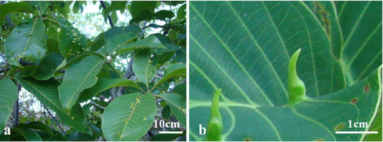
B. dracunculifoliaeinduces leaf-folding, glabrous, and green to reddish-brown galls on leaves of B. dracunculifolia
(Fig. 1). Young galls are defined as the initial lateral leaf folding along the midrib. Mature galls are defined by the complete marginal rolling with an increase in thickness and size, whereas the senescent galls are unfolded.
The leaves ofB.dracunculifolia (Fig.2a) have uniseriate
epidermis, isobilateral mesophyll with 2–3 layers of palisade
Fig. 1 Branch with mature and senescent galls induced by B. dracunculifoliaeonB. dracunculifolia
748 T.A. Magalhães et al.
Author's personal copy
parenchyma in the abaxial and adaxial positions, 2–3 layers of
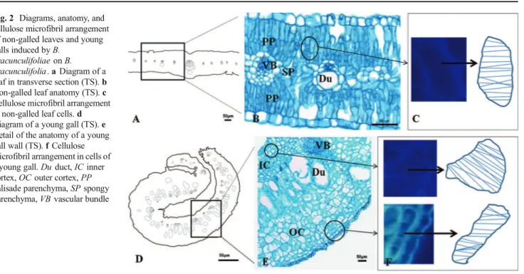
spongy parenchyma, and collateral vascular bundles associated with secretory ducts toward the abaxial position (Fig. 2b). The cellulose microfibrils in the epidermal and parenchymatic cell walls of non-galled leaves are perpendicular to the longest cell axis in elongated cells (Fig.2c) and randomly arranged in isodiametric cells.
The young galls (Fig.2d) have uniseriate epidermis in the inner and outer surfaces, multilayered outer cortex with secretory ducts, hypertrophied vascular bundles, and 4–5
layered parenchymatic inner cortex (Fig. 2e). Due to cell elongation, the cellulose microfibrils are oriented perpendicularly to the longest cell axis in inner and outer cortices (Fig.2f). The number of layers of the epidermis and outer cortex does not change toward gall maturation (Fig.3a) and senescence. The secretory ducts hypertrophy and the cells of the inner cortex divide up to 8–9 layers, remaining
parenchymatic (Fig. 3b). The cellulose microfibrils are oriented perpendicularly to the longest cell axis of the elongated cells either in the inner or the outer cortex
Fig. 2 Diagrams, anatomy, and cellulose microfibril arrangement of non-galled leaves and young galls induced byB.
dracunculifoliaeonB. dracunculifolia.aDiagram of a leaf in transverse section (TS).b Non-galled leaf anatomy (TS).c Cellulose microfibril arrangement in non-galled leaf cells.d Diagram of a young gall (TS).e Detail of the anatomy of a young gall wall (TS).fCellulose microfibril arrangement in cells of a young gall.Duduct,ICinner cortex,OCouter cortex,PP
palisade parenchyma,SPspongy parenchyma,VBvascular bundle
Fig. 3 Diagrams, anatomy, and cellulose microfibril arrangement of mature and senescent galls induced byB. dracunculifoliae
onB. dracunculifolia.aDiagram of a mature gall in transverse section (TS).bMature gall anatomy (TS).cCellulose microfibril arrangement in cells of a mature gall.dDiagram of a senescent gall (TS).eDetail of the anatomy of a senescent gall wall.fCellulose microfibril arrangement in cells of a senescent gall.Duduct,ICinner cortex,OCouter cortex,VB
vascular bundle
Patterns of cell elongation in galls on leaves ofBaccharis dracunculifolia 749
(Fig.3c). In senescent galls (Fig.3d), cell division results in a homogeneous cortex (Fig. 3e) accompanied by the reorientation (randomly) of the microfibrils (Fig. 3f). The cellulose microfibrils show the same arrangement either in transverse or longitudinal sections.
Non-galled leaves and gall histometry
The cells of the abaxial epidermis in non-galled tissues are isodiametric (cell area=403̀m2). During gall development,
they divide anticlinally and elongate periclinally forming the gall outer epidermis (cell area in mature gall=1,231̀m2). The
cells of the abaxial palisade layer are anticlinally elongated (cell area=213̀m2), and the cells of the spongy parenchyma are isodiametric (cell area=199̀m2). These two tissue layers
originate the gall outer cortex with hypertrophied and periclinally elongated or isodiametric cells (cell area in mature gall=7,016 ̀m2). The cells of the adaxial palisade layer are
anticlinally elongated (cell area=236̀m2) and originate the
inner cortex with hypertrophied and periclinally elongated cells in young (cell area=452 ̀m2) and mature galls (cell
area = 1,640 ̀m2). These cells become isodiametric in
senescent galls (cell area=2,560̀m2). The cells of the adaxial epidermis are isodiametric (cell area=366̀m2) and elongate
periclinally in the inner gall epidermis (cell area in mature gall=750 ̀m2). The cell area decreases in the abaxial and adaxial epidermis and outer cortex but increases in the inner cortex in the senescent gall.
Tissue redifferentiation vs. tissue histometry
The non-galled leaf tissues redifferentiate into neoformed gall tissues, with significant modifications in cell area. The main alterations responsible for gall shape determination are general tissue hyperplasia and the great hypertrophy of the cells of the abaxial epidermis, abaxial palisade parenchyma layer, and spongy parenchyma. The hyperplasia of the abaxial leaf epidermis results in a 50 % reduction in cell area in young galls. During gall maturation, this tissue layer elongates periclinally and becomes 3-fold larger when compared to non-galled leaf epidermis (Fig.4a, b). The gall cortex has a mixed origin. The outer layers come either from the abaxial palisade layers or from the spongy parenchyma. Its cells are anticlinally elongated or isodiametric, 33-fold larger in mature galls than in their precursor non-galled leaf cells (Fig.4c, d). The secretory ducts hypertrophy (45-fold larger in mature galls) and anticlinally elongate in the galls (Fig. 4e, f). The gall inner cortex originates from the adaxial palisade parenchyma and has periclinally elongated cells, 2-fold larger in young galls and 7-fold larger in mature galls, when compared to their correspondent non-galled leaf cells. The cells of the gall inner cortex are isodiametric and 11-fold larger in senescent galls (Fig. 4g, h), when the gall opens. The adaxial leaf
epidermis, whose cells are originally isodiametric in non-galled leaves, originates the 2-fold larger (mature galls) and periclinally elongated cells of the inner epidermis (Fig.4i, j).
Discussion
The process of gall induction by Baccharopelma dracunculifoliae on the leaves of B. dracunculifolia
completely alters the fates of its host leaf tissues. The microfibril orientation in gall tissues accompanies the new patterns of cell expansion and elongation. The epidermal cells elongate periclinally as a consequence of the reorientation of the microfibrils, which are perpendicular to the longest cell axis in gall cortex. Together with these new patterns, new cell sizes, the establishment of the inner and outer cortices, and the hypertrophy of the ducts determine the final gall shape.
The reorganization of tissues during gall development occurs due to tissue hyperplasia and cell hypertrophy, especially observed in the 35-fold larger cortical cell layers of the leaf-folding gall of B. dracunculifolia. These two
processes are common in arthropod-induced galls (Souza et al. 2000; Oliveira et al. 2006; Moura et al. 2008, 2009; Álvarez et al.2009; Raman2011; Oliveira and Isaias2010b; Isaias et al.2011), which indicates a pattern of cell responses to such biotic stimuli. During the development of the gall on
B.dracunculifolia, hyperplasia and hypertrophy are equally
evidenced in the young stage, while the great hypertrophy predominates at maturation. While on non-galled organs, cell hypertrophy follows cell divisions (Obroucheva 2008), our results demonstrate that both cell division and expansion contribute to the determination of gall structure, similarly to the assumption of Meijer and Murray (2001). In addition to hyperplasia and hypertrophy, changes in cell elongation were observed in the cells of epidermis and cortex. Cytometric data clearly show that the patterns of cell expansion and elongation are modified during gall development, mainly in the inner cortex, and are determinant for the new shape assumed by the gall. These new cell developmental patterns are directly linked to the reorientation of cellulose microfibrils redirected by the microtubule cytoskeleton, as described by Baskin (2001) and Wasteneys (2004).
Microfibril orientation influences the isotropic or anisotropic patterns of cell expansion. Anisotropic cell expansion requires cellulose synthesis enough to achieve parallel microfibril ordering and microtubules aligning in the same direction as nascent microfibrils, (Wasteneys 2004) especially in rapidly elongating cells (Sugimoto et al.2000). These redirections of microfibrils should occur in short periods of time since fast cell hypertrophy has been observed in several gall systems (Kraus et al.1996; Isaias1998; Souza et al. 2000; Oliveira et al. 2006; Moura et al. 2008, 2009; Oliveira and Isaias2010b; Isaias et al.2011). The alterations
750 T.A. Magalhães et al.
Author's personal copy
in the patterns of microfibrils deposition in cells of B.
dracunculifolia elegantly indicate the formation of relatively
short microfibrils, which directs the isodiametric expansion of cortical cells observed in the non-galled tissues and in senescent galls. Our results also demonstrated the anisotropic perpendicular organization of the elongated ones. This behavior corroborates Baskin’s (2005) proposal that the
direction of maximal expansion rate is generally perpendicular to the net orientation among microfibrils. This net reorientation requires the acidification of cell walls and the loosening of the cellulose microfibrils, which allow cell growth and the increase of reactive oxygen species (ROS) in the cell walls (Del Río and Puppo2009; Swanson and Gilroy
2010). This proposal was reinforced by the histochemical detection of ROS in chlorophyllous parenchyma of gall cortices and in tissue layers next to the larval chamber (Oliveira et al.2010,2011a,b; Isaias et al.2011).
The stimuli ofB.dracunculifoliae within the host tissues
ofB.dracunculifoliaresult in the folding of the host leaf and
the development of a morphologically simple gall morphotype. The distinct tissue layers are product of cell redifferentiation, with the reorientation of microfibrils in expected patterns either for isodiametric or axially elongated cells. We can conclude that the folding of the host leaves onB. dracunculifolia was defined mainly by the periclinal
elongation of cells in all tissue layers from induction to
Fig. 4 Histometric analysis (a,c, e,g,i) and diagrams (b,d,f,h, j) evidencing the patterns of cell expansion on non-galled leaves of
B. dracunculifoliaand on galls
induced byB. dracunculifoliae.
a,bAbaxial epidermis of non-galled leaves and gall outer epidermis.c,dAbaxial palisade and spongy parenchyma of non-galled leaves and gall outer cortex.e,fDucts on non-galled leaves and galls.g,hAdaxial palisade parenchyma of non-galled leaves and gall inner cortex.i,jAdaxial epidermis of non-galled leaves and gall inner epidermis.Lleaf,YGyoung gall,
MGmature gall,SGsenescent
gall. Tukey’s test was applied to
the cell area measurements. Results followed by different letters differ statistically at 0.05 %
Patterns of cell elongation in galls on leaves ofBaccharis dracunculifolia 751
maturation and by the hypertrophy of the cells of the outer cortex and of the outer epidermis. The differentially expanded cells in each tissue layer together with the low number of cell layers in the inner cortex were determinant for the final shape of the gall.
The current investigation demonstrates the role of cellulose microfibril reorientation for gall development. As many factors, other than this reorientation, can act on gall development, it should be interesting to check the possible relationship of the new cell elongation patterns with the pectic composition of the cell walls. The dynamics of pectin in the cell wall has been proved to influence cell expansion (Willats et al. 2001). More, specifically, arabinans and galactans influence the rigidity of cell wall (Jones et al.
1997) and should be associated to the mechanical properties of cell wall for expansion (McCartney et al. 2000; McCartney and Knox 2002) as well as on its elasticity (McCartney et al. 2003). Nevertheless, Wolf and Greiner (2012) proposed that the variation of methyl esterification in homogalacturonans is associated to reduced cell wall extensibility and cessation of growth. Gall development, due to its repetitive and determined patterns, has been proved to be elegant models for helping in the elucidation of the dynamics of pectin domains in the mechanisms of cell growth and expansion (Formiga et al.2013).
Acknowledgments The authors thank the Conselho Nacional de Desenvolvimento Científico e Tecnológico—CNPq (303352/2010-8, 307488/2009-8), Coordenação de Aperfeiçoamento de Pessoal de Nível Superior—CAPES, and the Fundação de Amparo à Pesquisa do Estado de Minas Gerais—FAPEMIG (APQ-04105-10; APQ-01801-09) for scholarships and financial support
Conflict of interest The authors declare no conflicts of interest in this manuscript.
References
Albersheim P, Darvill A, Roberts K, Sederoff R, Staehelin A (2011) Plant cell walls. Garland Science, Taylor and Francis Group, New York Álvarez R, Encina A, Pérez Hidalgo N (2009) Histological aspects of
threePistacia terebinthusgalls induced by three different aphids:
Paracletus cimiciformis,Forda marginataandForda formicaria. Plant Sci 176:303–314
Araújo AM, Fernandes GW, Bede LC (1995) Influência do Sexo e Fenologia deBaccharis dracunculifolia D.C. (Asteraceae) Sobre Insetos Herbívoros. Rev Bras Entomol 39:347–353
Arduin M, Fernandes GW, Kraus JE (2005) Morphogenesis of gall induced by Baccharopelma dracunculifoliae (Hemiptera: Psyllidae) onBaccharis dracunculifolia(Asteraceae) leaves. Braz J Biol 65:559–571
Baskin TI (2001) On the alignment of cellulose microfibrils by cortical microtubules: a review and a model. Protoplasma 215:150–171 Baskin TI (2005) Anisotropic expansion of the plant cell wall. Annu Rev
Cell Dev Biol 21:203–222
Bukatsch F (1972) Bermerkungen zur Doppelfärbung Astrablau-Safranin. Mikrokosmos 61:255
Cosgrove DJ (1998) Cell wall loosening by expansins. Plant Physiol 118: 333–339
Cosgrove DJ (1999) Enzymes and other agents that enhance cell wall extensibility. Annu Rev Plant Physiol Plant Mol Biol 50:391–417
Del Río LA, Puppo A (2009) Reactive oxygen species in plant signaling. Springer, Berlin
Espírito-Santo MM, Fernandes GW (2002) Host plant effects on the development and survivorship of the galling insect Neopelma baccharidis(Homoptera: Psyllidae). Aust Ecol 27:249–257
Formiga AT, Oliveira DC, Ferreira BG, Magalhães TA, Castro AC, Fernandes GW, Isaias RMS (2013) The role of pectic composition of cell walls in the determination of the new shape-functional design in galls ofBaccharis reticularia (Asteraceae). Protoplasma 250: 899–908
Herth W, Sghnepf E (1980) The fluorochrome, Calcofluor White, binds oriented to structural polysaccharide fibrils. Protoplasma 105:129–133 Isaias RMS (1998) Galhas entomógenas emMachaerium (Leguminosae-Papilionoidae): anatomia e histoquímica. PhD thesis. Departament of Botany, Universidade de São Paulo
Isaias RMS, Oliveira DC, Carneiro RGS (2011) Role ofEuphalerus ostreoides(Hemiptera: Psylloidea) in manipulating leaflet ontogenesis ofLonchocarpus muehlbergianus(Fabaceae). Botany 89:581–592 Isaias RMS, Carneiro RGS, Oliveira DC, Santos JC (2013) Annotated
and illustrated checklist of Brazilian gall morphotypes. Neotrop Entomol 42:230–239
Johansen DA (1940) Plant microtechnique. McGraw-Hill Book, New York Jones L, Seymour GB, Knox JP (1997) Localization of pectic galactan in tomato cell walls using a monoclonal antibody specific to (1→4)β
-D-galactan. Plant Physiol 113:1405–1412
Kraus JE, Sugiura HC, Cutupri S (1996) Morfologia e ontogenia em galhas entomógenas deGuarea macrophylla subsp.tuberculata
(Meliaceae). Fitopatol Bras 21:349–356
Kraus JE, Arduin M (1997) Manual básico de métodos em morfologia vegetal. EDUR, Seropédica
Lara ACF, Fernandes GW (1994) Distribuição de Galhas deNeopelma baccharidis(Homoptera: Psyllidae) emBaccharis dracunculifolia
(Asteraceae). Rev Bras Biol 54:661–668
Lev-Yadun S (2003) Stem cells plants are differentiated too. Curr Top Plant Biol 4:93–100
Mani MS (1964) Ecology of plant galls. Dr. W. Junk Publish, The Hague McCartney L, Knox JP (2002) Regulation of pectic polysaccharide domains in relation to cell development and cell properties in the pea testa. J Exp Bot 53:707–713
McCartney L, Ormerod AP, Gidley MJ, Knox JP (2000) Temporal and spatial regulation of pectic (1-4)-D-galactan in cell walls of
developing pea cotyledons implications for mechanical properties. Plant J 22:105–113
McCartney L, Steele-King CG, Jordan E, Knox JP (2003) Cell wall pectin (1-4)-D-galactan marks the acceleration of cell elongation in theArabidopsisseedling root meristem. Plant J 33:447–454
Meijer M, Murray JAH (2001) Cell cycle controls and the development of plant form. Curr Opin Plant Biol 4:44–49
Meyer J, Maresquelle HJ (1983) Anatomie des galles. Gebrüder Borntraeger, Berlin
Motulsky H (1992-2009) Analyzing data with Graph Pad Prism software. San Diego, California, USA, GraphPad Software Inc.
Moura MZD, Soares GLG, Isaias RMS (2008) Species-specific changes in tissue morphogenesis induced by two arthropod leaf gallers in
Lantana camaraL. (Verbenaceae). Aust J Bot 56:153–160
Moura MZD, Soares GLG, Isaias RMS (2009) Ontogênese da folha e das galhas induzidas porAceria lantanaeCook (Acarina: Eriophyidae) emLantana camaraL. (Verbenaceae). Rev Bras Bot 32:271–282 O’Brien TP, McCully ME (1981) The study of plant structure principles
and selected methods. Termarcarphi Pty, Melbourne
752 T.A. Magalhães et al.
Author's personal copy
Obroucheva NV (2008) Cell elongation as an inseparable component of growth in terrestrial plants. Russ J Develop Biol 39:13–24
Oliveira DC, Christiano JCS, Soares GLG, Isaias RMS (2006) Reações de defesas químicas e estruturais deLonchocarpus muehlbergianus
Hassl. (Fabaceae) à ação do galhadorEuphalerus ostreoidesCrawf. (Hemiptera: Psyllidae). Rev Bras Bot 29:657–667
Oliveira DC, Isaias RMS (2010a) Cytological and histochemical gradients induced by a sucking insect in galls ofAspidosperma australeArg. Muell (Apocynaceae). Plant Sci 178:350–358
Oliveira DC, Isaias RMS (2010b) Redifferentiation of leaflet tissues during midrib gall development in Copaifera langsdorffii
(Fabaceae). S Afr J Bot 76:239–248
Oliveira DC, Magalhães TA, Carneiro RGS, Alvim MN, Isaias RMS (2010) Do Cecidomyiidae galls ofAspidosperma spruceanum
(Apocynaceae) fit the pre-established cytological and histochemical patterns? Protoplasma 242:81–93
Oliveira DC, Carneiro RGS, Magalhães TA, Isaias RMS (2011a) Cytological and histochemical gradients on two Copaifera langsdorffii Desf. (Fabaceae)—Cecidomyiidae gall systems. Protoplasma 248:829–837
Oliveira DC, Moreira ASFP, Magalhães TA, Lemos-Filho JP (2011b) Is the oxidative stress caused byAspidospermaspp. galls capable of altering leaf photosynthesis? Plant Sci 180:489–495
Paiva JGA, Fank-de-Carvalho SM, Magalhães MP, Graciano-Ribeiro D (2006) Verniz vitral incolor 500®: uma alternativa de meio de montagem economicamente viável. Acta Bot Bras 20:257–264 Raman A (2011) Morphogenesis of insect-induced plant galls: facts and
questions. Flora 206:517–533
Ridley B, O’Neil MA, Mohnen D (2001) Pectins: structure, biosynthesis,
and oligogalacturonide-related signaling. Phytochemistry 57:929–967
Rohfritsch O (1992) Patterns in gall development. In: Shorthouse JD, Rohfritsch O (eds) Biology of Insect-Induced Galls. Oxford University, Oxford, pp 60–86
Rose JC (2003) The plant cell wall. Blackwell, Garsington Road, Oxford Souza SCPM, Kraus JE, Isaias RMS, Neves LJ (2000) Anatomical and ultrastructural aspects of leaf galls in Ficus microcarpa LF (Moraceae) induced by Gynaikothrips ficorum Marchal (Thysanoptera). Acta Bot Bras 14:57–69
Sperber CZ, Collevatti RG (1996) The gall makerNeopelma baccharidis
Burck. (Homoptera: Psyllidae) on Baccharis dracunculifolia
(Asteraceae): success and parasitoidism density dependence. An Soc Entomol Bras 25:59–63
Sugimoto K, Williamson RE, Wasteneys GO (2000) New techniques enable comparative analysis of microtubule orientation, wall texture, and growth rate in intact roots ofArabidopsis. Plant Physiol 124: 1493–1506
Swanson S, Gilroy S (2010) ROS in plant development. Physiol Plant 138:384–392
Wasteneys GO (2004) Progress in understanding the role of microtubules in plant cells. Curr Opin Plant Biol 7:651–660
Willats WGT, Mccartney L, Mackie L, Knox P (2001) Pectin: cell biology and prospects for functional analysis. Plant Mol Biol 47:9–27 Wolf S, Greiner S (2012) Growth control by cell wall pectins.
Protoplasma 249:169–175
Zeiss C (2008) Carl Zeiss Imaging Systems–32 Software Release 4.7.2. USA. Carl Zeiss MicroImaging Inc
Patterns of cell elongation in galls on leaves ofBaccharis dracunculifolia 753
CAPÍTULO II
Are the sites of reactive oxygen species production and programmed
cell death related during the development
of Baccharopelma dracunculifoliae (Psyllidae) gall
on Baccharis dracunculifolia (Asteraceae)?
Draft
! !
" # $ ! % !& ' ! % ( ) * + $ , '
-. + ' ( ) * + $ * / ' $ - %
) ' - ( * + ) , ' - /
' ( ) * + $ * / ' $ - %
0 ' )1 ( ) * + $ * / ' $
- %
' 2 ( * + ) , ' - /
3 4 " $$ ' 4 ' % ' 5" ' *67#
Draft
Thiago Alves Magalhães1, Denis Coelho de Oliveira2, Bruno Garcia Ferreira1, Jarina Pena
DaMata3, Maria de Fátima Martins Horta3, Rosy Mary dos Santos Isaias1
1
Universidade Federal de Minas Gerais (UFMG), Instituto de Ciências Biológicas,
Departamento de Botânica, Av. Antônio Carlos 6627, caixa postal 486, Pampulha, 312707
901, Belo Horizonte, MG, Brasil.
2
Universidade Federal de Uberlândia (UFU), Instituto de Biologia (INBIO), Campus
Umuarama, 384007902, P.O. Box: 593, Uberlândia, MG, Brasil.
3
Universidade Federal de Minas Gerais (UFMG), Instituto de Ciências Biológicas,
Departamento de Bioquímica e Imunologia, Av. Antônio Carlos 6627, caixa postal 486,
Pampulha, 312707901, Belo Horizonte, MG, Brasil.
Rosy Mary dos Santos Isaias (rosy@icb.ufmg.br). Phone:
+553134092687. Fax: +553134092671. Universidade Federal de Minas Gerais, Instituto de
Ciências Biológicas, Departamento de Botânica. Av. Antônio Carlos 6627, caixa postal 486,
Pampulha, 312707901, Belo Horizonte, MG, Brazil.
Page 1 of 24
Draft
Galls develop under the process of cell redifferentiation, in which neither new cellular nor
metabolic processes are distinct from those occurring on the normal pathways of the host
plant morphogenesis. Common events to normal morphogenesis such as generation of
Reactive Oxygen Species (ROS) and Programmed Cell Death (PCD) may occur in similar or
distinct sites and are determinant from induction until gall senescence. Current research deals
with DC. (Asteraceae) –
(Psyllidae) system to check if the accumulation of ROS and events of PCD are concomitant or
consecutive during gall development, especially in mature and senescent gall tissues. The
hypothesis herein proposed is rejected for PCD and ROS did not strictly accumulate in similar
sites of gall tissues. The PCD is not crucial for gall development but it is a consequence of the
end of cell cycles during gall senescence. Due to the death of the companion cells and the
collapse of the sieve tube elements, the nutrition of the gall tissues ceases, and, as a
consequence, the chloroplasts, the membrane systems, and the cell walls also enter
senescence process.
Cell redifferentiation, cell wall responses, cytology, PCD, TUNEL
Draft
3 Galling herbivores alter the morphogenetic patterns of the host plant organ (Mani
1964, Meyer and Maresquelle 1983, Rohfritsch and Anthony 1992, Oliveira and Isaias 2010a)
in a process of cell redifferentiation ( Lev7Yadun 2003, Oliveira and Isaias 2010a,
Vecchi et al. 2013). The redifferentiated cells and tissues have functions and characteristics
distinct from those of the non7galled organs (Oliveira and Isaias 2010b), which reinforce the
assumption that galls are true organs attached to the original host plant organ (Shorthouse et
al. 2005, Ferreira and Isaias 2014). Among the most common processes involved in the gall
formation, the cell hypertrophy and the tissue hyperplasia can be highlighted (Kraus et al.
1996, Isaias 1998, Souza et al. 2000, Oliveira et al. 2006, Oliveira and Isaias 2009, 2010a,
Magalhães et al. 2014), but no new metabolic or cytological processes occur (Vecchi et al.
2013). Nevertheless, recurrent events such as Programmed Cell Death (PCD) in specific sites
within gall tissues may occur. These PCD sites are crucial to sculpt the gall shape, by
eliminating some cells, avoiding the hypersensitive responses (HR), and improving the health
and quality of the remaining cells (Breusegem and Dat 2006). These authors presented a
review on the events of ROS homeostasis and cell death in plants, highlighting the importance
of ROS signaling during plant PCD. However, the relation between PCD and ROS production
during gall development is yet to be addressed.
One of the causes of cell death may be the overproduction of reactive oxygen species
(ROS) (Doke et al. 1996), as a possible plant response to pathogen attack or to mechanical
injuries (Doke et al. 1996, Maffei et al. 2007, Bolwell and Daudi 2009, Torres 2010). In galls,
ROS have been already histochemically detected by Oliveira and Isaias (2010b), Oliveira et
al. (2010, 2011a), and Isaias et al. (2011). These authors indicated that ROS can trigger gall
induction and development, and not simply induce cell death. PCD can be detected by
Page 3 of 24
Draft
ultrastructural features such as the formation of large autophagic vacuoles containing the
organelles and chromatin condensation (Van Doorn et al. 2011). The cytological responses
can also include DNA fragmentation detected by The DeadEnd™ Fluorometric TUNEL assay
(Promega, DeadEndTM Fluorometric TUNEL System, Madison, WI, USA), and the presence
of arabinogalactan proteins (AGPs) in cell walls (Gao and Showalter 1999, Mastroberti and
Mariath 2008). The histochemical detection of the precise sites of ROS accumulation
associated with the sites of fragmented DNA may elucidate the functionality and regulation of
the PCD during insect7plant interactions. These associated analyses in gall tissues may set
light on the comprehension of defensive mechanisms of plants, the establishment of the gall
inducing organisms within plant tissues, and the steps of gall development, which are herein
addressed in (Psyllidade) – DC
(Asteraceae) system. This is one of the best studied host plant7galling herbivore systems of
the Neotropical region, contemplating structural (Arduin et al. 2005, Magalhães et al. 2014),
immunocytochemical (Oliveira et al 2014), and ecological studies (Lara and Fernandes 1994,
Araújo et al. 1995, Sperber and Collevatti 1996, Espírito7Santo and Fernandes 2002).
The sites of accumulation of ROS are supposedly linked to the sites of PCD in this
gall, and should be crucial for the sculpturing of the kidney7shaped gall morphotype. To
validate this proposal, (i) the histochemical detection of sites of ROS accumulation, and (ii)
the detection of PCD by the TUNEL assay in non7galled and galled tissues were performed.
The evaluation of cytological alterations during gall formation complements these analyses.
! !
Samples of non7galled leaves (NGL) of (Asteraceae) (n ≥
15) and galls induced by (Psyllidae) in distinct
Draft
5 developmental stages (n ≥ 15) were collected at Serra do Cipó, between the kilometer 1107
112 of the MG 10 highway (19º17'57 "S / 43 º35'58" W), Minas Gerais state, Brazil.
The galls were separated in young, mature and senescent stages (Magalhães et al.
2014). Young galls (YG) are discrete leaf intumescences, folded along the midrib and open
along the leaf margins, with one or few individuals of inside the chamber.
In mature galls (MG), the leaf margins touch each other. Senescent galls (SG) are empty and
totally open by the separation of the leaf margins.
Hand7made sections of fresh material (NGL, YG, MG and SG) (n = 5) were incubated
in 0.5% 3,3'diaminobenzidine (DAB 7 Sigma®), according to manufacturer instructions and
kept in the dark (Rosseti and Bonatti 2001). The intensity of the reaction was examined every
15 minutes in a light microscope.
! " !"
The kit used for the TUNEL assay (Promega, DeadEndTM Fluorometric TUNEL
System, Madison, WI, USA) detects nuclear DNA fragmentation. The protocol for
labeling of PCD was performed according to the manufacturer with some alterations, herein
indicated. NGL, YG, MG and SG (n = 5) were fixed in 2.5% glutaraldehyde and 4.5%
formaldehyde in phosphate buffer (0.1 M; pH 7.2) (Karnovsky 1965), embedded in
Paraplast®, sectioned in a rotary microtome, deparafinized, post7fixed in 4% formalin, rinsed
with PBS buffer, and permeate with Proteinase K (2 µg/ml) for 15 minutes at 37°C. The
slides were incubated with Equilibration Buffer [potassium cacolidate (200 mM, pH 6.6),
Tris7HCl (25mM, pH 6.6), DTT (0.2mM), BSA (0,25 mg/ml) and cobalt chloride (2.5mM)]
for 10 minutes, then moved to a solution containing rTdT Enzyme and dUTP conjugated with
Page 5 of 24