Quim. Nova, Vol. 33, No. 5, 1044-1046, 2010
Artigo
*e-mail: [email protected]
AUSTIN, DEHYDROAUSTIN AND OTHER METABOLITES FROM Penicillium brasilianum
Betânia T. M. Schürmann
Escola de Farmácia, Universidade Federal de Ouro Preto, Rua Costa Sena, 171, 35400-000 Ouro Preto - MG, Brasil William S. T. Sallum e Jacqueline A. Takahashi*
Departamento de Química, Universidade Federal de Minas Gerais, Av. Antonio Carlos, 6627, 31270-901 Belo Horizonte - MG, Brasil
Recebido em 23/6/09; aceito em 19/1/10; publicado na web em 3/5/10
A culture of P. brasilianum, isolated from soil collected at the Serra do Cipó National Park, in Minas Gerais State (Brazil), was grown for 25 days on a dextrose-peptone-salts medium. The corresponding ethylacetate extract was column chromatographed and four compounds were isolated: austin, dehydroaustin, D-mannitol and penicillic acid. This is, in the best of our knowledge, the irst time that the meroterpenes austin and dehydroaustin have been isolated from this species. Activity of the extract and isolated compounds was tested against six bacteria and for acetylcholinesterase inhibition. Penicillic acid showed high activity in both tests.
Keywords: Austin; dehydroaustin; Penicillium brasilianum.
INTRODUCTION
Penicillium brasilianum Batista is a fungal species commonly found in soil. Several bioactive secondary metabolites were reported from this species, such as viridicatum toxin, verrucologen, fumitremorgin A, B and penicillic acid.1 The latter compound presents antibacterial, antiviral, cytotoxic, carcinogenic and antifungal activities2,3 while verrucologen and fumitremorgins have in vitro citotoxic activity against Ehrlich carcinoma tumor cells.4 Isolation of novel compounds with convulsive activity from this species has been recently reported and named brasiliamides A, B,5 C, D and E.6 Brasiliamides A, B, meroterpenoids preaustinoid A1, preaus-tinoid B2, austinolide along with, brasiliamide F, were also reported as metabolites from Penicillium brasilianum, isolated as an endophyte from Melia azedarach.7 Brasiliamide A presented bacteriostatic activity against B. subtilis.7
Penicillium brasilianum Batista was one of the 150 fungal species isolated from soil of the Serra do Cipó National Park (MG, Brazil) and its ethylacetate extract was identiied as one of the most active against Gram-positive and Gram-negative bacteria on a screening for antibacterial compounds, thus motivating the study of the metabolites responsible for the antibacterial activity in this extract.8
Meroterpenes are secondary metabolites found in plants, marine invertebrates and microorganisms that have been frequently isolated in fungi from Penicillium and Aspergillus genera.9 Austin is a good representative of this class, having been isolated for the irst time in 1976 by Chexal et al. from a culture of Aspergillus ustus.10 Later, in 1994, it was isolated, along with ive other meroterpenes, from Penicillium sp.11 Several Austin-like compounds have been reported from an endophyte species of Penicillium cultivated in rice: preaus-tinoid A and B,12 7-β-acetoxydehydroaustin, neoaustin, dehydro-austin, austinoneol,13 preaustinoid A1, A2 and B1.14 Some of these derivatives showed activity against Escherichia coli, Bacillus sp and Pseudomonas aureginosa.12
Therefore, the aim of the present work was to investigate the chemical composition of the ethylacetate extract of P. brasilianum in order to determine the structure of the compound responsible for its high antibacterial activity. Since P. brasilianum metabolites have never been tested as acetylcholinesterase inhibitors, this bioassay was also carried out for the isolated compounds.
EXPERIMENTAL
Source and maintenance of Penicillium brasilianum and bacterial strains P. brasilianum (LAB 34) was isolated from soil collected at the Serra do Cipó National Park, in Minas Gerais State (Brazil)8 and belongs to the Biotechnology and Bioassays Laboratory at the Chemistry Department from the Universidade Federal de Minas Gerais, Brazil. This isolate was identiied by L. M. de Abreu and then maintained on malt agar at -86 °C on an ultra-freezer. Bacterial strains used for antibacterial assays were: Staphylococcus aureus ATCC 29213, Listeria monocytogenes ATCC 15313 and Bacillus cereus ATCC 1177 (Gram positive) and Salmonela typhimurium ATCC 14028, Escherichia coli ATCC 25922 and Citrobacter freundii ATCC 8090 (Gram negative).
Reagents and equipment
Culture medium components were purchased from Biobrás (Montes Claros, Brazil). Solvents and eluants were purchased from Grupo Química (São Paulo, Brazil). Blanc and positive control discs were obtained from CECON (São Paulo, Brazil). Column grade silicagel (article 7734) and thin layer chromatography silicagel (ar-ticle 7731) were purchased from Merck (Darmstadt, Germany). The Vortex apparatus used was an AP 56-Phoenix (Araraquara, Brazil). Melting points were determined on a Mettler FP80HT apparatus and are uncorrected. Infrared spectra were obtained on a Spectrum One FTIR Spectrometer (Perkin-Elmer). 1H and 13C Nuclear Magnetic Resonance spectra were obtained from Bruker DRX-400 and DRX 200 Advance Spectrometers.
Metabolites production
Austin, dehydroaustin and other metabolites from Penicillium brasilianum 1045 Vol. 33, No. 5
Cultivation and extract preparation
The mycelium was separated from the culture broth by vacuum iltration. Both mycelium and broth were extracted separately three times with ethyl acetate (300.0 mL for each extraction). The resulting layers were combined and the solvent was eliminated by evaporation by using a rotator vacuum distilling apparatus. TLC comparison of mycelium and broth extracts showed the same proile and they were combined leading to 12.0 g of crude extract.
Metabolites puriication
The extract was fractionated by chromatography in a silica gel column (10.0 x 40.0 cm) using, as eluents, hexane, ethyl acetate and methanol 100% and in mixtures of growing polarities leading to 190 fractions (250.0 mL each), which were combined in 46 groups, ac-cording to thin layer chromatography proiles. Group 36 comprised fractions eluted by a mixture of EtOAc/MeOH 1:1 and presented a solid that was iltered off and recrystallized from methanol furnishing white crystals (1, 56.0 mg) that was tentatively identiied by 1H and 13C NMR and DEPT as D-mannitol (1). D-mannitol. m.p. 162-164 °C (rec. methanol), [α] D20 +4.5 (c 0.475, H
2O). IR (KBr): 3300 (OH). 15
Group 18 (687.9 mg), constituted by fractions eluted from hexane/ EtOAc 1:1, was again column chromatographed on silica gel using a gradient of hexane, dichloromethane and methanol; the group of frac-tions eluted with CH2Cl2 /MeOH 98:02 (345.0 mg) from this column was iltered on a neutral alumina column using hexane/EtOAc 1:1 as the eluent to remove a polar undesirable compound and the resulting material was again submitted to silica gel column chromatography resulting on a white solid (34.0 mg), that was identiied by infrared, uni- and bidimensional NMR as austin12. Austin (2) m.p. 296–300 °C. IR (KBr) 3487 (OH), 2991 and 2946 (CH), 1773 (C=O), 1745 (γ-lactone C=O), 1704 (δ-lactone C=O), 1226 and 1209 cm-1 [–C(=O)–O– ].
Another group of fractions eluted from the irst column of the extract with hexane/EtOAc 2:8 (Group 19) was subjected to a further silica gel column using a hexane, ethyl acetate and methanol gradient, from which a white solid was isolated (9.0 mg) and identiied as de-hydroaustin (3) (m.p. 281-284 °C) by uni and bidimensional NMR.14 Still from the initial column of the crude extract, group 33 (633.9 mg), eluted from EtOAc/MeOH 7:3, was rechromatographed using a hexane, ethyl acetate and methanol gradient to furnish, in the frac-tions eluted from hexane/ethyl acetate 1:1, a white solid (4, 12.5 mg), identiied as penicillic acid, m.p. 81–84°C (4).2
Antimicrobial activity assays by disc difusion16
The amount of 2.0 mg of each substance was solubilized in 1.0 mL of chloroform and 50.0 µL of each solution were quantitatively loaded on a disc paper to make a inal concentration of 100.0 µg/disc. The solvent was removed by dry air. The test plates were prepared with 7.5 mL of Number 1 Antibiotic Medium and sterilized in autoclave for 15 min at 121 oC. An aliquot of 0.4 mL of the diluted bacteria inoculum (500.0 µL of stock culture in 2.0 mL of BHI left for 18 h at 37 oC) was transferred to 4.5 mL of saline solution) and homogenized using a vortex. A negative control was set using a disc impregnated with 50.0 µL of chloroform, and chloramphenicol (30.0 µg/disc) was used as positive control of antibacterial test. Experiments were run in duplicate. The result was ridden after incubation for 24 h at 37 oC. Minimum inhibitory concentration by broth dilution test17
Minimum inhibitory concentration (MIC) was evaluated by the broth dilution test using standard inoculums of 10-5 CFU mL-1. Serial
dilutions of test compounds and extracts, previously dissolved in dimethylformamide (DMF, 2.0 mg/mL), were prepared to inal con-centrations of 512, 256, 128, 64, 32, 16, 8, 4, 2, and 1 µg in 1 mL of BHI (Brain Hearth Infusion) medium. To each tube, 100 µL of the inoculum were added. MIC, deined as the lowest concentration of the test compound which inhibits the visible growth after 18 h, was determined visually after incubation for 18 h at 37 °C. Tests using DMF as negative control and chloramphenicol (bacteria) or micona-zole (fungus) as positive control were carried out in parallel. All tests were performed in duplicate with full agreement between both results. Acetylcholinesterase inhibition on thin layer chromatography18
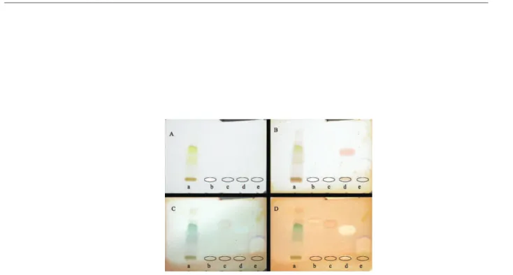
Crude ethylacetate extract and isolated compound austin (2), dihydroaustin (3) and penicillic acid (4) were spotted on a TLC plate. A positive control (Huperzine-A) was also spotted. After elution, the plate was sprayed with acetylcholinesterase solution and incubated at 37 °C. After 20 min, a solution containing α-naphthyl acetate solution and Fast Blue B salt was sprayed onto the plate. Active compounds showed a purple spot just after spraying and it turned white after a few hours (Figure 1S, supplementary material).
RESULTS AND DISCUSSION
Compound 1 that has spontaneously precipitated in some polar fractions was tentatively identiied as D-mannitol. Its 1H NMR spectrum showed signals in the region of δ 3.60-3.83 and only three signals were observed in the 13C NMR spectrum. Sub-spectrum DEPT revealed one of them to be a methylene group. A broad band, typical of hydroxylated compounds was observed in the IR spectrum at 3300 cm-1. These spectroscopic data were consistent with the presence of the symmetrical polyalcohol D-mannitol, considered the most abundant of all the soluble polyalcohols found in fungi.19 It has been reported that D-mannitol inhibits the angiotensin conversion enzyme (ECA) having also anti-hypertensive activity.20 Acyclic polyalcohols like mannitol are usually produced by fungi belonging to the phylos Ascomycota and Basidiomycota.21 Their biological function is still unknown but some authors suggest that these compounds act as energy and carbon source, having also a protecting activity, as for trealose, a compound that is responsible for spores germination when the fungi are under stressing conditions.19,21 Polyalcohols proile has been pointed out as a quimiotaxonomic marker for fungi.19
Schürmann et al.
1046 Quim. Nova
three bands of carbonyl groups. 1H NMR spectrum was typical of terpenoids with a concentration of signals between δH 1.1 and 2.0. A careful analysis of uni- and bidimensional spectra of this com-pound allowed to construct parts of the molecule, including an α,β
unsaturated δ-lactone fragment, which led, together with the other spectroscopic data, to its identiication as austin (2).12
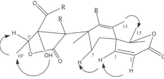
Due to their high structural complexity, NMR data for mero-terpenoids are somehow confusing in the literature. In this way, there is a disagreement between data reported for austin by Hayashi et al.,11 concerning to the chemical shifts of C14 and C15. In our study, there were found spatial correlations in NOESY correlations map among H1 x H2, H1 x H7α, H5’ x H10’, OH x H10’ and H13 x H15 (Figure 2). Based on these correlations, β coniguration was adopted for methyl group at δC 25.7, while Hayashi et al. attributed
α coniguration for the same carbon.
The third compound isolated showed several NMR spectra simi-larities with austin spectra. The methyl group signal at δH 1.65 showed to be coupled to the hydrogen at δH 5.29 (J 6.8Hz). Full NMR data indicated this compound to be dehydroaustin (3), which presents a second exocyclic double bound and formation of an oxygen bridge from C6` hydroxyl group and C9, in relation to austin (Figure 1). De-hydroaustin was irst reported as a metabolite of Aspergillus sp.22, but it has also been reported from Penicillium species.12,14 This is, in the best of our knowledge, the irst time that the meroterpenes austin and dehydroaustin have been isolated from this species. Compound 4 was isolated as a white solid and identiied by 1H and 13C NMR as well as by comparison with the original sample as penicillic acid22 (Figure 1).
In the antibacterial assay by disc difusion, the crude extract (100 µg/mL) was active against all tested bacteria but neither austin (100 µg/mL) nor dehydroaustin (100 µg/mL) presented activity towards any of the tested bacteria. Penicillic acid (100 µg/mL) was active against S. aureus (IZ= 18 mm), L. monocytogenes (IZ= 26 mm), B. cereus (IZ= 15 mm), S. typhimurium (IZ= 15 mm), E. coli (IZ= 15 mm) and C. freundii (IZ= 16 mm); for E. coli, penicillic acid was more active than the control, chloramphenicol, which presented IZ= 13 mm. MIC values were determined penicillic acid that showed MIC value of 512 µg/mL against S. aureus and S. typhimurium and 256 µg/mL against L. monocytogenes, B. cereus, S. typhimurium, E. coli and C. freundii. Crude extract, austin (2), dihydroaustin (3) and penicillic acid (4) were also tested for acetylcholinesterase inhibition by TLC assay (Figure 1S, supplementary material).18 Penicillic acid was again the only active compound, showing a clear inhibition zone. When analyzing the activity of a series of closely related compounds for acethylcholinesterase inhibition, Toda et al.23 reported that structurally lexible compounds would be more active than rigid ones due to the steric hindrance caused by the later in acetylcholinesterase active site. These indings were corroborated by kovarik et al.24 and may be correlated to the inactivity of austin and dehydroaustin. Penicillic acid, a lexible small molecule was, therefore, more likely expected to achieve a suitable conformation to it the enzyme.
SUPPLEMENTARY MATERIAL
TLC proile of the tested material in acetylcholinesterase assay can be found in the supplementary material (Figure 1S), as a pdf ile, at http://quimicanova.sbq.org.br, free of charge.
ACKNOWLEDGMENTS
The authors thank International Foundation for Science (IFS, grant F/3564-2), Fundação de Amparo à Pesquisa do Estado de Minas Gerais (FAPEMIG) for inancial support and Conselho Nacional de Desenvolvimento Cientíico e Tecnológico (CNPq) for a grant to J. A. Takahashi and Universidade Federal de Ouro Preto for a scholarship for B. T. M. Schürmann. We thank G. S. Ataliba and E. L. de Souza for technical assistance and A. A. P. Bracarense for preparing the extract. REFERENCES
1. Tuthill, D. E.; Frisvad, J. C.; Christensen, M.; Mycologia2001, 93, 298. 2. Kang, S. W.; Kim, S. W.; Biotechnol. Lett. 2004, 26, 695.
3. Frisvad, J. C.; Smedsgaard, J.; Larsen, T. O.; Samson, R. A.; Studies in Mycology2004, 49, 201.
4. Aiyatullov, S. S.; Kalinovskii, A. I.; Pivkin, M. V.; Dmitrenok, P. S.; Kuznetsova, T. A.; Chem. Nat. Compd.2004, 40, 615.
5. Fujita, T.; Makishima, D.; Akiyama, K.; Hayashi, H.; Biosci. Biotechnol. Biochem.2002, 66, 1697.
6. Fujita, T.; Hayashi, H.; Biosci. Biotechnol. Biochem.2004, 68, 820. 7. Fill, T. P.; Santos, R. M. G.; Barrisson, A.; Rodrigues-Filho, E.; Souza,
A. Q. L.; Z. Naturforsch. C2009, 64, 355.
8. Takahashi, J. A.; Castro, M. C. M.; Souza, G. G.; Lucas, E. M. F.; Bracarense, A. A. P.; Abreu, L. M.; Marriel, I. E.; Oliveira, M. S.; Floreano, M. B.; Oliveira, T. S.; J. Mycologie Med.2008, 18, 198. 9. Cueto, M.; Macmillan, J. B.; Jensen, P. R.; Fenical, W.; Phytochemistry
2006, 67, 1826.
10. Chexal, K. K.; Springer, J. P.; Clardy, J.; Cole, R. J.; Kirksey, J. W.; Dorner, J. W.; Cutler, H. G.; Strawter, B. J.; J. Am. Chem. Soc.1976, 98, 6748.
11. Hayashi, H.; Mukaihara, M.; Murao, S.; Arai, M.; Lee, A. Y.; Clardy, J.; Biosci., Biotechnol. Biochem. 1994, 58, 334.
12. Santos, R. M. G.; Rodrigues–Filho, E.; Phytochemistry2002, 61, 907. 13. Santos, R. M. G.; Rodrigues–Filho, E.; J. Braz. Chem. Soc. 2003, 14,
722.
14. Santos, R. M. G.; Rodrigues–Filho, E.; Z. Naturforsch.2003, 58, 663. 15. Hagiwara, S.; Takahashi, M.; Shen, Y.; Kaihou, S.; Tomiyama, T.;
Yazawa, M.; Tamai, Y.; Sin, Y.; Kazusaka, A.; Terazawa, M.; Biosci. Biotechnol. Biochem. 2005, 69, 1603.
16. Bauer, S. W.; Kirby, W. M.; Sherris, J. C.; Thurck, M.; Am. J. Pathol. 1966, 45, 493.
17. Takahashi, J. A.; Pereira, C. R.; Pimenta, L. P. S.; Boaventura, M. A. D.; Silva, L. G. F.; Nat. Prod. Lett.2006, 20, 21.
18. Di Giovanni, S. D.; Borloz, A.; Urbain, A.; Marston, A.; Hostettmann, K.; Carrupt, P. A.; Reist, M.; Eur. J. Pharm. Sci.2008, 33, 109. 19. Solomon, P. S.; Waters, O. D. C.; Oliver, R. P.; Trends Microbiol.2007,
15, 257.
20. Feoilova, E. P.; Appl. Biochem. Microbiol.2001, 37, 124.
21. Ramstedt, M.; Jirjis, R.; Soederhaell, K.; New Phytologist 1980, 105, 281.
22. Simpson, T. J.; Stenzel, D. J.; Bartlett, A. J.; O’Brien, E.; Holker, J. S. E.; J. Chem. Soc., Perkin Trans. 11982, 11, 2687.
23. Toda, N.; Iwata, Y.; Tago, K.; Kogen, H.; Kaneko, T.; Miyamoto, S.; Chem-Bio Informatics J. 2003, 3, 46.
24. Kovarik, Z.; Calic, M.; Sinko, G.; Bosak, A.; Arhiv Za Higijenu Rada i Toksikologiju 2007, 58, 201.
Quim. Nova, Vol. 33, No. 5, S1, 2010
Material Suplementar
*e-mail: [email protected]
AUSTIN, DEHYDROAUSTIN AND OTHER METABOLITES FROM Penicillium brasilianum
Betânia T. M. Schürmann
Escola de Farmácia, Universidade Federal de Ouro Preto, Rua Costa Sena, 171, 35400-000 Ouro Preto - MG, Brasil William S. T. Sallum e Jacqueline A. Takahashi*
Departamento de Química, Universidade Federal de Minas Gerais, Av. Antonio Carlos, 6627, 31270-901 Belo Horizonte - MG, Brasil