FARMS IN BRAZIL
Lidiane Roberta Cruz da Silva1*; Odacy Camilo de Souza1; Maria José dos Santos Fernandes1; Débora Maria Massa Lima1; Rosalie Reed Rodrigues Coelho2; Cristina Maria Souza-Motta1
1
Departamento de Micologia, Universidade Federal de Pernambuco, Recife, PE, Brasil; 2Departamento de Microbiologia Geral,
Instituto de Microbiologia, Universidade Federal do Rio de Janeiro, RJ, Brasil.
Submitted: July 01, 2010; Approved: August 26, 2010.
ABSTRACT
Litopenaeus vannamei, which is the most common shrimp species cultivated in the northeast of Brazil, is very
susceptible to microbial diseases, and this consequently affects productivity. There are reports of bacteria,
viruses and protozoa in these shrimp, but not fungi. This study aims to isolate and identify fungi present in
shrimp Litopenaeus vannamei, and in their nursery waters, at two breeding farms in Brazil. The pathogenic
potential of the isolates was assessed through the qualitative detection of proteases and aflatoxin B
production. The 146 isolated fungi comprised 46 species. Aspergillus, Penicillium and Furarium were the
three most relevant genera and Aspergillus flavus was the predominant species with a total of 33 isolates.
Most of the isolated species are known as potentially pathogenic to humans and other animals. Eighteen
isolates of A. flavus and two of A. parasiticus were able to produce aflatoxin B and 33 out of the 46 species
produced protease, indicating that these fungi may also become pathogenic to shrimp and their consumers.
Key words: shrimp, fungi, water, protease, aflatoxin B.
INTRODUCTION
Shrimp grown in captivity are very susceptible to diseases
caused by microorganisms, and this dramatically affects
productivity. Penaideae is a common shrimp family used in
nurseries, and in northeastern Brazil, Litopenaeus vannamei
(Boone) is the most cultivated species (8). Studies investigating
the agents that affect this invertebrate are rare and only a few
viruses, bacteria and protozoa are known to cause disease in
these crustacea (32). No articles dealing with the presence of
fungi in healthy cultivated Litopenaeus vannamei shrimp have
been found in literature. Among the various enzyme groups
produced by fungi, proteinases are a class with an important
role in physiological and biotechnological processes (16).
Some fungi secrete proteinases primarily to provide nutrients to
the cells, however in pathogenic fungi these enzymes facilitate
the adhesion and invasion in host tissue, acquiring a significant
role in the destruction of cell membranes, which are composed,
mainly, of proteins and lipids (15). Here, in particular, they
may also act on shrimp carapace, which consists primarily of
chitin, proteins and lipids (8).
Some species of the Aspergillus genus, especially A.
flavus and A. parasiticus, can produce aflatoxins, mycotoxins
derived from their secondary metabolism, which can
contaminate grains, especially corn, peanut and wheat (5, 38).
There are several types of aflatoxin, but aflatoxin B has the
highest toxicity (17).
On most farms the shrimp are fed with artificial diet or
artificial feed, which is mainly composed of corn grains and
ground wheat with added proteins, vitamins and salts (27).
However, in a farm in northeastern Brazil, shrimp are not fed
with artificial feed but are fed with natural resources such as
algae and small crustaceans, reproducing the natural habitat of
these animals (10), and in this case the diet is called organic
feed.
This study aims to isolate and identify culturable fungi in
Litopenaeus vannamei (Boone) shrimp and in the nursery
waters of two farms in northeast Brazil, one with the artificial
feeding and the other with an organic feeding system.
Furthermore, we screened the ability of the isolated fungi for
proteolytic activity, and in a preliminary test, we detected the
production of aflatoxin by some isolated strains.
MATERIALS AND METHODS
Collection of shrimp and nursery water samples
Adult shrimp showing the characteristics of the species
and without external injuries were collected from nurseries.
Water samples were collected from both adult and juvenile
shrimp nurseries. Two breeding farms were chosen for this
study, one which uses the artificial feeding system (Farm I) and
other which uses the organic feeding system (Farm II). Both
farms are located in Rio Grande do Norte, Brazil. Six shrimp
samples and 36 water samples from both farms were collected
in previously sterilized containers, transported to the
laboratory, and processed on the same day.
Isolation of fungi from shrimp and nursery water
The fungal isolation from shrimp surfaces was based on
the method described by Lacaz et al. (23). Twenty five grams
of whole shrimp were weighed and suspended in 225 ml of
sterile distilled water. After hand shake rotations (two minutes)
0.1 ml of the suspension was seeded in triplicate on the surface
of Sabouraud agar medium plus chloramphenicol (50 mg/L),
contained in Petri dishes. Fungi from nursery waters were
isolated based on on the method described by Ali-Shtayeh et
al.(2), by seeding one ml of each water sample, in triplicate, on
the surface of the same medium. In both cases the plates were
incubated at 28ºC ± 2ºC until fungal growth, approximately 10
days. Then, fragments of fungal colonies were transferred,
separately, to the same medium and incubated under the same
conditions. After confirmation of purity, fungi were transferred
to specific media for species-level identification.
Identification of fungi
For identification of the isolated fungi Czapeck-agar,
potato-dextrose-agar (PDA) and malt extract-agar (23) were
used. Macroscopic (colony color, appearance and size) and
microscopic (somatic and reproductive microstructures)
characteristics were observed in accordance with specific
literature (3, 9, 12, 13, 14, 18, 22, 24, 30, 31, 33, 34, 35).
The Aspergillus genus was studied using Aspergillus
Differential Medium (ADM) (tryptone 15 g, yeast extract 10 g,
ferric citrate 5 g, agar 16g) (6), which differentiates species of
the flavus group. The cultures were incubated for 72 hours at
28ºC ± 2ºC, in the dark, so the presence of yellow-orange to
yellow-olive pigmentation on the back of the colony could
confirm the isolates as belonging to the flavus group.
Detection of the proteolytic activity
Fragments of each fungal culture were transferred to the
center of casein medium (skimmed milk 50 g, agar 10 g and
distilled water 1000 ml), contained in Petri dishes, and
incubated at 28ºC ± 2°C for 10 days. Proteolytic activity was
detected by the formation of a transparent zone around the
clarification of the medium due to acids or alkaline metabolites
present in milk, an acidified solution of mercury chloride
(mercury chloride 12 g, 16 ml concentrated hydrochloric acid
and distilled water 100 ml) was poured into the plates. After 10
minutes, the reduction of the transparent zone indicated that
casein was not digested (23).
Detection of aflatoxin B production
An agar plug (3 mm in diameter) obtained from the
culture growth of each fungus in ADM medium (2-3 days,
28ºC ± 2ºC) was center inoculated in Coconut Agar Medium
(CAM) (“Socôco” Coconut Milk 200 ml, agar 16.0 g, distilled
water 1000 ml, pH 6.9) (25) and incubated for 6-7 days at 28ºC
± 2ºC. The presence of aflatoxin B was detected by observing a
blue to violet fluorescent zone on the back of the growthwhen
subjected to UV light (365nm) in a dark chamber (Tecnal,
model: TE-540).
RESULTS
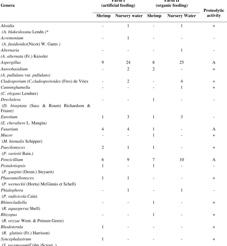
One hundred and forty six fungal isolates were obtained
from adult shrimp (51 isolates) and nursery water (95 isolates),
comprising 20 genera and 46 species (Tables 1, 2 and 3).
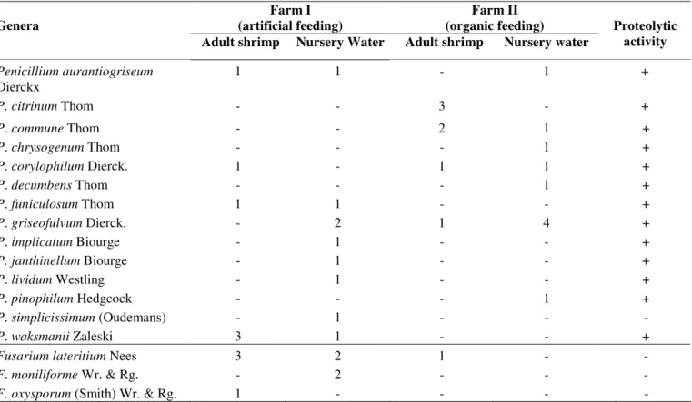
Aspergillus (Table 2) and Penicillium (Table 3) were the most
common genera, comprising 12 and 14 species, respectively. In
addition, three different species of the Fusarium genus were
also isolated (Table 3). These three genera comprised 73% of
total isolates. All other 17 genera were isolated as a single
species (Table 1).
Aspergillus flavus was the predominant species and was
present in all samples, i.e., in adult shrimp and in nursery
waters on both farms, comprising 33 isolates (Table 2). Three
other species were also relevant: Aspergillus parasiticus, with
seven isolates, and Aspergillus fumigatus and Penicillium
griseofulvium, with six isolates each. Species with four (A.
niger, A. ochareceus, and P. waksmanii) three (A. terreus, P.
aurantiogriseum, P. citrinum, P. commune and P.
corylophilum) and two isolates (A. japonicus, A. oryzae, A.
sydowii and P. finiculosum) were also obtained (Tables 2 and
3).
When casein was used as substrate, proteinase was
detected in 33 of the 46 isolated species, comprising 72% of
proteolytic strains (Tables 1, 2 and 3). Considering the 26 most
prevalent species, belonging to the genera Aspergillus and
Penicillium, only A. fumigatus, A. japonicus and P.
simplicissimum were not capable of producing proteinase under
these conditions (Table 2 and 3). The three Fusarium species
isolated didn’t show any hydrolysis zone in the presence of
casein either (Table 3).
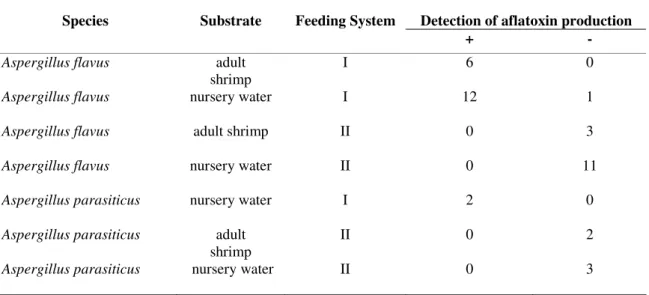
The 33 Aspergillus flavus and seven A. parasiticus isolates
were tested for their ability to produce aflatoxin B. Twenty
were positive, 18 (55%) belonging to A. flavus and 2 (28%) to
A. parasiticus (Table 4). It is worth noting that only one isolate,
out of the 18 Aspergillus flavus obtained from Farm I, which
uses artificial feed, did not produced aflatoxin. Furthermore,
the 15 Aspergillus flavus isolates obtained from shrimp and/or
nursery water from the farm that uses organic feed (Farm II)
showed no toxin production. The same trend occurred with A.
parasiticus, although the number of tested isolates was much
lower: the two isolates obtained from Farm I nursery waters
showed aflatoxin production, while all five isolates obtained
from Farm II nursery waters or shrimp did not show any
aflatoxin production.
After identification and detection of proteinase production
a representative of each isolated species, which retained its
sporulation ability, was preserved in the URM Micoteca
Culture Collection of the Federal University of Pernambuco,
Table 1. Occurrence of different fungi genera isolated from shrimp and nursery waters from two farms, one with an artificial feeding
system (I) and the other with an organic feeding system (II) and their proteolytic activity. (*) In parentheses the sole isolated species of
the genus.
Farm I (artificial feeding)
Farm II (organic feeding) Genera
Shrimp Nursery water Shrimp Nursery Water
Proteolytic activity
Absidia
(A. blakesleeana Lendn.)*
- 1 - 1 +
Acremonium
(A. fusidioides(Nicot) W. Gams )
- 1 - - -
Alternaria
(A. alternata (Fr.) Keissler
- - - 1 -
Aspergillus 9 24 8 25 A
Aureobasidium
(A. pullulans var. pullulans)
- 2 2 - +
Cladosporium (C.cladosporioides (Fres) de Vries - 2 - 4 +
Cunninghamella
(C. elegans Lendner)
- - - 1 +
Drechslera
(D. biseptata (Sacc & Roum) Richardson &
Frazer)
- - 1 - -
Eurotium
(E. chevaliere L. Mangin)
1 3 1 3 -
Fusarium 4 4 1 - A
Mucor
(M. hiemalis Schipper)
- - 1 - +
Paecilomyces
(P. variotii Bain.)
2 1 1 - +
Pencicillium 6 9 7 10 A
Pestalotiopsis
(P. guepini (Desm.) Steyaert)
1 - 1 - -
Phaeoanellomyces
(P. werneckii (Horta) McGinnis et Schell)
1 1 - - +
Phialophora
(P. radicicola Cain)
- 1 - 1 -
Rhinocladiella
(R. aquaspersa Shell)
- - 1 - +
Rhizopus
(R. oryzae Went. & Prinsen Geere)
- - 1 - +
Rhodotorula
(R. glutinis (Fr.) Harrison)
1 - - - +
Syncephalastrum
(S. racemosum(Cohn.)Scroet. )
1 - - - +
Table 2. Occurrence of different species of Aspergillus isolated from shrimps and nursery water from farms with artificial feeding
system (I) and organic feeding system (II) and its proteolytic activity.
Table 3. Occurrence of different species of Penicillium and Fusarium isolated from shrimp and nursery waters from two farms, one with an artificial feeding system (I) and the other with an organic feeding system (II) and their proteolytic activity
Farm I (artificial feeding)
Farm II (organic feeding) Genera
Adult shrimp Nursery Water Adult shrimp Nursery water
Proteolytic activity
Penicillium aurantiogriseum
Dierckx
1 1 - 1 +
P. citrinum Thom - - 3 - +
P. commune Thom - - 2 1 +
P. chrysogenum Thom - - - 1 +
P. corylophilum Dierck. 1 - 1 1 +
P. decumbens Thom - - - 1 +
P. funiculosum Thom 1 1 - - +
P. griseofulvum Dierck. - 2 1 4 +
P. implicatum Biourge - 1 - - +
P. janthinellum Biourge - 1 - - +
P. lividum Westling - 1 - - +
P. pinophilum Hedgcock - - - 1 +
P. simplicissimum (Oudemans) - 1 - - -
P. waksmanii Zaleski 3 1 - - +
Fusarium lateritium Nees 3 2 1 - -
F. moniliforme Wr. & Rg. - 2 - - -
F. oxysporum (Smith) Wr. & Rg. 1 - - - -
Farm I (artificial feeding)
Farm II (organic feeding)
Genera Shrimp Nursery water Shrimp Nursery water
Proteolytic activity
Aspergillus caespitosus Raper & Thom - - - 1 -
A. flavipes (Bain. and Sart.)Thom - - 1 - +
A. flavus Link 6 13 3 11 +
A. fumigatus Fresenius - 3 - 3 -
A. japonicus Saito - 1 - 1 -
A. niger V. Thieg. 1 1 - 2 +
A. niveus Blochwitz - - 1 +
A. ochraceus Wilhelm - 2 1 1 +
A. oryzae (Ahlb.) Cohn 1 1 - - +
A. parasiticus Speare - 2 2 3 +
A. sydowii (Bain. & Start.)Thom and Church - 1 - 1 +
Table 4. Detection of aflatoxin production by Aspergillus flavus and Aspergillus parasiticus isolated from adult shrimp and
nursery waters from two farms, one with an artificial feeding system (I) and the other with an organic feeding system (II).
Detection of aflatoxin production
Species Substrate Feeding System
+ -
Aspergillus flavus adult
shrimp
I 6 0
Aspergillus flavus nursery water I 12 1
Aspergillus flavus adult shrimp II 0 3
Aspergillus flavus nursery water II 0 11
Aspergillus parasiticus nursery water I 2 0
Aspergillus parasiticus adult
shrimp
II 0 2
Aspergillus parasiticus nursery water II 0 3
DISCUSSION
In the present study 20 genera and 46 species of culturable
fungi were identified in Litopenaeus vannamei shrimp and their
cultivation waters at two farms in Brazil. The three most
relevant genera were Aspergillus, Penicillium and Furarium.
There are several reports on the diversity of viruses (37),
bacteria (11, 28) and protozoa (20) that affect Litopenaeus
vannamei grown in nurseries, however papers dealing with the
isolation of conidial fungi, Ascomycota in healthy shrimp of
this species, or water used for their cultivation, have not been
reported so far. As to other shrimp species, Colorni (7) reported
a fungal infection in Penaeus semisulcatus, cultivated in Israel.
The author observed a large lesion in the muscle of a specimen
of the shrimp, from which the fungus Fusarium solani was
isolated, and concluded that the handling of infected shrimp
may represent risk to farmers. Also Khoa et al. (21) isolated
Fusarium incarnatum from gill lesions of the black tiger
shrimp Penaeus monodon, grown in Vietnam, and found these
shrimp to present high mortality. In our study Fusarium species
were also detected, but in healthy specimens (F. lateritium,
F.oxysporum), or in adult nursery water (F. lateritium and F.
moniliforme).
Most of the species isolated in the present research have
been reported as mycoses agents in humans, causing
otomycosis, keratitis, skin infections, lung infections and even
systemic infections. These are opportunistic fungi, which affect
only humans and other animals that have some type of immune
suppression, or are immunocompromised (18). Three
pathogens well known to man may be cited,
Phaeoannellomyces werneckii, which causes Tinea nigra,
Syncephalastrum racemosum, responsible for skin infection
and Rhinocladiella aquaspersa, which is the etiologic agent of
chromoblastomycosis (18, 23). Although many of the isolated
species are potential pathogens, it must be stressed that these
environmental isolates might miss the genes responsible for
pathogenesis when in contact with the host.
Aspergillus flavus was the most prevalent specie,
comprising 22% of all isolates. A. fumigatus, A. niger and A.
of pulmonary aspergillosis. Furthermore, A. flavus is known as
the most powerful fungal species able to produce aflatoxins,
which are mycotoxins with carcinogenic potential (23). In this
study all 18 isolates of A. flavus and two of A. parasiticus
which produced aflatoxin B were isolated from the farm where
shrimp are feed with artificial feed, in contrast to the isolates
obtained from the farm with organic feeding system, where
none of them were able to produce this mycotoxin. Although
the number of the samples is small, and therefore a statistical
study could not be performed, our results indicate that,
possibly, the artificial feed in the nurseries potentiates aflatoxin
B production by these fungi, contaminating the shrimp, and
therefore impairing their production, and also transferring the
toxin to the consumers. Bintvihok et al. (4) analyzed 150
samples of black tiger shrimp Penaeus monodon in Thailand,
fed with feed contaminated with aflatoxin B, and concluded
that this type of system may reduce production, causing
economic losses.
In our study 33 out of the 46 isolated species were able to
degrade casein, comprising 72% of proteolytic strains. This
number may be higher, if one considers that different isolates
of the same species can present different proteolytic
characteristics. Although casein proteolysis is not a direct
indication of pathogenesis to shrimp and their consumers, it is
well known that proteinases are important enzymes for both,
fungal growth and host tissue invasion (23). Also Horng-Der et
al. confirmed the pathogenic nature of this enzyme to trigger
allergic processes when they analyzed the effects of the
production of serine protease by Penicillium chrysogenum and
Aspergillus fumigatus in mechanisms of allergic diseases in
humans (19).
It is important to emphasize that this is the first report of
the isolation of conidial fungi and Ascomycota in shrimp
cultivation water, and that there are few reports of diseases in
shrimp caused by fungi. Furthermore, as far as we know, there
is no description in literature about the diversity of species of
culturable fungi present in healthy cultivated shrimp
Litopenaeus vannamei.
So, in conclusion, the results of the present study
demonstrate a significant diversity of culturable fungi. Most of
the isolated species were obtained from adult shrimp artificially
cultivated and from their nursery waters, and they presented
aflatoxin production and proteolytic activity, suggesting that
they may possibly be pathogens for both, the shrimp and the
consumers.
ACKNOWLEDGMENTS
This work was supported by CNPq. The authors are
grateful to Nogueira, E.B.S. and Torres, K.B. for technical
support.
REFERENCES
1. Alexopoulos, C.J.; Mims, C.W.; Blackwell, M. (1996). Introductory mycology. John Wiley & Sons, Inc, New York.
2. Ali-shtayed; Khaleel, T.K.M.; Jamous, R.M.F. (2003). Ecology of dermatophytes and other keratinophilic fungi in swimming pools and polluted and unpolluted streams. Mycopathologia. 156 (3), 51- 59. 3. Barnett, J.A.; Payne, R.W.; Yarrow, D. (2000). Yeasts characteristics
and identification. Cambridge University Press, Spain.
4. Bintvihok, A.; Ponpornpisit, A.; Tangtrongpiros, J.; Panichkriangkrai, W.; Rattanapanee, R.; Doi, K.; Kumagai, S. (2003). Aflatoxin contamination in shrimp feed and effects of aflatoxin addition to feed on shrimp production. J. Food Prot, 66 (5), 882- 888.
5. Bok, J.W.; Keller, N.P.; Lae, A. (2004). A regulator of secondary metabolism in Aspergillus spp. Euk Cell. 3, 527- 535.
6. Bothast, R.J.; Fennel, D.I. (1974). A medium for rapid identification and enumeration of Aspergillus flavus and related organisms. Mycologia. 66, 365-369.
7. Calorni, A. (1989). Fusariosis in the shrimp Penaeus semisulcatus
cultured in Israel. Mycophatologia. 108, 145- 147.
8. Campos, A.A.B.; Maia, E.P.; Costa, W.M.; Brito, L.O.; Oliveira, A. (2007). Descrição dos principais grupos fitoplanctônicos do afluente e efluente em fazenda de criação do camarão marinho Litopenaeus vannamei com sistema de recirculação parcial de água. B. Inst. Pesca. 33, 113- 119.
9. Carmichael, J.W.; Kendrick, W.B.; Conners, I.L.; Singler, L. (1980).
10. Chellapa, N.T.; Lima, A.K.A.; Câmara, F.R.A. (2007). Riqueza de microalgas em viveiros de cultivo orgânico de camarão em Tibau do Sul, Rio Grande do Norte. Rev. Bras. Bioc. 5 (2), 120- 122.
11. Cheng, W.; Wang, L.; Chen, J. (2005). Effect of water temperature on the immune response of with shrimp Litopenaeus vannamei to Vibrio alginolyticus. Aquaculture. 250, 592- 601.
12. Domsch, K. H.; Gams, W.; Anderson, T. (1993). Compendium of soil fungiv. I. IHW – Verlag, San Francisco.
13. Ellis, M.B. (1971). Dematiaceous hyphomycetes. Commonwealth Mycological Institute, Kew.
14. Ellis, M.B. (1976). More dematiaceous Hyphomycetes. Commonwealth Mycological Institute, Kew.
15. Ghannom, M.A. (2000). Potential role of phospholipase in virulence and fungal pathogenesis. Clin. Microbiol. Rev. 13, 122- 143.
16. Gouka, R.J.; Punt, P.J.; van den Hondel, C.A.M.J.J. (1997). Efficient production of secreted proteins by Aspergillus progress, limitations and prospects. App. Microbiol. Biotechnol. 47, 1- 11.
17. Groopman, J.D.; Cain, L.G.; Kensler, T.W. (1992). Exposure in human populations: measurements and relationship to cancer. CRC Crit. Rev. Toxicol. 19, 113-145.
18. Hoog, G.S; Guarro, J.; Gene, J. (2000). Atlas of clinical fungi. Centraalbureua voor Schimmelcultures, 2nd Edition., Utrecht, The Netherlands.
19. Horng-Der, S.; Ming, F.T.; Ren-Bin, T.; Hong, C. (2007). Aspergillus
and Penicillium allergens: focus on proteases. Curr. Allergy Asthma Rep. 7, 351- 356.
20. Jiménez, R.; Barniol, L.; Machuca, M. (2002). Nematopsis marinus n. sp., a new septate gregarine from cultured penaeoid shrimp Litopenaeus vannamei (Boone), in Ecuador. Aquaculture res. 33 (4), 231- 240. 21. Khoa, L.V.; Hatai, K.; Aoki, T. (2004). Fusarium incarnatum isolated
from black tiger shrimp, Penaeus monodon Fabricius, with black gill disease cultured in Vietnam. J. Fish. Disease. 27, 507- 515.
22. Klick, M.A. (2002). Identification of Common Aspergillus Species.
Centraalbureau voor Schimmelcultures, Utrecht,. the Netherlands. 23. Lacaz, C.S.; Porto, E.; Martins, J.C; Heins-Vaccari, E.M.; Melo, N.T.
(2002). Tratado de micologia médica. Sarvier, São Paulo.
24. Leslie, J.F.; Summerell, B.A. (2006). The Fusarium laboratory manual. Blackwell Publishing, Malden MA, USA.
25. Lin, M.T.; Dianese, J.C. (1976). A coconut-agar medium for rapid detection of aflatoxin production by Aspergillus spp. Phytopathology. 66, 1466- 1469.
26. Mangiarotti, A.M.; Careta, G. (1984). Keratinophilic fungi isolated from a small pool. Mycopathologia. 85, 9- 11.
27. Mendes, P.P.; Albuquerque, M.L.L.T.; Queiroz, D.M.; Santos, B.L.S.; Lima, A.C.; Lopes, Y.V.A. (2006). Aclimatação do camarão marinho
Litopenaeus vannamei (Boone, 1931) à água doce com diferentes estratégias de alimentação e calagem. Acta Sci. Anim. Sci. 28 (1), 89- 95. 28. Moss, S.M.; LeaMaster, B.R.; Sweeney, J.N. (2007). Relative abundance and species composition of Gram-negative, aerobic bacteria associated with the gut of juvenile white shrimp Litopenaeus vannamei reared in oligotrophic well water and eutrophic nursery water. J. World Aquac. Soc. 31 (2), 225- 263
29. Pinheiro, W.C.; Filho, J.A.; Maracajá, P.B. (2007). Efeitos climáticos e físico-químicos sobre a biologia do Litopenaeus vannamei cultivado em viveiro. Rev. Verde. 2, 142- 150.
30. Pitt, J.I. (1988). A laboratory Guide to Common Penicillium Species. Commonwealth scientific and industrial research organization – Division of food processing, North Wales.
31. Pitt, J.I. (1991). A laboratory Guide to Common Penicillium Species. Commonwealth scientific and industrial research organization – Division of food processing, North Wales.
32. Pontes, C. S.; Arruda, M. F. (2005). Comportamento de Litopenaeus vannamei (Boone) (Crustacea, Decapoda, Penaeidae) em função da oferta do alimento artificial nas fases clara e escura do período de 24 horas. Rev. Bras. Zool. 22, 648- 652.
33. Raper, K.B.; Fennel, D.I. (1977). The genus Aspergillus. Robert & Krieger, Florida.
34. Riddell, R.W. (1950). Permanent stained mycological preparation obtained by slide culture. Mycologia. 42, 265- 270.
35. Samson, R.A.; Frisvad, J.C. (2004). Penicillium subgenus Penicillium: new taxonomics schemes, mycotoxins and other extrolites. Stud. Mycol. 49, 1- 266.
36. Shakeri, J.; Foster, H.A. (2006). Proteolytic activity and antibiotic production by Trichoderma harzianum in relation to pathogenicity to insects. Enz. Microb.Technol. 40 (4), 961- 968.
37. Vidal, O.M.; Granja, C.B.; Aranguren, F.; Brock, J.A.; Salazar, M. (2007). A profound effect of hyperthermia on survival of Litopenaeus vannamei juveniles infected with White Spot Syndrome Virus. J. World Aquac. Soc. 32 (4), 364-372.
38. Yu, J.; Cleveland, T.E.; Nierman, W.C.; Bennett, J.W. (2005).
Aspergillus flavus genomics: gateway to human and animal health, food safety, and crop resistance to diseases. Rev. Iberoam. Micol. 22, 194- 202.