Case 8427
A placental site trophoblastic tumour
Marques V, Cunha TM, Coutinho S, Oliveira P
Genital (Female) Imaging Section:
2010, May. 18 Published:
33 year(s), female Patient:
Clinical History
A 33-year-old woman with amenorrhoea since her last pregnancy 13 months earlier.
Imaging Findings
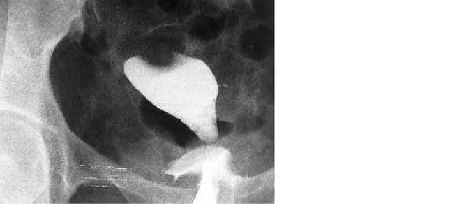
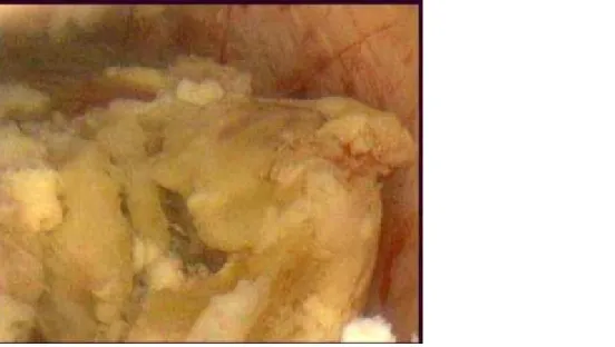
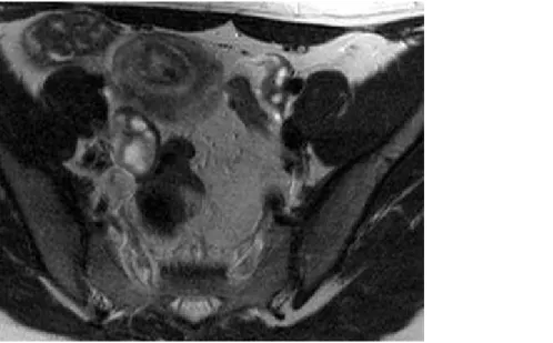
A 33-year-old woman (gravida 2, para 2) presented with 13-month amenorrhoea since her last pregnancy. The hysterosalpyngography showed a regular filling defect in the uterine fundus, interpreted as a leyomyoma (Fig. 1). Transvaginal ultrasound (TVUS) revealed a 3 cm submucous tumour in the uterine fundus (Fig. 2). The hysteroscopy revealed a soft, necrotic-looking mass, occupying most of the posterior endometrial cavity (Fig. 3). Multiple biopsies were undertaken. Histology revealed intermediate trophoblastic tissue with myometrial invasion, raising the possibility of a placental site trophoblastic tumour (Fig. 4). The serum HCG level was 7, 8 UI/L (N<5). A magnetic resonance (MR) examination was performed. A heterogeneous tumour, submucous in location, in the right fundal region, was detected, superficially invading the
myometrium 5 mm. On contrast-enhanced T1-weighted images there was little central enhancement of the mass relative to the surrounding myometrium (Figs. 5 a, b, c). On T2-weighted MR images the mass had peripheral low signal with central high signal intensity relative to normal
Discussion
Placental site trophoblastic tumour (PSTT) is the rarest variant of Gestational trophoblastic disease (GTD).
GTD always derives from an abnormal fertilisation and, although these tumours represent less than 1% of gynaecological malignancies, it is important to know their natural history and approach, not only because they can threat the lives of fertile women, but also for their high curability if treated on time.
GTD can be defined as a group of gestational and neoplastic conditions derived from the trophoblast, and is usually subdivided, according to the International Classification of Diseases (ICD), in molar gestations [complete hydatidiform mole (CHM), partial hydatidiform mole (PHM) and invasive mole (IM)] and trophoblastic tumours [choriocarcinoma, placental site trophoblastic tumour (PSTT) and epithelioid trophoblastic tumour). The trophoblast begins as the outer covering of the early blastocyst and provides the nutrition path between the maternal endometrium and the developing embryo. It will become the placental functional unit. It is formed by three components: cytotrophoblast, syncytiotrohoblast and the intermediate trophoblast. A common characteristic of all GTD is the abnormal proliferation of trophoblast, but different components predominate in different tumours.
PSTT can develop after a full-term delivery, a non-molar abortion or a molar gestation. Time to presentation varies between 1 week and 14 years, usually through abnormal vaginal bleeding. Their biological behaviour is variable, ranging from benign lesions confined to the uterus, to highly aggressive malignant disease with systemic metastases. The main negative prognostic variables are time to presentation from last known pregnancy and mitotic index. Relative to other forms of GTD, in PSTT the levels of serum HCG are usually lower (due to lack of syncytiotrophoblast
proliferation).
The radiologic manifestations vary, including solid and cystic lesions, with or without a central component, that usually invade the myometrial wall. Because of this, PSTT can have an appearance similar to choriocarcinoma or invasive mole. On MR, the myometrial mass is isointense to the normal myometrium on T1-weighted images and isointense to slightly hyperintense on T2-weighted images. Endometrial masses are heterogeneous. The junctional zone is disrupted. In the majority of the cases there are cystic spaces and vascular structures.
The treatment is surgical (total abdominal hysterectomy with or without bilateral oophorectomy) due to relative resistance to chemotherapy. Accurate localisation by MR may allow hysterotomy to be performed, preserving the uterus for future pregnancies. Unfortunately, 30% of the patients with PSTT present with metastases at the time of diagnosis.
Final Diagnosis
Placental site trophoblastic tumor.
Figures
Filling defect with a regular contour in the uterine fundus.
Figure 2 Transvaginal ultrasound (TVUS)
Submucous fundal tumour isoechogenic to myometrium. We can detect a small amount of
fluid in the uterine cavity.
Soft, necrotic mass occupying most of the posterior uterine wall.
Figure 4 Histology of the biopsies done during hysteroscopy
Intermediate trophoblastic tissue with myometrial invasion.
Figure 5 MR examination
Axial contrast-enhanced fat-sat T1. On contrast enhanced T1-weighted images there was
sparse central enhancement of the mass relative to the surrounding myometrium. The tumour
superficially invaded the myometrium (arrow).
Axial T2. On T2-weighted images the mass has peripheral low signal with a central high
signal intensity area relative to normal myometrium.
Sagittal T2. On T2-weighted images the mass has peripheral low signal with a central high
signal intensity area relative to normal myometrium.
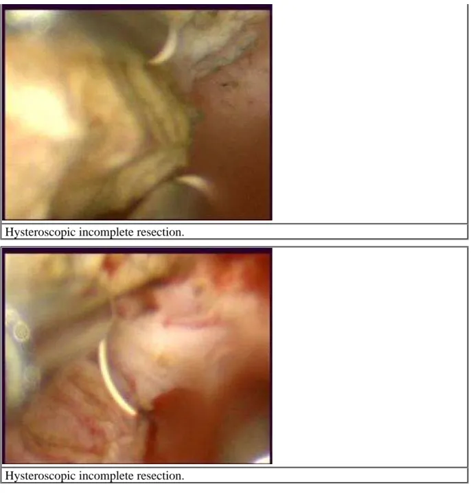
Hysteroscopic incomplete resection.
Hysteroscopic incomplete resection.
Figure 7 Surgical specimen of hysterectomy
We can see an haemorragic area in the uterine fundus related to previous resection. There is
no evidence of macroscopic tumour.
MeSH
[C04.850.908.416] Gestational Trophoblastic Neoplasms
A group of interrelated trophoblastic diseases arising from pregnancy. They are commonly associated with hyperplasia of trophoblasts (TROPHOBLAST) and markedly elevated human CHORIONIC GONADOTROPIN. They include HYDATIDIFORM MOLE, invasive mole (HYDATIDIFORM MOLE, INVASIVE), placental-site trophoblastic tumor (TROPHOBLASTIC TUMOR, PLACENTAL SITE), and CHORIOCARCINOMA. These neoplasms have varying propensities for invasion and spread.
[C04.850.908.416.186.875] Trophoblastic Tumor, Placental Site
An uncommon variant of CHORIOCARCINOMA. It is composed almost entirely of mononuclear cytotrophoblasts (TROPHOBLASTS). Because its secretion of hCG (CHORIONIC
GONADOTROPIN) is low, a large tumor may develop before the hCG can be detected.
References
[1] Wagner BJ, Woodward PJ, Dickey GE (1996) From the Archives of AFIP. Gestational Trophoblastic Disease: Radiologic-Pathologic Correlation Radiographics 16:131-48
[2] Machtinger R, et al (2005) Placental site trophoblastic tumor: outcome of five cases including fertility preserving management Gynecologic Oncology 96:56-61
[3] Vitellas KM, et al (2000) case of the day Case 2: Placental site trophoblastic tumor AJR 175:898-900
[4] Brandt KR, Coakley KJ (1998) MR appearance of placental site trophoblastic tumor: a report of three cases AJR 170:485-7
[5] Elsayes KM, et al (2009) Imaging the placenta: a multimodality pictorial review Radiographics 29:1371-91
[7] Leyendecker JR, et al (2004) MR imaging of maternal diseases of the abdomen and pelvis during pregnancy and the immediate postpartum period Radiographics 24:1301-16. Review
[8] Nagayama M, et al (2002) Fast MR imaging in obstetrics Radiographics 22(3):563-82. Review
Citation
Marques V, Cunha TM, Coutinho S, Oliveira P (2010, May. 18) A placental site trophoblastic tumour {Online}