396
Bastos AL et al. Pulmonary artery aneurysms in Behçet’s disease
Radiol Bras. 2011 Nov/Dez;44(6):396–398
Pulmonary artery aneurysms in Behçet’s disease: case
report
*
Aneurismas da artéria pulmonar na doença de Behçet: relato de caso
Andréa de Lima Bastos1, Isabela Lage Alves de Brito2
Behçet’s disease is an inflammatory disease that may involve the chest, manifesting itself by the presence of pulmonary artery aneurysms. The authors report a case of Behçet’s disease where findings at chest radiography and computed tomography have suggested the possibility of the diagnosis.
Keywords: Behçet’s disease; Radiology; Vasculitis.
A doença de Behçet é uma doença inflamatória que pode envolver o tórax manifestando-se pela presença de aneuris-mas das artérias pulmonares. Relatamos um caso de doença de Behçet cujas alterações observadas em radiografias e tomografia computadorizada do tórax sugeriram a possibilidade do diagnóstico.
Unitermos: Doença de Behçet; Radiologia; Vasculite.
Abstract
Resumo
* Study developed at Hospital Júlia Kubitschek – FHEMIG, Belo Horizonte, MG, Brazil.
1. Master, Fellow PhD degree in Health Sciences, Physician Assistant at Diagnostic Imaging Service, Hospital Júlia Kubits-chek – FHEMIG, Belo Horizonte, MG, Brazil.
2. MD, Specialist in Pneumology, Physician Assistant at Pneu-mology Service, Hospital Júlia Kubitschek – FHEMIG, Belo Hori-zonte, MG, Brazil.
Mailing Address: Dra. Andréa de Lima Bastos. Hospital Júlia Kubitschek – Fundação Hospitalar do Estado de Minas Gerais (FHEMIG), Unidade de Diagnóstico por Imagem. Rua Doutor Cris-tiano Rezende, 2745, Bairro Flávio Marques Lisboa (Barreiro). Belo Horizonte, MG, Brazil, 30620-470. E-mail: andblima@ yahoo.com.br
Received February 24, 2011. Accepted after revision June 21, 2011.
Bastos AL, Brito ILA. Pulmonary artery aneurysms in Behçet’s disease: case report. Radiol Bras. 2011 Nov/Dez;44(6):396–398.
0100-3984 © Colégio Brasileiro de Radiologia e Diagnóstico por Imagem CASE REPORT
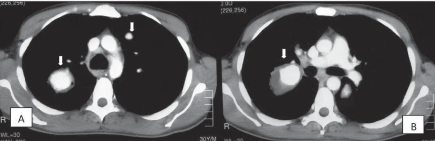
with corticoid and immunosuppressive drugs (Figure 1). Chest computed tomog-raphy, before and after intravenous admin-istration of iodinated contrast medium, demonstrated aneurysmal dilatation of the right interlobar artery, measuring approxi-mately 50 mm in its greater axial axis, with diffuse contrast enhancement of the lu-men, and with the presence remarkable parietal thickening (Figure 2). Other smaller aneurysmal formations with simi-lar morphological characteristics were bi-laterally observed, presenting varied sizes and predominance in central regions and in the upper and middle thirds of the lungs (Figures 1 and 3). Anatomical alterations were not observed in the mediastinal re-gion.
The diagnosis of Behçet’s disease was established on the basis of the presence of oral and recurrent genital ulcers in associa-tion with acneiform and cicatricial skin lesions.
DISCUSSION
According to the classification devel-oped by the International Study Group for Behçet’s Disease, the presence of recurrent oral ulcers and any two of the following alterations – genital ulcers, typical ocular lesions (uveitis, retinal vasculitis), charac-teristic skin lesions (nodule-like erythema, acneiform and/or cicatricial lesions) –, or Behçet’s disease, this is the greatest cause
of mortality related to the disease(4,5).
The chest involvement manifests as sev-eral findings that may be demonstrated by imaging studies. The knowledge on such findings is useful in the disease diagnosis. The present report is aimed at describ-ing the radiological and clinical finddescrib-ings observed in a case of Behçet’s disease. This paper was approved by the Research Eth-ics Committee of the authors’ institution (opinion CEP/FHEMIG: 069/2010).
CASE REPORT
A male, 30-year-old patient was referred to the authors’ institution because of find-ings observed at chest radiography, for spe-cialized diagnosis. The patient reported the onset of dry cough, weight loss, fever, hyporexia, adynamia and some episodes of hemoptysis for two months. Later, he pre-sented progressive dyspnea. Additionally, he reported the presence of recurrent oral and genital ulcerations since his adoles-cence. At clinical examination, acneiform nodules and cicatricial lesions were ob-served on the patient’s skin surface.
Posteroanterior chest radiography dem-onstrated the presence of a well defined mass with soft tissue density and regular contour in the region of the right pulmo-nary hilum, whose dimensions were sig-nificantly reduced after specific treatment INTRODUCTION
Behçet’s syndrome is an inflammatory, chronic and systemic disease whose etiol-ogy remains unknown, with higher inci-dence in men aged between 30 and 40 years(1,2). This disease is frequently found
in Mediterranean countries and in Asia, with highest prevalence in Turkey, and was characterized by Hulusi Behçet through the triad: recurrent oral, genital ulcerations and uveitis.
Manifestations in several other organs have been described, with vasculitis being the main pathological finding(1). The
dis-ease diagnosis is based on the criteria pro-posed by the International Study Group for Behçet’s Disease(3). The prognosis is
deter-mined by the involvement of the cardiovas-cular, gastrointestinal and central nervous systems(2). Although the vascular
397
Bastos AL et al. Pulmonary artery aneurysms in Behçet’s disease
Radiol Bras. 2011 Nov/Dez;44(6):396–398
Figure 1. Posteroanterior chest radiographs demonstrating a hilar mass at right (arrow on A), and the mass with decreased dimensions after treatment (arrow on B).
Figure 3. Contrast-enhanced chest computed tomography, on mediastinal window, at the level of the upper lung lobes (A) and of the bronchial bifurcation (B), demonstrating aneurysmal dilatation of the right interlobar artery and of segmental branches at left (arrows).
398
Bastos AL et al. Pulmonary artery aneurysms in Behçet’s disease
Radiol Bras. 2011 Nov/Dez;44(6):396–398
a positive pathergy test define the diagno-sis of Behçet’s disease(3).
Chest involvement is observed in 8% of patients and may manifest through a wide range of alterations in the mediastinum, leading to complications such as fibrosing mediastinitis; in the pulmonary parenchyma, where infarction, atelectasis, hemorrhage, diffuse airspace nodules, pneumonia and fibrosis may be observed; in the pleura and mainly in the vascular system(4–6).
Pulmonary vasculitis observed in Behçet’s disease compromises great and medium caliber vessels (according to the Chapel Hill classification criteria for vas-culitis), most frequently affecting the venous system in 85% of cases, in the form of thrombophlebitis(4,6). The main venous
complications in Behçet’s disease are su-perior vena cava syndrome and Budd-Chiari syndrome(4). Arterial alterations are
less frequently observed. The destruction of elastic fibers caused by the inflammatory process of the vasa vasorum results in vas-cular lumen dilatation and development of aneurysms in 65% of cases(4). Behçet’s
dis-ease is the main cause o pulmonary artery aneurysms which may occur either in the main segments or in segmental branches, and that can be seen at computed
tomog-raphy, indicating a poor prognosis(4,5). Such
aneurysms may regress with specific me-dicamentous therapy utilizing corticoid and cyclophosphamide, but disease progression and recurrence are commonly observed in such patients(4,7).
In this case, the main differential diag-nosis is Hughes-Stovin syndrome that pre-sents quite similar radiological and histo-pathological findings; in this syndrome, however, the absence of oral and genital ulcerations is observed (4,6).
Chest radiography is the primary imag-ing method in the evaluation of Behçet’s disease, demonstrating vascular and pul-monary alterations. Such a method is also important in the evaluation of the therapeu-tic response(4,5). Computed tomography
plays a relevant role in the imaging diag-nosis of Behçet’s disease, particularly through the multidetector technology and therefore the study of vascular, mediastinal and parenchymal structures. Computed tomography angiography performed with such a technology is superior to magnetic resonance imaging, because of its greater spatial resolution, less imaging artifacts, besides the possibility of simultaneous evaluation of the pulmonary parenchyma
(4,5). The knowledge on the alterations
re-lated to Behçet’s disease is useful in the diagnosis, aiding in the determination of the cause of symptoms, particularly hemop-tysis, and guiding in the choice of the ap-propriate treatment.
CONCLUSION
The presence of pulmonary artery aneu-rysms suggests the diagnosis of Behçet’s disease as a primary hypothesis, besides being a sign of poor prognosis.
REFERENCES
1. Erkan F, Gül A, Tasali E. Pulmonary manifestations of Behçet’s disease. Thorax. 2001;56:572–8. 2. Iscan ZH, Vural KM, Bayazit M. Compelling
na-ture of arterial manifestations in Behcet disease. J Vasc Surg. 2005;41:53–8.
3. Criteria for diagnosis of Behçet’s disease. Interna-tional Study Group for Behçet’s Disease. Lancet. 1990;335:1078–80.
4. Ceylan N, Bayraktaroglu S, Erturk SM, et al. Pul-monary and vascular manifestations of Behcet dis-ease: image findings. AJR Am J Roentgenol. 2010; 194:158–64.
5. Hiller N, Lieberman S, Chajek-Shaul T, et al. Tho-racic manifestations of Behçet disease at CT. Radiographics. 2004;24:801–8.