Fusarium Head Blight Resistance in Wheat
Xianghui Zhang1,4, Jianming Fu2, Yasuaki Hiromasa3, Hongyu Pan1, Guihua Bai2*
1Jinlin University, Changchun, Jilin, People’s Republic of China,2United States Department of Agriculture/Agricultural Service, Hard Winter Wheat Genetics Research Unit, Kansas State University, Manhattan, Kansas, United States of America,3Department of Biochemistry, Kansas State University, Manhattan, Kansas, United States of America,4Department of Plant Pathology, Kansas State University, Manhattan, Kansas, United States of America
Abstract
Background:Fusarium head blight (FHB), mainly caused byFusarium graminearum, substantially reduces wheat grain yield and quality worldwide. Proteins play important roles in defense against the fungal infection. This study characterized differentially expressed proteins between near-isogenic lines (NILs) contrasting in alleles ofFhb1, a major FHB resistance gene in wheat, to identify proteins underlining FHB resistance ofFhb1.
Methods:The two-dimensional protein profiles were compared between theFusarium-inoculated spikes of the two NILs collected 72 h after inoculation. The protein profiles of mock- andFusarium-inoculatedFhb1+NIL were also compared to identify pathogen-responsive proteins.
Results:Eight proteins were either induced or upregulated in inoculatedFhb1+NIL when compared with mock-inoculated Fhb1+NIL; nine proteins were either induced or upregulated in the Fusarium-inoculated
Fhb1+NIL when compared with Fusarium-inoculated Fhb12NIL. Proteins that were differentially expressed in the Fhb1+NIL, not in the
Fhb12NIL, after Fusariuminoculation included wheat proteins for defending fungal penetration, photosynthesis, energy metabolism, and detoxification.
Conclusions:Coordinated expression of the identified proteins resulted in FHB resistance inFhb1+NIL. The results provide insight into the pathway ofFhb1-mediated FHB resistance.
Citation:Zhang X, Fu J, Hiromasa Y, Pan H, Bai G (2013) Differentially Expressed Proteins Associated with Fusarium Head Blight Resistance in Wheat. PLoS ONE 8(12): e82079. doi:10.1371/journal.pone.0082079
Editor:Wengui Yan, National Rice Research Center, United States of America ReceivedJune 26, 2013;AcceptedOctober 29, 2013;PublishedDecember 20, 2013
This is an open-access article, free of all copyright, and may be freely reproduced, distributed, transmitted, modified, built upon, or otherwise used by anyone for any lawful purpose. The work is made available under the Creative Commons CC0 public domain dedication.
Funding:This work was supported by the National Research Initiative Competitive Grants CAP project 2011- 68002-30029 from the USDA National Institute of Food and Agriculture US Wheat and Barley Scab Initiative. The funders had no role in study design, data collection and analysis, decision to publish, or preparation of the manuscript.
Competing Interests:The authors have declared that no competing interests exist. * E-mail: [email protected]
Introduction
Wheat (Triticum aestivum) Fusarium head blight (FHB), mainly caused by Fusarium graminearum, is a destructive wheat disease in warm and humid regions worldwide [1,2]. FHB causes premature spike death or blighting and substantially reduces grain yield and quality [1]. A recent FHB outbreak erupted in the major wheat-growing areas in China and caused yield losses from 10 to 100% in 2012 [X-H. Zhang, 2012, unpublished data]. In the U.S., FHB has spread south and is becoming more frequent and severe in the Great Plains. Infected grains are contaminated with mycotoxins that are harmful to human and animal health when they are used as food or feed [2]. The most common toxin associated with FHB is deoxynivalenol (DON); DON-contaminated wheat grains have undesirable end-use quality, thus low grain sale price.
Although certain cultural practices or timely application of fungicides can reduce FHB damage, the most economically effective and environmentally friendly approach to reducing the losses caused by this disease is to grow resistant cultivars [1]. To date, a number of germplasm lines from China, Europe, and the U.S. have been identified with a high level of FHB resistance [1,3].
Among them, ‘Sumai3’ and its derivatives, such as ‘Ning7840’, showed the best resistance to FHB.Fhb1 from Sumai3 has been used in breeding programs worldwide because it has shown the largest effect on FHB resistance identified so far [2–4]. Detailed wheat defense mechanisms against FHB infection, however, remain poorly characterized.
genes, peroxidase and PR-1 to -5, accumulated as early as 6 to 12 h after wheat spikes were inoculated with F. graminearum. Gottwald et al. [9] suggested that Jasmonate and ethylene dependent defense and suppression of fungal virulence factors are major mechanisms of FHB resistance in wheat. Lemmens et al [10] hypothesize thatFhb1resistance is due to a DON-glucosyl-transferase that detoxicify DON in Sumai3 or its derivatives; but several other more recent gene expression studies did not support the hypothesis [11–13]. Therefore, the genes involved in perceiving the pathogen attack signal and the gene expression cascade for FHB resistance remain to be elucidated.
The proteomic approach is a powerful tool to study mechanisms of plant resistance to fungal infection. An initial proteomic study on the interaction between F. graminearum and wheat was conducted to identify FHB infection response proteins by comparing protein profiles of F. graminearum- inoculated with mock-inoculated wheat spikelets of ‘Ning7840’, an Fhb1carrier, and gel-based proteomic analysis of the resistant cultivar revealed accumulation of plant proteins involved in oxidative stress, PR responses, and nitrogen metabolisms [14]. A further study revealed upregulation of proteins in the antioxidant and jasmonic acid-signaling pathway and PR responses and amino acid synthesis after 3 days of inoculation [15]. A similar study was done for an FHB-resistant Chinese landrace ‘Wangshuibai’ [16]. Protein profiles in these studies were compared between a mock- and
Fusarium-inoculated cultivar, however, which provides information only on how a plant responds to pathogen attack, not how the plant resisted the pathogen infection. BecauseFhb1has shown the largest effect on FHB resistance among FHB resistance genes reported to date, comparative analysis of protein profiles of near-isogenic lines (NILs) contrasting inFhb1alleles should shed light on wheat resistance mechanisms to FHB. Only one study compared protein profiles between NILs that were developed from two backcrosses and the resistant NIL contains 89% of recurrent genome [11]. A resistant NIL with a higher proportion of recurrent genome will minimize background effect on the expression of the resistance gene. We have developed such a set of NILs by transferring theFhb1resistance allele to a susceptible cultivar (‘Clark’) through backcrossing for seven times [17] and used the NILs to profile differentially expressed Fhb1 related proteins.
Materials and Methods
Pathogen inoculum preparation
The pathogen inoculum was a field isolate ofF. graminearumthat originated in Kansas. Mung bean broth medium was used to grow
F. graminearum conidia, and was made by boiling 40 g of mung beans in a 1-l flask for 10 min, then removing the beans by filtering the liquid through a piece of cheesecloth. About 100 ml of the broth in each 250-ml Erlenmeyer flask was autoclaved, inoculated with the mycelium of F. graminearumwhen the liquid was cooled, and then placed on a shaker running at 220 RPM for 4 days at 25uC to grow conidia. Conidial suspensions were diluted with autoclaved water to a final concentration of 100 spores/ml and stored at 4uC for inoculation.
Plant materials and disease inoculation
Two NILs, NIL75 (Fhb1+
NIL) and NIL98 (Fhb12NIL), were developed by backcrossing ‘Clark’ (a highly FHB susceptible parent) to ‘Ning 7840’ (Fhb1donor) seven times [17]. Fhb1+
NIL contains less than 0.5% of donor genome. After seedlings of both lines were vernalized for 6 weeks at 4uC in a growth chamber, they were transplanted into 10.8-cm Dura-pots containing Metro-Mix
360 soil mix (Hummert Int, Earth City, MO) and grown in a greenhouse with 12 h supplemental light. For each treatment, 3 pots were transplanted with 5 plants per pot.
At anthesis, 10ml of F. graminearum conidial suspension (100 spores/ml) was injected into a spike with a syringe. Mock
inoculation used the same amount of mung bean broth served as a negative control. For each treatment, 9 plants in three pots were inoculated with 3 spikes per pot. All inoculated spikes were misted with distilled water and bagged after inoculation to maintain inoculated spikes at 100% relative humidity. Previous reports indicated that most proteins related to FHB resistance expressed at 72 h after inoculation [11,15], therefore all inoculated and control spikes were harvested 72 h after inoculation. The harvested tissues were placed immediately into liquid nitrogen and then stored in a280uC freezer until protein extraction.
The NILs were also evaluated for FHB resistance in a separated experiment using the same protocol as previously described. In each experiment, 5 pots per NIL with 5 plants per pot were inoculated with conidia at anthesis. FHB was scored at 18 days after inoculation and calculated as mean percentage of symptom-atic spikelets per spike (PSS). The experiments were repeated twice.
Protein extraction and quantification
Frozen mock- and Fusarium-inoculated spikes collected from each replication were weighed, transferred into a pre-chilled mortar, and ground into fine powder in liquid nitrogen. The powder was added with three times (g/ml) extraction buffer (50 Mm Tris-HCL, 2 mM EDTA, 10 mM b-mercaptoethanol, 10% glycerol (v/v), 1% protease inhibitor cocktail (v/v), mixed, and transferred into a 2.0-ml tube. The tube was centrifuged at 16,000 g for 20 min. The supernatant was transferred into a new tube for centrifuging again at 16,000 g for another 20 min. The supernatant in the new tube was aliquoted and frozen at280uC. All the above procedures were operated at 4uC. Proteins were cleaned up using a kit from Bio-Rad Laboratories Ltd. (Hercules, CA), and the pellets were re-suspended in a rehydration buffer (7M urea, 2M thiourea, 1% ASB-14, 40 mM Tris, 0.001% bromophenol blue, 1% DTT, and 1% Bio-lyte buffer). The procedure for protein quantification followed Fu et al. [18]. Protease inhibitor cocktail for plant cell and tissue extracts was purchased from Sigma-Aldrich (Milwaukee, WI). All other reagents used for 2D-gel were purchased from Bio-Rad Labora-tories Ltd.
Isoelectric focusing (IEF) and SDS–PAGE
Isolated proteins from three biological replications of each treatment were pooled and approximately 120 ug of each protein sample was mixed with the rehydration buffer (Bio-Rad Labora-tories Ltd.) to a total volume of 350ml. All proteins were passively
with 1% (w/v) agarose gel. The gels were run at 100 V for 60 min followed by 200 V for 6 h on PROTEAN II XI Cell (Bio-Rad Laboratories Ltd.).
Gel staining and image analysis
SDS-PAGE gels were stained using the colloidal CBB G250 staining method [19]. The stained gels were scanned on an EPSON 1680 scanner (EPSON, Long Beach, CA). Triplicate images from three independent gels (technical replications) for each treatment were obtained, and the normalized volumes of the three images were derived using Image Master 2D Platinum 6.0 DIGE software (GE Healthcare Biosciences, Pittsburgh, PA) and were averaged for quantification of each spot. Protein spots with at least 1.5-fold change in spot density and volume between infected and mock-inoculated samples or between inoculated NILs contrasting inFhb1alleles were selected for further analysis. The selected protein spots were manually excised for sequencing.
In gel digestion
Excised gel pieces were incubated in 100ml of 50% acetonitrile
at 30uC for 10 min. After de-staining, the gel pieces were shrunk by adding 50ml of 100% acetonitrile for 10 min. After the solvent
was discarded, the gel plugs were dried by a speed vacuum concentrator and incubated with 200 ng sequencing-grade trypsin (Trypsin Gold, Promega, Madison, WI) in 20ml of 20-mM ammonium bicarbonate. Upon rehydration, the gel plugs were incubated with an additional 20ml of 20-mM ammonium bicarbonate and 10% acetonitrile at 30uC for 17 h. Tryptic peptides were recovered from the gel plugs using 100ml of 50%
acetonitrile in 2% trifluoroacetic acid (TFA) at 30uC for 30 min. Extracted peptides were concentrated in a speed vacuum concentrator and added with 100ml of 2% acetonitrile in 0.1% formic acid.
Protein identification
Nano-HPLC was performed automatically using a micro-column switching device, Switchos (LC Packings, Amsterdam, the Netherlands), coupled to an autosampler, Famos (LC Packings), and a nanogradient generator, UltiMate Nano HPLC (LC Packings). Peptide solution (30ml) was loaded to a C18 reversed-phase capillary column (75mm ID615 cm, PepMap: Dionex) in conjunction with an Acclaim C18 PepMap trapping column (300mm ID610 mm, Dionex). Peptides were separated by a nanoflow linear acetonitrile gradient using buffer A (0.1% formic acid, 2% acetonitrile) and buffer B (0.1% formic acid, 80% acetonitrile), starting from 5% to 60% buffer B over 45 min at a flow rate of 200 nl/min. The column was then washed in 95% buffer B for 5 min. System-control software Hystar 3.2 (Bruker Daltonics Inc., Billerica, MA) was used to control the entire process. The eluted peptides were injected into an HCT Ultra Ion Trap Mass Spectrometer (Bruker Daltonics Inc., Billerica, MA). The mass spectrometer was set up in a data-dependent MS/MS mode to acquire full scans (m/z acquisition range from 300 to 1500 Da). The four most intense peaks in any full scan were selected as precursor ions and fragmented by collision energy. MS/MS spectra were interpreted and peak lists were generated by DataAnalysis 3.4 and Biotools 3.0 software (Bruker Daltonics Inc., Billerica, MA).
Bioinformatics
Peptide masses were compared to the NCBInr (April, 2012) database and Triticum aestivum EST database (downloaded in November 2012 from NCBI EST database) using MASCOT 2.3
(http://www.matrixscience.com) and Proteinscape 2 (Bruker Daltronics). The EST database included 858,408 EST sequences. The following parameters were used in all searches: maximum number of missed cleavages allowed was 2; mass tolerance was 1.2 Da for MS and 1.0 Da for MS/MS; and taxonomy was
Triticum aestivums. Fixed modification was set on cystein with carbamidomethylation. Variable modification was done on methionine with oxidation and asparagine/glutamine with deamidation. The expectation values for accepting individual MS/MS spectra were 32 for protein database and 52 for EST database, which represent identity or extensive homology of probability lower than 0.05 (P,0.05). Peptides scoring less than 20 were rejected automatically to ensure that all protein identifica-tions were reliable. Homology searches against matched EST sequences were conducted using the BLAST program (http:// www.ncbi.nlm.nih.gov/).
Results and Discussion
Unique wheat materials for profiling wheat defense-related proteins
Although many studies have been done using gene expression assays [8,9,12,20], a limited number of protein-profiling studies on FHB resistance have been conducted to date. In most of these protein studies, protein profiles were compared between pathogen-inoculated and mock-pathogen-inoculated plants from a single genotype [14,16], or from two unrelated genotypes that contrast in both disease resistance and genetic backgrounds [15]. In these cases, many differentially expressed proteins between two treatments could be basal defense proteins or due to difference in genetic backgrounds; thus, most of them may not be the proteins underlying plant resistance to the pathogen. Only in one study,
Fhb1NILs containing 89% of recurrent genome were used [11]. In that study, Fhb1 was derived from HC374 and might originate from either Wuhan (a unknown source of FHB resistance) from China or Nyubai from Japan. In this study, we developed a pair of NILs by backcrossing ‘Clark’ (a U.S. winter wheat cultivar) to ‘Ning7840’ (aFhb1 carrier derived from Sumai3) seven times to minimize the background difference between the NILs; thus, the selected NILs contrast inFhb1alleles but share more than 99.5% recurrent genome and are ideal genetic materials for identifying proteins for FHB resistance related toFhb1.
To confirm the difference in FHB resistance between the two NILs, single-floret inoculation was conducted in the greenhouse. At 21 days after inoculation, theFhb1+
NIL showed infection either in inoculated spikelets only or spreading to one to several uninoculated spikelets (Fig. 1); whereas the Fhb12NIL showed infection spread from the inoculated spikelet to most or all of uninoculated spikelets in the inoculated spikes and all infected spikelets were completely bleached. The mean PSS was 22.8% for theFhb1+
NIL and 76.0% for theFhb12NIL over three greenhouse experiments, showing a significant contrast (53%) in PSS between the two contrasting NILs. The differentially expressed proteins in the Fhb1+NIL, not in the Fhb12NIL, after inoculation with F.
graminearumare most likely related to FHB resistance regulated by
Fhb1.
Wheat proteins responsive toF. graminearuminoculation
Protein profiles of spikes collected 72 h after inoculation with
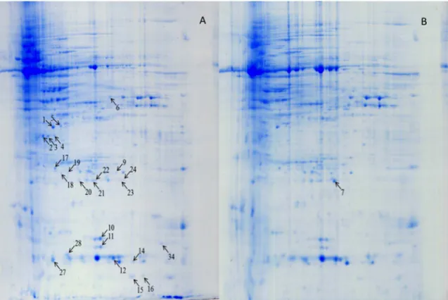
F. graminearumwere compared with 72 h mock-inoculated spikes. Four proteins from seven spots were induced (qualitative difference) byF. graminearuminoculation, and they presented only inF. graminearum-inoculated Fhb1+
NIL spikes, not in the mock-inoculated Fhb1+
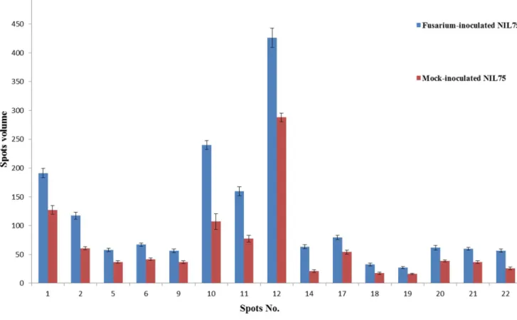
chloroplast oxygen-evolving enhancer protein 1 (OEE1, spots 3 and 4), PR-4 (spots 15 and 16), OEE 2 (spots 23 and 24), and single stranded nucleic acid binding (SSB) protein (spot 28) (Fig. 2; Table 1). Eleven proteins from 17 spots were significantly upregulated (quantitative difference) in Fhb1+NIL spikes after F. graminearum inoculation when compared with the mock-inoculated spikes (Fig. 3); they were Rossmann-fold NAD(P)(+ )-binding proteins (Fig. 2, spots 1 and 5), chloroplast OEE1 (spot 2), glyceraldehyde-3-phosphate dehydrogenase (GAPDH, spot 6), superoxide dismutase (SOD, spots 9, 10 and 11), nucleoside diphosphate kinase (NDPK, spots 12 and 34), 20 kDa chaperonin (spot 17), OEE 2 (spots 18, 19, 20, 21), SSB protein (spot 27), and an unknown protein (spot 22) (Fig. 2; Table 1). These proteins, either pathogen-induced or upregulated, were mainly involved in stress response, PR response, resistance to fungal penetration, plant photosynthesis, and energy metabolism. Some, such as
GAPDH, SOD [14,15], and EEO [16], have been reported previously asFusarium-responsive proteins. Only two protein spots were upregulated in Fhb12NIL spikes after F. graminearum
inoculation when compared with the mock-inoculated spikes of the same NIL (Data not shown). The results suggest thatFhb1+
NIL had a majority of genes that were either induced or upregulated in response to inoculation ofF. graminearum thanFhb12NIL, which agrees with previous reports [11,15,16,21].
Wheat proteins associated with FHB resistance
To identify wheat proteins associated with resistance to
F. graminearum, the protein profiles ofFhb1+NIL
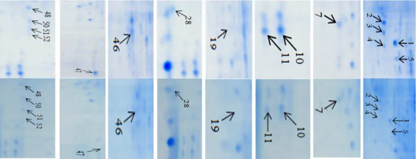
and Fhb12 NILs were compared after inoculation with F. graminearum. Fig. 4 presents representative gel images that were selected from three independent experiments showing differentially expressed proteins between the two inoculated NILs. Eight protein spots presented only inFhb1+NIL, but not in theFhb12NIL after inoculated with
F. graminearum; they were chloroplast OEE1 (spot 4) and OEE 2 (spot 19), Rossmann-fold NAD(P)(+)-binding proteins (spot 5), single-stranded nucleic acid binding protein (spot 28), beta-cyanoalanine synthase (CAS, spot 46), chitinase (spot 47), Cu/ Zn SOD (spot 51), and actin depolymerisation factor (ADF)/ cofilin-like (spot 52) (Fig. 4; Table 1). Nine protein spots showed significantly higher levels of expression in Fhb1+NIL than in Fhb12NIL (Fig. 5): Rossmann-fold NAD(P)(+)-binding proteins (spot 1), chloroplast OEE1 (spots 2 and 3), OEE2 (spot 7), SODs (spots 10, 11 and 50), PR-10 (spot 48), and NDPKI-like protein (spot 49). Most of these differentially expressed proteins in
Fhb1+
NIL were induced byF. graminearum, except CAS, chitinase, and ADF/cofilin. These proteins are mainly for oxidative stress responses, resistance to fungal penetration, plant photosynthesis, plant detoxification, etc. Some proteins such as SOD and chitinase have been reported previously asFusarium-responsive proteins or resistance related proteins [11,14,15,16,21], but most are newly identified proteins related to FHB resistance in this study including an ADF/cofilin protein, CAS, NAD(P)(+)-binding protein, and NDPKI-like protein.
Resistance to pathogen penetration at the plant cell surface by formation of cell wall apposition (CWA), a physical and chemical barrier to cell penetration by a pathogen, is a key mechanism for plants. The actin cytoskeleton plays an important role in formation of CWA [22], and actin-binding proteins, such as ADF/cofilins, regulate the dynamic behavior of actin filaments during forming CWA. Actin dynamics demonstrated a role in the activation of gene-for-gene resistance ofArabidopsis thalianatoPseudomonas syringae
pv tomato [23]; abiotic stresses also induced significant expression of ADF/cofilins in cereal plants [24]. Thus, ADF/cofilins might be important proteins to protect plants against biotic and abiotic stresses. A high level of ADF/cofilin protein presented in only
Fhb1+
NIL suggests that CWA formation is an important initial step for wheat resistance toFusariumearly penetration in cell walls of spikelets in resistant wheat.
Reactive oxygen species (ROS) such as H2O2 also play an
important role in plant-pathogen interaction [25]. Pathogen-induced H2O2is required for peroxidase-dependent lignification
that hinders the penetration of a pathogen [26,27]. H2O2is very
important for resistance to fungal basal penetration because enzymatic removal of H2O2enhances the fungal penetration on
leaf epidermal cells [26]. H2O2is also required for protein
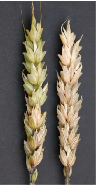
cross-linking in cell wall at the site of pathogen contact to produce a stress; the host cells’ generation of an oxidative burst also stresses the pathogen [26,27,28]. The cross-linking of proline-rich proteins in the cell wall makes plant cells more resistant to cell wall– degrading enzymes produced by a pathogen and may entrap the Figure 1. Wheat spikes of Fhb1+NIL (NIL75, left spikes) and
Fhb12NIL (NIL75, right spike) at 18th day after single spikelet inoculation with F. graminearum. F. graminearuminfection did not spread or spread to only several uninoculated spikelets in Fhb1+NIL, but entire spike
fungal penetration peg in a CWA [28]. In the barley–B. graminis
interaction system, the presence of H2O2in CWAs can be used as
a biochemical marker to identify nonpenetrated cells [29]. Because H2O2is membrane-permeable, it may also act as a diffusible signal
that leads to systemic acquired resistance [27]; thus, H2O2might
be a potent messenger in cell wall–associated defense. In plant cells, SOD can rapidly convert the O22to H2O2that accumulates
at the site of pathogen contact where CWA is formed, which restricts pathogen movement and reproduction, and prevents the spread of the pathogen to other parts of the plants [27,30]. In this study, four spots of SODs were induced or upregulated in the inoculated Fhb1+
NIL, not in the inoculated Fhb12NIL (Fig. 4; Table 1); therefore, the SODs played a critical role in resistance to FHB penetration by strengthening cell walls. Several other studies also presented evidences to support that cell wall thickening is a major mechanism of FHB resistance [11,22,16].
Plants may actively defend against pathogen infection by producing enzymes that digest fungal cell walls to stop fungal penetration. Because all true fungi contain chitin as a primary structural component of their cell walls, the chitinase family of PR proteins is of particular importance [31]. Chitin in fungal cell walls can be hydrolyzed by chitinases into smaller oligomers or monomers [31,32], so chitinases are considered to play a major role during plant–fungus pathogenic interactions [33–35]. Chit-inase was reported to be upregulated in FHB-resistant wheat ‘Ning7840’ [15,20]. Transgenic plants that overexpressed chit-inases exhibited enhanced resistance to pathogens [36,37]. Transgenic wheat that overexpressed a barley class II chitinase gene significantly increased Type II FHB resistance [37]. In this study, differentially expressed chitinase (Fig. 4, spots 47 and 48) that presented only in the Fhb1+
NIL provides another line of
evidence that the degradation of fungal cell wells by chitinases enhances FHB resistance in wheat.
Oxygen-evolving enhancer proteins (OEEs), consisting of three subunits [OEE1 (33 kDa), OEE2 (23 kDa), and OEE3 (16 kDa)], are nuclear-encoded chloroplast proteins and are peripherally bound to photosystem II (PSII) on the luminal side of the thylakoid membrane [38]. Photosynthetic oxygen evolution requires the interaction of several different yet closely coupled biochemical reactions. The light-capturing and charge-separating capacities of PSII must work in close cooperation with an oxygen-evolving complex capable of utilizing this oxidizing power to split water into oxygen and hydrogen. Electrons stripped from water during this reaction are funneled back into photochemical reaction center II, then transported through the electron transport chain to photosystem I, eventually to be used for the reduction of NADP [39]. Wang et al. [16] found that OEE2 of PSII was upregulated in FHB resistant cultivar ‘Wangshuibai’ after inoculation with
F. graminearum. In barley, PSII oxygen-evolving complex protein 2 precursor was expressed in response toF. graminearum[40]. In the current study, both OEE1 (Fig. 4, protein spot 4) and OEE2 (spot 19) were detected in theF. graminearum-inoculated Fhb1+
NIL, but not in the Fhb12NIL, suggesting that the two OEEs played an important role in maintaining PSII activity when wheat was inoculated with F. graminearum. Mizobuchi and Yamamoto [41] demonstrated that OEE1 was essential for oxygen evolving activity and PSII stability. In wheat FHB, the most obvious visual disease symptom on the spike of a susceptible plant infected by
F. graminearum starts with chlorosis to bleached spikes; thus, photosynthesis in the infected spikes is significantly reduced or stopped completely in a susceptible genotype [42]. Therefore, the recovery or turnover of OEEs in the inoculated FHB-resistant Figure 2. Protein profiles of wheat spikes ofFhb1+NIL (NIL75) inoculated with
F. graminearum(A) and mung bean broth (B) after 72 h.
spikes may be attributed to maintaining the capacity of PSII for enhanced photosynthesis afterF. graminearumattacks.
A higher level of NAD(P)(+)-binding proteins detected in inoculated Fhb1+NIL than inFhb12NIL also supported the idea that enhanced photosynthesis is related to FHB resistance. NAD(P)(+)-binding proteins bind nicotinamide dinucleotide (NAD) to catalyze reactions central to energy production, storage, and transfer. These reactions are essential to nearly all core metabolic pathways including photosynthesis [43]. Glycerade-hyde-3-phosphate dehydrogenase (GAPDH) is a NAD(P)(+) -binding protein [43] and has been reported to be involved in photosynthetic metabolism and responses to abiotic [44,45] and biotic stresses [46].
Nucleoside diphosphate kinases (NDPKs) are primary metabolic enzymes that maintain the balance between cellular ATP and other nucleoside triphosphates and play regulatory roles in response to multiple stresses [47]. In rice, NDPK1was reported to be involved in the defense against bacterial infection [43].NDPK
transcript was upregulated in response to wounding in tomato [48], and transgenic Arabidopsis that overexpressed AtNDK1
exhibited tolerance to paraquat (N,N9-dimethyl-4,49-bipyridinium dichloride), suggesting a role forAtNDK1in ROS response [49]. In this study, F. graminearum induced a higher level of NDPK1 expression in the Fusarium-inoculated Fhb1+NIL thanFhb12NIL, which suggests that NDPK1 protein may be involved in defense against FHB infection in wheat.
Many biotic and abiotic stress conditions can induce ethylene production in plants, while HCN is a byproduct in the ethylene pathway [50]. HCN is extremely toxic to plant cells, but
beta-cyanoalanine synthase (CAS) can rapidly detoxify HCN and recycle the reduced nitrogen of cyanide for amino acid synthesis [51]. Cell wall protein fractions from pathogens induced significant ethylene production in plants, and trace amounts of ethylene can elicit many physiological responses [50,52,53]; thus, the induced expression of CAS detoxified HCN resulting from the elevated level of ethylene production in cell wall protein–treated plants and the high level of CAS activity were caused by the general response to ethylene production. A previous study reported that wheat spikes challenged with cell wall proteins from
Pythium oligandrumhad a significantly reduced number of infected spikelets compared with the control afterF. graminearuminoculation and demonstrated that CAS induced by fungal elicitors reduced the level ofFusarium infection in wheat [54]. Several proteins in ethylene signal pathway have been associated with FHB resistance [9,21,55]. In this study, a high level of CAS (spot 46) (Fig. 4; Table 1) was detected only in theFhb1+
NIL, not in theFhb12NIL, indicating the important role of CAS and ethylene signaling in preventing FHB spread within a spike afterFusariuminfection in wheat.
Single stranded nucleic acid binding (SSB) protein is essential for DNA replication and repair in nearly all organisms, so it is crucial to genome maintenance [56]. SSB proteins bind with high affinity and specificity to ssDNA intermediates to protect them from degradation and destabilize inhibitory secondary structures within the ssDNA, and they regulate the activities of other proteins by direct binding to bring them to their sites of action on DNA [56]. In Arabidopsis, organellar single-stranded DNA binding protein 1 was shown to be required for mtDNA stability [57]. In Figure 3. Histograms show the volume changes of 15 upregulated spots inF. graminearum-inoculated NIL75 (Fhb1+NIL) and mock-inoculated NIL75.
Spot No. gi number description/function Frame MW pI
Mascot Protein Score
unique
peptides coverage
1 gi|383674360|gb|HX134554.1|HX134554 Rossmann-fold NAD(P)(+)-binding proteins* 3 58.9 6.9 529 7 14*
gi|383674331|gb|HX134525.1|HX134525 Rossmann-fold NAD(P)(+)-binding proteins* 4 70.3 7 262 3 9*
2 gi|383633628|gb|HX052023.1|HX052023 Photosystem II oxygen-evolving enhancer protein 1* 2 61.1 6.9 809 14 60*
gi|73912433 Aspartic proteinase [Triticum aestivum] 54.3 5 289 5 14
3 gi|383633628|gb|HX052023.1|HX052023 Photosystem II oxygen-evolving enhancer protein 1* 2 61.1 6.9 664 12 53*
gi|73912433 Aspartic proteinase [Triticum aestivum] 54.3 5 163 3 10
4 gi|383633628|gb|HX052023.1|HX052023 Photosystem II oxygen-evolving enhancer protein 1* 2 61.1 6.9 693 11 53*
5 gi|383741321|gb|HX179787.1|HX179787 Rossmann-fold NAD(P)(+)-binding proteins* 2 57.4 7 433 6 46*
gi|383674331|gb|HX134525.1|HX134525 Rossmann-fold NAD(P)(+)-binding proteins 4 70.3 7 274 7 40*
6 gi|253783729 Glyceraldehyde-3-phosphate dehydrogenase 36.5 6.8 309 5 18
gi|383741321|gb|HX179787.1|HX179787 Phenylcoumaran benzylic ether reductase (PCBER) like* 2 57.4 7 284 4 33
7 gi|131394 Oxygen-evolving enhancer protein 2, chloroplastic 27.3 9.5 541 7 37
9 gi|125663927 Manganese superoxide dismutase 19.3 6.9 266 5 40
10 gi|226897529 Superoxide dismutase 15.1 5.7 244 5 29
11 gi|226897529 Superoxide dismutase 15.1 5.7 192 3 28
12 gi|383599548|gb|HX251834.1|HX251834 Nucleoside diphosphate kinase Group I (NDPk_I)-like* 4 40.8 7 436 7 45
gi|383670986|gb|HX111086.1|HX111086 Ribulose bisphosphate carboxylase/oxygenase (Rubisco), small subunit* 2 56.5 7 261 7 20
14 gi|82619 Ribulose-bisphosphate carboxylase (EC 4.1.1.39) small chain precursor 15.3 9.9 131 4 21
15 gi|6048567 Pathogenesis-related protein 4 13.1 6.3 208 2 30
16 gi|6048569 Pathogenesis-related protein 4 13.1 7.8 146 3 28
17 gi|383595701|gb|HX164281.1|HX164281 20 kDa chaperonin, chloroplastic-like* 4 63.9 7 847 13 57
18 gi|131394 Oxygen-evolving enhancer protein 2, chloroplastic 27.3 9.5 445 6 26
19 gi|383675855|gb|HX136948.1|HX136948 Oxygen-evolving enhancer protein 2* 2 61.9 7 372 6 27
20 gi|131394 Oxygen-evolving enhancer protein 2, chloroplastic 27.3 9.5 354 5 24
21 gi|131394 Oxygen-evolving enhancer protein 2, chloroplastic 27.3 9.5 114 3 15
22 n.d.
23 gi|131394 Oxygen-evolving enhancer protein 2, chloroplastic 27.3 9.5 393 5 23
24 gi|131394 Oxygen-evolving enhancer protein 2, chloroplastic 27.3 9.5 531 7 26
27 gi|974605 Single-stranded nucleic acid binding protein 16.2 5 372 6 81
gi|11990893 Ribulose-1,5-bisphosphate carboxylase/oxygenase small subunit 19.5 9.8 169 4 26
28 gi|974605 Single-stranded nucleic acid binding protein 16.2 5 180 3 35
gi|11990897 Ribulose-1,5-bisphosphate carboxylase/oxygenase small subunit 19.4 9.7 140 4 20
34 gi|383743921|gb|HX172565.1|HX172565 Nucleoside diphosphate kinase Group I (NDPk_I)-like* 5 58.6 7 293 5 25
46 gi|383608201|gb|HX252082.1|HX252082 WD40 domain* 2 53.7 7 692 10 59
gi|295917894 Beta-cyanoalanine synthase 40.2 6.4 266 5 18
Proteins
Associate
d
with
Wheat
FHB
Resistance
ONE
|
www.ploson
e.org
7
December
2013
|
Volume
8
|
Issue
12
|
this
study,
a
significantly
higher
level
of
SSB
protein
was
detected
in
inoculated
Fhb1
+
NIL
than
in
Fhb1
2
NIL,
suggesting
the
SSB
protein
may
be
actively
involved
in
protection
of
ssDNA
in
mitochondrial
or
chloroplast
from
degradation
in
FHB-resistant
genotypes.
Table 1.Cont.
Spot No. gi number description/function Frame MW pI
Mascot Protein Score
unique
peptides coverage
47 gi|18146825 Chitinase 1 27.1 9.6 778 10 66
48 gi|1568639 Cu/Zn superoxide dismutase 20.3 5.3 263 4 31
gi|196051131 Pathogenesis related protein 10 17.1 5.1 169 4 34
49 gi|383739814|gb|HX143464.1|HX143464 Nucleoside diphosphate kinase Group I (NDPk_I)-like* 4 45.9 7 177 3 16
50 gi|1572627 Cu/Zn superoxide dismutase 20.2 5.3 235 3 37
51 gi|1572627 Cu/Zn superoxide dismutase 20.2 5.3 144 3 37
52 gi|383606411|gb|HX251502.1|HX251502 Actin depolymerisation factor/cofilin -like* 6 54.7 6.9 248 4 41
*indicates that matched sequence was found in the EST and its function was the same as indicated by BLAST search. doi:10.1371/journal.pone.0082079.t001
Figure
4.
Enlarged
partial
gel
images
showin
g
differentially
expresse
d
protein
spots
between
inoculated
resistant
line
Fhb1
+
NIL
(NIL75)
and
inoculated
susceptible
line
Fhb1
2
NIL
(NIL98).
doi:10.1371
/journal.pone
.0082079.g00
4
Proteins
Associate
d
with
Wheat
FHB
Resistance
ONE
|
www.ploson
e.org
8
December
2013
|
Volume
8
|
Issue
12
|
Conclusions
Comparisons of protein expression profiles between the unique pair of NILs contrasting in Fhb1 alleles identified nine types of either induced or upregulated proteins that were associated with wheat FHB resistance in the Fhb1+
NIL. These differentially expressed proteins may be involved in complicated processes to defend against fungal infection in FHB-resistant genotypes by degrading fungal cell walls and strengthening plant cell walls at the site of pathogen contact to hinder pathogen penetration; detoxifying toxic cyanide in the ethylene pathway; and maintain-ing photosynthesis and energy metabolism. Although theFhb1was previously located on chromosome 3BS [4], whether the genes encoding these FHB resistance-related proteins are located on the same chromosome remains unknown. It is possible that Fhb1
gene(s) on the chromosome 3BS trans-regulate the expression of some of these genes in downstream to provide FHB resistance;
therefore, further cloning ofFhb1may elucidate the functions of
Fhb1in the wheat FHB system.
Acknowledgments
Disclaimer: USDA is an equal opportunity provider and employer. Mention of trade names or commercial products in this article is solely for the purpose of providing specific information and does not imply recommendation or endorsement by the U.S. Department of Agriculture. Contribution number 13-288-J from the Kansas Agricultural Experiment Station.
Author Contributions
Conceived and designed the experiments: GB. Performed the experiments: XZ JF. Analyzed the data: XZ JF YH. Contributed reagents/materials/ analysis tools: GB HP YH. Wrote the paper: XZ GB JF.
References
1. Bai G-H, Shaner GE (1994) Scab of wheat: prospects for control. Plant Dis 78: 760–766.
2. Bai G-H, Shaner GE (2004) Management and resistance in wheat and barley to Fusarium head blight. Annu Rev Phytopathol 42: 135–161.
3. Anderson JA, Stack RW, Liu S, Waldron BL, Field AD, et al. (2001). DNA markers for Fusarium head blight resistance QTL in two wheat populations. Theor Appl Genet 102: 1164–1168.
4. Cuthbert PA, Somers DJ, Thomas J, Cloutier S, Brule’-Babel A (2006) Fine mapping Fhb1, a major gene controlling Fusarium head blight resistance in bread wheat (Triticum aestivumL.). Theor Appl Genet 112: 1465–1472. 5. Bowles DJ (1990) Defense-related proteins in higher plants. Annu Rev Biochem
59: 873–907.
6. Veronese P, Ruiz MT, Coca MA, Hernandez-Lopez A, Lee H, et al. (2003) In defense against pathogens. Both plant sentinels and foot soldiers need to know the enemy. Plant Physiol 131: 1580–1590.
7. Boddu J, Cho S, Kruger WM, Muehlbauer GJ (2006) Transcriptome analysis of the barley-Fusarium graminearuminteraction. Mol Plant Microbe Interact 19: 407– 417.
8. Pritsch C, Muehlbauer GJ, Bushnell WR, Somers DA, Vance CP (2000) Fungal development and induction of defense response genes during early infection of wheat spikes byFusarium graminearum. Mol Plant-Microbe Interact 13: 159–169. 9. Gottwald S, Samans B, Lu¨ck S, Friedt W (2012) Jasmonate and ethylene dependent defence gene expression and suppression of fungal virulence factors: two essential mechanisms of Fusarium head blight resistance in wheat? BMC Genomics 13: 369.
10. Lemmens M, Scholz U, Berthiller F, Asta CD, Koutnik A, et al. (2005) The ability to detoxify the mycotoxin deoxynivalenol colocalizes with a major
quantitative trait locus for Fusarium head blight resistance in wheat. Mol Plant-Microbe Interact 18: 1318–1324.
11. Gunnaiah R, Kushalappa AC, Duggavathi R, Fox S, Somers DJ (2012) Integrated metabolo-proteomic approach to decipher the mechanisms by which wheat QTL (Fhb1) contributes to resistance againstFusarium graminearum. PLoS ONE 7(7): e40695.
12. Jia HY, Cho S, Muehlbauer GJ (2009) Transcriptome analysis of a wheat near-isogenic line pair carrying Fusarium head blight–resistant and –susceptible alleles. Mol Plant-Microbe Interact 22: 1366–1378.
13. Zhuang Y, Gala A, Yen Y (2013) Identification of functional genic components of major Fusarium head blight resistance quantitative trait loci in wheat cultivar Sumai 3. Mol Plant Microbe Interact 26: 442–50.
14. Zhou W-C, Kolb FL, Riechers DE (2005) Identification of proteins induced or upregulated by Fusarium head blight infection in the spikes of hexaploid wheat (Triticum aestivum). Genome 48: 770–780.
15. Zhou W-C, Eudes F, Laroche A (2006) Identification of differentially regulated proteins in response to a compatible interaction between the pathogenFusarium graminearumand its host,Triticum aestivum. Proteomics 6: 4599–4609. 16. Wang Y, Yang LM, Xu HB, Li QF, Ma ZQ, et al. (2005) Differential proteomic
analysis of proteins in wheat spikes induced by.Fusarium graminearum. Proteomics 5: 4496–4503.
17. Bernardo NA, Ma HX, Zhang DD, Bai GH (2011) Single nucleotide polymorphism in wheat chromosome region harboringFhb1for Fusarium head blight resistance. Mol Breed DOI 10.1007/s11032-011-9565-y.
18. Fu J, Momcilovic I, Clemente T, Nersesian N, Trick H, et al. (2008) Heterologous expression of a plastid EF-Tu reduces protein thermal aggregation
Figure 5. Histograms show the volume changes of 9 upregulated protein spots in the resistant NIL75 (Fhb1+NIL) and the susceptible NIL98 (Fhb12NIL) after inoculation withF. graminearum.
and enhances CO2 fixation in wheat (Triticum aestivum) following heat stress. Plant Mol Biol 68: 277–288.
19. Candiano G, Bruschi M, Musante L (2004) Blue silver: A very sensitive colloidal Coomassie G-250 staining for proteome analysis. Electrophoresis 25: 1327– 1333.
20. Bernardo A, Bai GH, Guo PG, Xiao K, Guenzi AC, et al. (2007) Fusarium graminearum-induced changes in gene expression between Fusarium head blight-resistant and susceptible wheat cultivars. Funct Integr Genomics 7: 69–77. 21. Ding L, Xu H, Yi H, Yang L, Kong Z, et al (2011) Resistance to hemi-biotrophicF. graminearuminfection is associated with coordinated and ordered expression of diverse defense signaling pathways. PLoS ONE 6(4): e19008. 22. Hardham AR, Jones DA, Takemoto D (2007) Cytoskeleton and cell wall
function in penetration resistance. Curr Opin Plant Biol 10, 342–348. 23. Tian MY, Chaudhry F, Ruzicka DR, Meagher RB (2009) Arabidopsis
actin-depolymerizing factor AtADF4 mediates defense signal transduction triggered by thePseudomonas syringaeeffector AvrPphB. Plant Physiol 150: 815–824. 24. Ali GM, Komatsu SJ (2006) Proteomic analysis of rice leaf sheath during
drought stress. Proteome Res 5: 396–403.
25. Levine A, Tenhaken R, Dixon R, Lamb C (1994) H2O2from the oxidative burst
orchestrates the plant hypersensitive disease resistance response. Cell 79: 583– 593.
26. Hu¨ckelhoven R (2007) Cell wall–associated mechanisms of disease resistance and susceptibility. Annu Rev Phytopathol 45: 101–27.
27. Noctor G, Foyer CH (1998) Ascorbate and glutathione: keeping active oxygen under control. Annu Rev Plant Physiol Plant Mol Biol 49: 249–279. 28. Brisson LF, Tenhaken R, Lamb C (1994) Function of oxidative cross-linking of
cell wall structural proteins in plant disease resistance. Plant Cell 6: 1703–12. 29. Hu¨ckelhoven R, Fodor J, Preis C, Kogel KH (1999) Hypersensitive cell death
and papilla formation in barley attacked by the powdery mildew fungus are associated with hydrogen peroxide but not with salicylic acid accumulation. Plant Physiol 119: 1251–60.
30. Jime´nez A, Hernandez JA, Del-Rio LA, Sevilla F (1997) Evidence for the presence of the ascorbate-glutathione cycle in mitochondria and peroxisomes of pea leaves. Plant Physiol 114, 275–284.
31. Wessels JGH (1994) Developmental regulation of fungal cell-wall formation. Annu Rev Phytopathol 32: 413–437.
32. Bishop JG, Dean AM, Mitchell-Olds T (2002) Rapid evolution in plant chitinases: molecular targets of selection in plant-pathogen coevolution. Proc Natl Acad Sci U S A 97: 5322–5327.
33. Collinge DB, Kragh KM, Mikkelsen JD, Nielsen KK, Rasmussen J, et al. (1993) Plant chitinases. Plant J 3: 31–40.
34. Kasprzewska A (2003) Plant chitinases –regulation and function. Cell Mol Biol Lett 8: 809–824.
35. Meins F, Fritig B, Linthorst HJM, Mikkelsen JD, Neuhaus JM, et al. (1994) Plant chitinase genes. Plant Mol Biol Rep 12, S22-S28.
36. Schlumbaum A, Mauch F, Vo¨geli U, Boller T (1986) Plant chitinases are potent inhibitors of fungal growth. Nature 324: 365–367.
37. Shin S, Mackintosh CA, Lewis J, Heinen SJ, Radmer L, et al. (2008) Transgenic wheat expressing a barley class II chitinase gene has enhanced resistance against
Fusarium graminearum. J Exp Bot 59: 2371–2378.
38. Sugihara K, Hanagata N, Dubinsky Z, Baba S, Karube I (2000) Molecular characterization of cDNA encoding oxygen evolving enhancer protein 1 increased by salt treatment in the mangroveBruguiera gymnorrhiza. Plant Cell Physiol 41: 1279–1285.
39. Mayfieldl SP, Bennoun P, Rochaix JD (1987) Expression of the nuclear encoded OEE1 protein is required for oxygen evolution and stability of photosystem II particles inChlamydomonas reinhardtii. EMBO 6: 313–318.
40. Geddes J, Eudes F, Laroche A, Selinger LB (2008) Differential expression of proteins in response to the interaction between the pathogenFusarium graminearum
and its host,Hordeum vulgare. Proteomics 8: 545–554.
41. Mizobuchi A, Yamamoto Y (1989) Assembly of photosystem II polypeptides and expression of oxygen evolution activity in the chloroplasts ofEuglena gracilisZ during the dark-light transition. Biochim Biophys Acta 977: 26–32.
42. Ribichich KF, Lopez SE, Vegetti AC (2000) Histopathological spikelet changes produced byFusarium graminearumin susceptible and resistant wheat cultivars. Plant Dis 84: 794–802.
43. Bellamacina C (1996) The nicotinamide dinucleotide binding motif: a comparison of nucleotide binding proteins. FASEB J 10: 1257–1269. 44. Yang YJ, Kwon HB, Peng HP, Shih MC (1993) Stress responses and metabolic
regulation of glyceraldehyde-3-phosphate dehydrogenase genes in Arabidopsis. Plant Physiol 101: 209–216.
45. Jeong MJ, Park SC, Byun MO (2001) Improvement of salt tolerance in transgenic potato plants by glyceraldehyde-3 phosphate dehydrogenase gene transfer. Mol Cells 12: 189–185.
46. Laxalt AM, Cassia RO, Sanllorenti PM, Madrid EA, Andreu AB, et al. (1996) Accumulation of cytosolic glyceraldehyde-3-phosphate dehydrogenase RNA under biological stress conditions and elicitor treatments in potato. Plant Mol Biol 30: 961–72.
47. Cho SM, Shin SH, Kim KS, Kim YC, Eun MY et al. (2004) Enhanced expression of a gene encoding a nucleoside diphosphate kinase 1 (OsNDPK1) in rice plants upon infection with bacterial pathogens. Mol Cells 18: 390–395. 48. Harris N, Taylor JE, Roberts JA (1994) Isolation of an mRNA encoding a
nucleoside diphosphate kinase from tomato that is up-regulated by wounding. Plant Mol Biol 25: 739–42.
49. Fukamatsu Y, Yabe N, Hasunuma K (2003) Arabidopsis NDK-1 is a component of ROS signaling by interacting with three catalases. Plant Cell Physiol 44: 982– 989.
50. Yip WK, Yang SF (1988) Cyanide metabolism in relation to ethylene production in plant tissues. Plant Physiol 88: 473–476.
51. Takahashi H, Ishihara T, Hase S, Chiba A, Nakaho K, et al. (2006) Beta-cyanoalanine synthase as a molecular marker for induced resistance by fungal glycoprotein elicitor and commercial plant activators. Phytopathology 96: 908– 916.
52. Peiser GD, Wang TT, Hoffman NE, Yang SF, Liu HW, et al. (1984) Formation of cyanide from carbon 1 of 1-aminocyclopropane-1-carboxylic acid during its conversion to ethylene. Proc Natl Acad Sci USA 81: 3059–3063.
53. Toppan A, Esquerre-Tugate MT (1984) Cell surfaces in plant-microorganism interactions IV. Fungal glycopeptides which elicit the synthesis of ethylene in plants. Plant Physiol 75: 1133–1138.
54. Takenaka S, Nishio Z, Nakamura Y (2003) Induction of defense reactions in sugar beet and wheat by treatment with cell wall protein fractions from the mycoparasitePythium oligandrum. Phytopathology 93: 1228–1232.
55. Yang F, Yang F, Jacobsen S, Jørgensen HJ, Collinge DB, et al. (2013)Fusarium graminearumand its interactions with cereal heads: studies in the proteomics era. Front Plant Sci 4: 37.
56. Ha T, Kozlov AG. Lohman TM (2012) Single-molecule views of protein movement on single-stranded DNA. Annu Rev Biophys 41: 295–319. 57. Zaegel V, Guermann B, Le Ret M, Andre’s C, Meyer D, et al. (2006) The