Structural and Mechanistic Insight into DNA Unwinding
by
Deinococcus radiodurans
UvrD
Meike Stelter
1,2,3,4☯, Samira Acajjaoui
1☯, Sean McSweeney
1, Joanna Timmins
1,2,3,4*1 Structural Biology Group, European Synchrotron Radiation Facility, Grenoble, France, 2 University Grenoble Alpes, Institut de Biologie structurale, Grenoble, France, 3 Centre National de la Recherche Scientifique, Institut de Biologie structurale, Grenoble, France, 4 Commissariat à l’énergie atomique et aux énergies alternatives, Département du Science du Vivant, Institut de Biologie structurale, Grenoble, France
Abstract
DNA helicases are responsible for unwinding the duplex DNA, a key step in many biological processes. UvrD is a DNA helicase involved in several DNA repair pathways. We report here crystal structures of Deinococcus radiodurans UvrD (drUvrD) in complex with DNA in different nucleotide-free and bound states. These structures provide us with three distinct snapshots of drUvrD in action and for the first time trap a DNA helicase undergoing a large-scale spiral movement around duplexed DNA. Our structural data also improve our understanding of the molecular mechanisms that regulate DNA unwinding by Superfamily 1A (SF1A) helicases. Our biochemical data reveal that drUvrD is a DNA-stimulated ATPase, can translocate along ssDNA in the γ′-5′ direction and shows ATP-dependent γ′-5′, and surprisingly also, 5′-γ′ helicase activity. Interestingly, we find that these translocase and helicase activities of drUvrD are modulated by the ssDNA binding protein. Analysis of drUvrD mutants indicate that the conserved -hairpin structure of drUvrD that functions as a separation pin is critical for both drUvrD’s γ′-5′ and 5′-γ′ helicase activities, whereas the GIG motif of drUvrD involved in binding to the DNA duplex is essential for the 5′-γ′ helicase activity only. These special features of drUvrD may reflect its involvement in a wide range of DNA repair processes in vivo.
Citation: Stelter M, Acajjaoui S, McSweeney S, Timmins J (β01γ) Structural and Mechanistic Insight into DNA Unwinding by Deinococcus radiodurans
UvrD. PLoS ONE 8(10): e77γ64. doi:10.1γ71/journal.pone.0077γ64 Editor: Sergey Korolev, Saint Louis University, United States of America
Received February 1, β01γ; Accepted September β, β01γ; Published October 15, β01γ
Copyright: © β01γ Stelter et al. This is an open-access article distributed under the terms of the Creative Commons Attribution License, which permits unrestricted use, distribution, and reproduction in any medium, provided the original author and source are credited.
Funding: This work was funded by the inhouse research program of the ESRF. MS and JT are funded by the CNRS via an ATIP-AVENIR grant and by the Ligue contre le Cancer. The funders had no role in study design, data collection and analysis, decision to publish, or preparation of the manuscript. Competing interests: The authors have declared that no competing interests exist.
* E-mail: [email protected]
☯ These authors contributed equally to this work.
Introduction
Many biological processes, such as DNA replication, transcription, recombination or repair, require access to the genetic information hidden within the duplex DNA of the genome and for this purpose the double-stranded DNA (dsDNA) needs to be transiently unwound. A diverse set of enzymes, known as DNA helicases, is responsible for catalyzing this process [1,β]. DNA helicases are ubiquitous enzymes and many different helicases are found in a single cell due to the diversity of structures adopted by duplexed DNA. Helicases are a subset of the translocase enzyme family that share a number of conserved signature motifs responsible for either NTP binding and hydrolysis, DNA binding or for coupling these two processes. Based on primary structure analyses and extensive biochemical studies, six superfamilies of helicases have so far been described, each of which possesses a different set of conserved signature motifs [γ,4]. Three of these
superfamilies (SF1, SFβ and SF6) have been further classified according to their polarity γ′-5′ (type A) or 5′-γ′ (type B) [4].
UvrD is classified as a SF1A helicase [γ] and plays important functions in DNA replication [5], recombinational repair [6-8], methyl-directed mismatch repair [9] and nucleotide excision repair [10]. UvrD consists of two RecA-like domains (1A and βA) that are responsible for nucleotide binding and hydrolysis and two additional domains (1B and βB) that are involved in dsDNA binding. UvrD has been shown to translocate along single-stranded DNA (ssDNA) as a monomer, while a number of studies indicate that oligomerization, and notably dimerization, of UvrD is required for helicase activity [11-15]. Over the past 15 years, several crystal structures of SF1A helicases have been determined. In 1996, the structure of
Geobacillus stearothermophilus PcrA (gsPcrA) was solved in its apo form [16] and in 1997, the first crystal structure of
complex with γ′-tailed DNA consisting of a 10 base pair DNA duplex and a seven base single-stranded γ′-tail were determined in apo- and AMPPNP-bound forms [18] and in β006, several structures of E. coli UvrD (ecUvrD) bound to γ′-tailed DNA were determined revealing the details underlying DNA unwinding by SF1A helicases [19]. These structures led to the proposal of a combined wrench-and-inchworm mechanism for DNA unwinding [19,β0]. In this model, a rotational movement regulated by ATP binding and hydrolysis acting as the ‘engine’ is combined with alternate tight and loose interactions at four protein-DNA contact points to produce a highly coordinated unidirectional movement along DNA.
In the radiation-resistant bacterium, Deinococcus radiodurans, unlike in E. coli, UvrD is involved in diverse DNA repair pathways [7]. In particular, UvrD has been shown to play a central role in double-strand break (DSB) repair and reconstitution of the genome following chromosome fractionation [7]. In E. coli, the RecQ, RecD and Helicase IV enzymes participate in DSB repair while in D. radiodurans, these three helicases have been shown to be dispensable [7]. The involvement and importance of a helicase in a given cellular pathway are not conserved from one bacterium to another.
Here we present crystal structures of full-length and a C-terminally truncated construct of D. radiodurans UvrD (drUvrD) in complex with γ′-tailed dsDNA. Our structures obtained in apo- and AMPPNP bound states provide us with several snapshots of this essential cellular process and reveal a large-scale spiral movement of UvrD around the duplexed DNA. Our structural data and biochemical analysis of wild-type and mutant drUvrD support the previously proposed wrench-and-inchworm model and provide further insight into the local conformational changes associated with DNA unwinding. A structural comparison of drUvrD with its E. coli homologue reveals that most of the differences reside in the inter-domain contacts and the ssDNA binding pocket and gating mechanism. Our biochemical studies reveal that drUvrD is an active DNA-stimulated ATPase that also possesses ATP-dependent translocase and helicase activities. Further investigations of these in vitro activities demonstrated that drUvrD translocates along ssDNA with a biased γ′-5′ directionality but, despite belonging to the SF1A protein family, can unwind duplexed DNA in both the γ′-5′ and 5′-γ′ directions. Interestingly, we find that these translocase and bipolar helicase activities of drUvrD are modulated by the ssDNA binding protein (SSB).
Materials and Methods
Cloning, expression and purification of drUvrD and drSSB
Full-length drUvrD (drUvrDFL) and a truncated construct of
drUvrD (drUvrD∆C), missing residues 666-745 (Figure 1A), were
cloned into pET151d (Invitrogen). drUvrDFL mutants were
prepared with the QuikChange mutagenesis kit (Agilent Technologies). All constructs were expressed in BLβ1 (DEγ) cells. Protein expression was induced by 1 mM IPTG at 15°C overnight. Cells were lysed by sonication and the protein was purified by Ni affinity chromatography (Macherey-Nagel) in 50
mM Tris-HCl pH 8.0, 150 mM NaCl, 5% glycerol and 5 mM MgClβ, followed by His-tag cleavage with TEV protease and
dialysis to remove the imidazole used for eluting the protein from the Ni column. The protein was further purified on a HiTrap Heparin column (GE Healthcare) and eluted in 50 mM Tris-HCl pH 8.0, 400 mM NaCl, 0.1 mM EDTA, 1 mM DTT, 5 mM MgClβ and 5% glycerol. The protein was concentrated to
7-8 mg/ml and stored at -80°C. Deinococcus radiodurans SSB (drSSB) was cloned into pET151d (Invitrogen) for expression with a cleavable N-terminal His-tag and expressed in BLβ1 (DEγ) Star at β0°C overnight. drSSB was purified on Ni-NTA (Qiagen) followed by a size-exclusion chromatography (Superdex 75 10/γ00 GL) in 50 mM Tris-HCl pH 8.0 and 100 mM NaCl and was stored at -80°C.
DNA oligonucleotides
All DNA oligonucleotides used in this study were purchased from Eurofins-MWG and their sequences are presented in Table S1. The DNA used for co-crystallization with drUvrD∆C
was composed of Forβ5 and its complementary strand Revβ5, while drUvrDFL was co-crystallized with DNA formed by Forβ8
and Revβ8 (Figure 1B). Annealed DNAs were purified by anion exchange chromatography (MonoQ; GE Healthcare). HPLC-purified oligonucleotides, Forβ5-β1F and Revβ5-β1F, containing a fluorescein-derivatized thymine (Fluo-dT) at position β1 were simply annealed prior to crystallization trials. For the helicase and DNA binding assays, the DNA substrates were made of a β5mer oligonucleotide containing a Fluo-dT at position 1β (H1T1β) and a complementary oligonucleotide containing no extension (H4), a 15 nucleotide (nt) or a 7nt polydT ssDNA extension at either the γ′ (Hγ-15 or Hγ-7) or 5′ end (H5-15 or H5-7). For the streptavidin-displacement assay, the γ′-tailed DNA substrate consisted of a γ′-fluorescein labeled β5mer oligonucleotide (H1-γF) annealed to its complementary oligonucleotide with a β5nt polydT ssDNA extension at its γ′ end and containing a biotin conjugated thymine in position 49 (Hγ-β5-B49), while the 5′-tailed DNA substrate was composed of a 5′-FAM labeled β5mer oligonucleotide (H1-5FAM) annealed to its complementary oligonucleotide with a β5nt polydT ssDNA extension at its 5′ end and containing a biotin conjugated thymine in position β (H5-β5-Bβ).
Crystallization
drUvrDFL and drUvrD∆C were mixed with their respective
DNAs at a β:1 molar ratio in 50 mM Tris-HCl pH 8.0, 150 mM NaCl, 5 mM MgClβ, 5% glycerol, 0.1 mM EDTA, 1 mM DTT and
1 mM AMPPNP (Sigma) and concentrated to 8-10 mg/ml. Crystals were obtained using the hanging-drop vapor diffusion method at β0°C. drUvrD∆C-Forβ5/Revβ5 form I crystals
appeared very rapidly (<1 day) in β0% PEG γγ50, 0.1 M Bis-Tris Propane pH 7.0 and 0.β M Na-Nitrate, while form II crystals were obtained after at least one week in ββ% PEG γγ50, 0.1 M Bis-Tris Propane pH 7.5 and 0.1 M Na-Fluoride. High quality crystals of drUvrDFL-Forβ8/Revβ8 suitable for data
liquid nitrogen. 5 mM AMPPNP was included in the cryoprotectants.
Data collection and Structure Determination
Diffraction data (Table 1) were collected at the European Synchrotron Radiation Facility (ESRF) in Grenoble, France and were processed with either XDS [β1] or iMosflm [ββ]. The structure of drUvrD∆C-Forβ5/Revβ5 form I was solved by
molecular replacement using Mr. Bump [βγ] and the gsPcrA helicase as a search model (PDB entry γPJR). After several rounds of substantial rebuilding of the protein chains in Coot
[β4], the DNA could be built and the AMPPNP molecules docked into the electron density. Subsequently, this model was used to solve the structures of drUvrD∆C-Forβ5/Revβ5 form II
and drUvrDFL-Forβ8/Revβ8 by molecular replacement with
Phaser [β5]. The drUvrD∆C-Forβ5/Revβ5 form I and form II
models were refined with Refmac [β6], while the drUvrDFL
-Forβ8/Revβ8 model, solved at lower resolution, was refined in
Phenix [β7] using drUvrD∆C-Forβ5/Revβ5 form I as a reference
model (Table 1). Fig.s of structures were prepared with Pymol
[β8] and the movie of the morph was created with Chimera
[β9].
ATPase activity
The rate of ATP hydrolysis by 100 nM drUvrDFL and drUvrD∆C
in the presence of a β5mer polydT oligonucleotide was measured using the spectrophotometric method [γ0] at β5°C in 50 mM Tris–HCl pH 8.0, 100 mM NaCl, 0.1 mM EDTA, 1 mM DTT, 5 mM MgClβ and 5% glycerol (buffer A). The KssDNA was
determined by measuring the rate of ATP hydrolysis in the presence of β mM ATP as a function of increasing concentrations of ssDNA (0-10 µM). Kinetic parameters (Vmax,
Km and Kcat) were determined by measuring the rate of ATP
hydrolysis in the presence of an excess of ssDNA (10xKssDNA)
at various ATP concentrations (0-1 mM). The measurements were made in triplicate and the average ATPase rates were plotted and fitted to a hyperbola using Origin.
Helicase assay
Helicase activity of drUvrDFL was assayed in 10 mM Tris-HCl
pH 8.0, 50 mM NaCl, 1% glycerol, 5 mM MgClβ and 0.1 mg/mL
BSA (buffer B). 80 µl reactions containing β0 nM DNA and β50 nM wild-type or mutant drUvrD were incubated at β5°C. The duplexed DNA was either blunt or contained 15nt or 7nt ssDNA extensions at either the γ′- or 5′-ends. The reactions were initiated by addition of β mM ATP. At indicated time points, 10
Figure 1. Domain organization of drUvrD and structure of the various DNA oligonucleotides used for crystallization. A. Schematic representation of the domain structures of drUvrDFL and drUvrD∆C. B. Structure of DNA oligonucleotides used for
crystallization with drUvrDFL and drUvrD∆C. The circles represent UvrD bound to the DNA as observed in our crystal structures. doi: 10.1γ71/journal.pone.0077γ64.g001
µl samples of the reaction were quenched with β.5 µl of a solution containing 0.8% SDS, 0.08% bromophenol blue, β4% glycerol, 80 mM EDTA and β0 µM unlabeled H1 oligonucleotide. The reactions were carried out in the absence and presence of β50 nM drSSB. Reaction products were run on a β0 % polyacrylamide TBE gel and the DNA bands were visualized and quantified using a ChemiDoc MP imaging system and the Image Lab software (Bio-Rad). Initial reaction rates were estimated using GraphPad Prism6 and averaged data from three independent experiments were plotted in GraphPad Prism6 with standard deviations represented as vertical error bars.
Table 1. Crystallographic data collection and refinement statistics.
Dataset drUvrDFL drUvrD∆C form I drUvrD∆C form II Data collection
Protein drUvrDFL drUvrD∆C drUvrD∆C
DNA Forβ8/Revβ8 Forβ5/Revβ5 Forβ5/Revβ5
Nucleotide AMPPNP AMPPNP AMPPNP
Space group Pβ1 Pβ1β1β1 Pβ1
Cell dimensions a, b, c (Å) α, , (°)
71.58, γ90.58, 71.65 90.00, 106.00, 90.00 67.57, 67.45, γ86.04 90.00, 90.00, 90.00 68.49, 89.79, β9γ.80 90.00, 89.97, 90.00
Beamline ESRF ID14-4 ESRF ID14-β ESRF IDβγ-1
Resolution (Å) 46.15 - 4.00 (4.ββ- 4.00)
47.40 - β.55 (β.69 - β.55)
47.6γ - γ.00 (γ.16 - γ.00) Rmerge (%) 10.6 (65.8) 7.1 (59.β) 6.4 (γβ.0) <(I)/σ(I)> 10.1 (β.γ) β0.4 (γ.6) 6.5 (1.9) Completeness (%) 99.6 (99.5) 100.0 (100.0) 89.0 (86.5) Refinement
N° of reflections (F > 0
σF ) γ0,051 56,088 59,507
Rfact/Rfree (%) β4.6/β7.1 β1.1/β6.6 ββ.8/β8.8
Mol/asu 4 chains UvrD β
chains dsDNA
β chains UvrD 1 chains dsDNA
4 chains UvrD β chain dsDNA
Ligands 4 AMP-PNP β AMP-PNP β AMP-PNP
Wilson B-factor 149.8 6γ.4 7β.γ
Average B-factor (Åβ)
Protein β0γ.0 6γ.9 10β.β
DNA β56.8 16γ.9 1γβ.0
AMPPNP 168.8 β9.7 84.6
Solvent N/A γ8.5 84.γ
Ramachandran
Favoured (%) 9γ.8 89.1 89.0
Allowed (%) 6.1 10.6 10.7
Disallowed (%) 0.β 0.γ 0.γ
Rms deviations
Bonds (Å) 0.006 0.017 0.01β
Angles (°) 1.1 1.7 1.5
PDB ID 4cβt 4cβu 4cγ0
Values in parentheses are for highest resolution shell. doi: 10.1γ71/journal.pone.0077γ64.t001
Streptavidin displacement assay
The translocase activity of drUvrD was assayed using the streptavidin-displacement assay [γ1,γβ]. DNA oligonucleotides used in this assay consisted of dsDNA duplexes with a β5 nt ssDNA extension at either the γ′- or 5′-end and a biotin label in positions 49 and β respectively. The DNA-streptavidin complexes were formed by incubating the biotinylated dsDNA (0.β µM) with streptavidin (γ.β µM, Sigma) in 10 mM Tris-HCl pH 8.0, 50 mM NaCl, 0.5 mM EDTA at β5°C for γ0 min, before addition of 180 µM biotin. Displacement reactions of 80 µl containing β0 nM streptavidin-loaded DNA and β50 nM
drUvrDFL were incubated in buffer B at β5°C. The reactions
were initiated by addition of β mM ATP. At indicated time points, 10 µl samples of the reaction were quenched with β.5 µl of a solution containing 0.48% SDS, 0.0γβ% bromophenol blue, β0% glycerol, 160 mM EDTA and β0 µM unlabeled H1 oligonucleotide. The reactions were carried out in the absence and presence of β50 nM drSSB. Reaction products were run on a 10 % polyacrylamide TBE gel and the DNA bands were visualized and quantified using a ChemiDoc MP imaging system and Image Lab software (Bio-Rad). Averaged data from three independent experiments were plotted in GraphPad Prism6 with standard deviations represented as vertical error bars.
DNA Binding
Equilibrium DNA binding assays were performed on a Synergy H4 Hybrid Microplate reader (BioTek), fitted with polarization filters to measure fluorescence anisotropy. The binding assays were conducted in γ84-well plates at room temperature in 80 µl reaction volumes in buffer A supplemented with 0.05% Tween β0, 0.1 mg/mL BSA and 1 mM AMPPNP. 0 to 8 µM wild-type and mutant drUvrD were titrated into β.5 nM fluorescently-labeled dsDNA containing 15nt ssDNA extensions at either the γ′- or 5′-end. Averaged data from three independent experiments were fitted to a standard binding equation (y=Bmax*x/(Kd+x)) assuming a single binding site [γγ] using GraphPad Prism6. The fits were very good, with Rβ values all above 0.98.
Results
Crystal structures of drUvrD-DNA complexes
A ternary complex containing β molecules of intact drUvrDFL,
a β1-mer DNA duplex with 7nt ssDNA extensions at each of its γ′-ends and the non-hydrolysable ATP analogue, AMPPNP, was crystallized in space group Pβ1 with four drUvrD chains
and two DNA duplexes per asymmetric unit (Figure 1B). These crystals diffracted X-rays to 4.0 Å (Table 1). Despite being present in the crystallized protein, residues 66γ-745 corresponding to the variable C-terminal region could not be traced in the electron density map, confirming that this region is particularly flexible [γ4]. Crystals containing the C-terminally truncated drUvrD∆C (Figure 1A), an 18-mer DNA duplex with
day) and belonged to space group Pβ1β1β1 with two protein
monomers and one DNA duplex per asymmetric unit, while crystal form II appeared after at least one week and belonged to space group Pβ1 with four molecules of protein and two DNA
duplexes per asymmetric unit. In all structures, each drUvrD monomer was bound to the ds-ssDNA junction at either end of the DNA duplex, thus forming an assembly of one DNA duplex with two UvrD monomers (Figure 1B). In the structures of
drUvrDFL and drUvrD∆C form I, each protein monomer contains
one bound AMPPNP molecule, whereas in drUvrD∆C form II
each assembly is composed of a DNA duplex with an AMPPNP-bound UvrD on one end and an apo-UvrD on the other.
In all three structures, the quality of the electron density corresponding to the bound DNA varied considerably over the molecule. In contrast to the very well defined map of the ssDNA tails, the duplex regions were less clear and exhibited significantly higher B-factors than the adjacent protein atoms. In drUvrD∆C form I and drUvrDFL, the nucleotides at the junction
between the dsDNA and the ssDNA are poorly defined, indicating that this region is relatively flexible.
As in previous structures of UvrD-like helicases [17-19],
drUvrD crystallized as a monomer, and no putative dimer interfaces were detected between adjacent protein molecules in our three structures. drUvrD displays γ6% sequence identity with E. coli Rep (ecRep) and UvrD (ecUvrD) helicases and 4β% sequence identity with G. stearothermophilus PcrA (gsPcrA) helicase, all of which are members of the SF1A helicase family (Figure S1). The overall structures of the
drUvrD monomers are very similar to those observed in the closed conformation of gsPcrA-DNA and ecUvrD-DNA complexes [18,19] formed by domains 1A, 1B, βA and βB (Figure βA and βB). When present, AMPPNP is bound at the interface between domains 1A and βA. ssDNA interacts with all four domains, a majority of contacts being with domain βA, and interactions with dsDNA involve domains 1B, βA and βB (Figure βA).
A close look at the residue conservation pattern (Figure S1) reveals that most of the non-conserved residues are found at domain interfaces. The buried surface areas and the nature of contacts at domain interfaces are indeed very different in ec -and drUvrD (Table Sβ). In drUvrD, the interface of domains
Figure 2. Structure of the drUvrD helicase. A. Crystal structure of one monomer of drUvrD∆C bound to duplex DNA with a
single-stranded extension at the γ′-end. The translocating strand is colored black and the complementary strand is colored red. The domains of drUvrD∆C are shown in ribbon and are colored green (1A), beige (1B), orange (a) and blue (βB). AMPPNP is shown in
sticks. B. Overlay of nucleotide-bound drUvrD (blue) and ecUvrD (grey) structures. The main structural difference is the linker between domains βB and βA that adopts a helical arrangement in drUvrD (αβ5) as opposed to a flexible coil in ecUvrD.
doi: 10.1γ71/journal.pone.0077γ64.g00β
1B/βB is significantly smaller than in ecUvrD and the interfaces between domains 1A/1B and 1B/βB involve many more ionic interactions. Such differences may impart increased plasticity and flexibility to drUvrD [γ5-γ7], but may also increase its sensitivity to stress-related changes in its local environment (e.g. pH, temperature, salt concentration).
Large-scale conformational changes of drUvrD
While the relative orientation of the components of the protein-DNA assembly observed in drUvrD∆C form II (Figure
γC) is similar to those observed in previous structures of SF1A helicases, our structures of drUvrDFL and drUvrD∆C form I
provide us with two new snapshots of helicase-DNA complexes (Figure γA and γB). In both drUvrDFL and drUvrD∆C form I, the
two drUvrD protomers are located on the same side of the DNA duplex and induce a bend in the DNA (Table Sγ). In the case of
drUvrDFL, one of the two assemblies in the asymmetric unit
forms a very bent assembly (β5° bend in the DNA axis) where domains βB of the two protomers come closer together (Figure γA). The second protein-DNA assembly obtained in drUvrDFL
crystals is more similar to the assembly obtained in drUvrD∆C
form I. In these structures, the two drUvrD protomers are still located on the same side of the DNA duplex (Figure γB), but the DNA duplex is less bent (16° bend in the DNA axis; Table Sγ). In drUvrD∆C, loss of one of the AMPPNP molecules leads
to the formation of crystal form II in which the apo molecule has rotated 1β5° around the DNA with respect to the position of
drUvrDFL or 105° with respect to crystal form I (Figure γD and
γE), resulting in an assembly with one drUvrD on either side of the DNA duplex (Figure γC). In this assembly, the
apo-drUvrD∆C molecule has twisted the γ′-ssDNA extension and
maintains the ssDNA in a bent orientation with respect to the DNA duplex axis. As a result, the DNA duplex itself shows a reduced helical twist and reduced bending (Table Sγ). The accompanying movie (Movie S1) presents a morph of drUvrD∆C
as it rotates around the duplexed DNA and undergoes this large spiral movement corresponding to the transition from crystal form I to crystal form II.
Local structural rearrangements of drUvrD
The loss of the bound nucleotide also results in major structural rearrangements within the protein monomer (Figure 4). Loss of the nucleotide induces a ~15° rotation of domain βB and an ~8° rotation of domains 1A and 1B relative to domain βA in the plane formed by the ss- and dsDNA (Figure 4A and 4B). It also leads to a ~15° twist of domains 1A and 1B relative to domain βA around the ssDNA axis (Figure 4C). Similar rotations have been observed previously in the structures of
gsPcrA [18] and ecUvrD [19]. In ecUvrD, however, all three domains (1A, 1B and βB) moved as a single unit as UvrD converted from a nucleotide-bound state to an apo-form and no significant changes in domain interactions were observed. In contrast, in drUvrD domains 1A and 1B move independently of domains βA and βB and these movements lead to a number of structural rearrangements and the disruption of several salt bridges between domains 1A/1B and 1B/βB (Table Sβ).
Most conformational changes (within the protomer) associated with ATP hydrolysis involve a number of conserved
sequence motifs (Figure S1) identified in ecUvrD [19] and described hereafter. In ecUvrD helix αβ4 from domain βB was referred to as the gating helix and was proposed to regulate the exiting of ssDNA. In drUvrD, this helix adopts a similar, closed conformation in both the nucleotide-bound and apo forms of
drUvrD (Figure 4D). This was also the case in the gsPcrA-DNA complexes [18] and in several of the nucleotide-bound and apo structures of ecUvrD [19]. In contrast, the linker that follows this helix and connects domain βB to domain βA (an unstructured loop in ecUvrD), adopts a different conformation depending on the nucleotide-bound state of drUvrD. In AMPPNP-bound
drUvrD, the linker forms a loop (residues 544-548) and a small helix, αβ5 (Figures βB and 4D), while in the nucleotide-free
drUvrD, this region is very flexible with part of the chain missing from the electron density maps, most likely to allow the ssDNA to exit the molecule. Helices αβ4 and αβ5 from domains βB and βA on one side and the conserved sequence motif Ia ( β and αγ) from domain 1A on the other side thus form an ssDNA gateway, which opens and closes like sliding doors (Figure 4D). In the AMPPNP bound form, the hydroxyl group of Ser546, on the linker between αβ4 and αβ5, is only 4.5 Å away from the carbonyl oxygen of Phe65 (motif Ia), thus blocking the ssDNA exit. In this form, the loop preceding αβ5 closes down on the γ′-end of the ssDNA and interacts directly via Ser546 with the phosphate of the terminal nucleotide. This conformation is stabilized by helix αβ5, which is missing in
ecUvrD and is unstructured in apo-drUvrD. Upon AMPPNP release, rotation of domains 1A, 1B and βB opens the gateway; in apo-drUvrD, the opening increases to nearly 10 Å to allow a single nucleotide to thread through. Additionally, in AMPPNP-bound drUvrD, the ssDNA gateway is plugged by the tip of the γ- α4 loop (Thr91 from motif Ib) and this plug also moves out of the way in apo-drUvrD to let the ssDNA through (Figure 4D).
Mechanistic insight into DNA unwinding by drUvrD
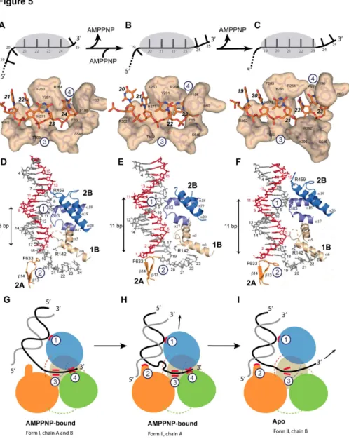
In drUvrD∆C form I, four nucleotides (ntβ1-β4) are tightly
bound in the ssDNA-binding pocket (Figure 5A). The terminal nucleotide (ntβ5) has already exited the helicase and is no longer visible in the electron density maps. Nucleotides β1 and ββ interact with Argγ6β and Asnγ64 (motif IVa) and stack against Pheβ6γ (motif III) that interferes with the regular stacking of the ssDNA bases and forces nucleotides βγ and β4 to adopt an orientation orthogonal to nucleotides β1 and ββ. Nucleotide β4 is stabilized in this conformation by π-stacking of the base between Argβ64 and Phe196 (motif Id) and of the deoxyribose ring against Phe65 (motif Ia). These residues are in turn stabilized by a series of stacking interactions involving notably Tyrβ61 (motif III) and His9γ (motif Ib). In the AMPPNP-bound molecule of drUvrD∆C form II, nucleotides β0 to βγ are
bound in the binding pocket (Figure 5B), indicating that drUvrD has translocated along the ssDNA by one nucleotide compared to form I and as a result both nucleotides β4 and β5 have become untraceable.
As in ecUvrD, the apo-drUvrD∆C observed in crystal form II,
sites. As a result, nucleotide βγ is now trapped on its way out. To allow the terminal nucleotide to exit, Phe196, Phe65 and His9γ from motifs Ia, Ib and Id have moved out of the way and the αβ4-αβ5 linker that interacts via Ser546 with the terminal nucleotide in the AMPPNP–bound forms, has maintained its grip on the γ′-end of the ssDNA and pulled it through the opened gateway driven by the rotation of domain βB (Figure 5C). Nucleotides β1 and ββ are now stabilized in their new binding sites by interactions with Tyrγ90 and Argγ9β from motif IVb.
Investigation of the dsDNA binding shows that it is also affected by the nucleotide-bound state of drUvrD (Figure 5D-F). Interactions between drUvrD and dsDNA involve four contact points: one helix-loop-helix (HLH) motif from domain 1B (α5-α6), two of the three HLH motifs from domain βB (α17-α18 and α19-αβ0) and the -hairpin motif ( 1γ- 14) from domain βA. In the AMPPNP-bound structures, three of these four sites are in contact with dsDNA; two of them are in common and the third differs between the two forms (Figure 5D and 5E). In both forms, Arg14β from the α5-α6 HLH motif interacts with the
Figure 3. Crystal structures of drUvrD-DNA complexes. A ribbon illustration of the AMPPNP-bound drUvrDFL is shown in A, the
AMPPNP-bound drUvrD∆C form I is shown in B, the mixed AMPPNP-bound (red) and apo- (blue) drUvrD∆C form II is shown in C. The
DNA and AMPPNP are shown in sticks. D-E. Large-scale conformational changes. D. Overlay of chains A (red) of drUvrDFL,
drUvrD∆C form I and apo-drUvrD∆C form II, illustrating the large spiral movement of chains B colored respectively yellow, grey and
blue. The DNA is shown as an orange ribbon. E. As in (D) but viewed down the DNA axis, and for clarity drUvrD∆C form I has been
removed.
doi: 10.1γ71/journal.pone.0077γ64.g00γ
unpaired nt19 at the ss-dsDNA junction and the α17-α18 HLH motif containing the conserved GIG sequence (motif IVc, Figure S1) interacts extensively with nt9-1β in form I and nt7-10 in form II (Figure 5D and 5E). In form I, the third binding site
involves Arg459 from the α19-αβ0 HLH motif, which interacts with the deoxyribose ring of nt1γ (opposite strand) in the minor groove of the DNA duplex (Figure 5D), while in form II, Phe6γγ from the -hairpin motif stacks against the first base-pair
Figure 4. Conformational changes associated with ATP hydrolysis and nucleotide release. A-C. Domain movements. The AMPPNP-bound form is colored in red, while the apo-form is colored in blue. A. Upon ATP hydrolysis and nucleotide release, domain βB along with the dsDNA rotates by ~15° and domain 1A and 1B by 8° relative to domain βA. B. Close up view of the rotation of domain βB and duplex DNA. C. Domains 1A and 1B undergo a 15° twist relative to domain βA around the ssDNA axis (orange). D. Conformational changes occurring at the ssDNA gateway (circled in green). The linker between domains βB and βA adopts a short helix (αβ5) and loop in the AMPPNP-bound form and interacts tightly with the γ′-end of the ssDNA via Ser546, while it consists of an unstructured loop (dashed line) in the apo-form. In the AMPPNP form, the ssDNA gateway is more closed: the distance between the carboxyl oxygen of Phe65 (motif Ia) and the hydroxyl group of Ser546 is 4.5 Å in the AMPPNP-bound form versus 9.9 Å in the apo-form. The represented DNA corresponds to the AMPPNP bound form.
Figure 5. DNA binding of drUvrD. Illustrations of drUvrD binding to dsDNA with a γ′-ssDNA tail in form I (A,D and G), form II with AMPPNP bound (B, E and H) and in the apo-form of form II (C, F and I). A-C. Schematic diagrams (top) illustrating the translocation of form I (A), form II with AMPPNP bound (B) and the apo-form of form II (C) of drUvrD∆C along the ssDNA. The ssDNA nucleotides
are illustrated as black bars and are numbered as in the crystal structures. The grey oval shape representing drUvrD covers the nucleotides bound in the ssDNA binding pocket. Surface representations of the ssDNA binding pockets of these three forms of
drUvrD∆C bound to ssDNA (orange sticks) are shown below. The important residues are labeled and the bases are numbered as in
the schematic diagrams. D-F. Binding of drUvrD∆C to dsDNA in form I (D), form II with AMPPNP bound (E) and in the apo-form of
form II (F). The dsDNA is illustrated in sticks with the translocated strand in grey. Domains of drUvrD are colored as in Figure βA. The helices belonging to the HLH motifs and the -hairpin structure (orange) are shown and labeled according to the secondary structure succession (Figure S1). The positively charged residues in contact with dsDNA are illustrated in sticks and the GIG motif is indicated. The number of base-pairs formed between the ss-dsDNA junction and the contact point with the drUvrD GIG motif is shown to the left of each panel. This number differs significantly between the two crystal forms. G-I. Schematic representation of
drUvrD's DNA binding in the different crystal structures as indicated below the models. The four protein-DNA contact points that are critical for the wrench-and-inchworm unwinding mechanism are indicated with circled numbers in all panels: HLH motifs interact with dsDNA (1), the -hairpin motif with the ss-dsDNA junction (β), motif III with the ssDNA (γ) and the ssDNA gateway with the exiting ssDNA (4). G. In AMPPNP bound Form I, contact points 1, γ and 4 are tight. H. In AMPPNP bound Form II, drUvrD's GIG motif (1) has slided along the DNA duplex and pushes the DNA junction against the -hairpin motif (β), which now stacks tightly against the first base-pair. I. In the apo molecule of Form II, the ssDNA gateway (4) has opened and ssDNA exited the helicase. Domains of
drUvrD are colored as in Figure βA.
doi: 10.1γ71/journal.pone.0077γ64.g005
(nt1=nt18) of the duplex (Figure 5E). In the absence of nucleotide, however, only two of these contacts remain: the GIG sequence in the α17-α18 HLH motif interacts with nt7-10 and Phe6γγ from the -hairpin motif stacks against the first base-pair (Figure 5F).
Analysis of the ss- and dsDNA binding in the different structures (Figure 5) indicate how local conformational changes and domain rotations are transformed into drUvrD's linear movement along the DNA via alternate loose and tight protein-DNA contact points, as proposed in the wrench-and-inchworm model for DNA unwinding. During ATP-binding-induced domain closing, binding to duplexed DNA through several HLH motifs (contact 1) and to ssDNA (contacts γ and 4) are tight, while contact with the ss-dsDNA junction (contact β) is loose (Figure 5D and 5G). UvrD then slides along the duplex away from the junction and thereby pushes the duplex DNA against the -hairpin (contact β). Phe6γγ located at the tip of the --hairpin now stacks against the first base-pair (Figure 5E and 5H). Since the ssDNA gateway is closed at this stage, this movement creates a tension on the ss-dsDNA junction, which distorts the first nucleotide at the ss-dsDNA junction (nt19), thus forming a bulge (Figure 5E and 5H). This tension is then released during ADP and Pi release: domain rotations open
drUvrD’s ssDNA gateway (contact 4) to allow the ssDNA to exit the helicase. Contacts with the ssDNA (contact γ) remain tight throughout the process (Fig.s 5A-5C) in order to guide and tether the ssDNA through the gateway and straighten the bulged out nucleotide (Figure 5I). During this step, contacts with the DNA duplex are restricted to the GIG motif in α17-α18 HLH (contact 1 is looser) and the -hairpin (contact β) that is stacked against the first base-pair and is now in a position to act as a solid separation pin for subsequent unwinding of the duplex DNA (Figure 5F and 5I).
drUvrD is an active, DNA-stimulated ATPase and an ATP-dependent helicase
To better understand how a structurally and mechanistically conserved protein such as UvrD may be involved in diverse repair pathways in different species, we investigated drUvrD’s catalytic activities in vitro. As other SF1A helicases, drUvrD displays a clear DNA-stimulated ATPase activity (Figure 6A). Analysis of the ATPase data measured on drUvrDFL and
drUvrD∆C allowed us to determine their apparent turnover rates
(Kcat) for ATP hydrolysis, along with their Km for ATP and their
KssDNA (corresponding to the concentration of ssDNA required
for half-maximal ATPase rate) (Figure 6A). These values are in agreement with those measured for wild-type and a C-terminally truncated form of ecUvrD [γ4,γ8]. When compared to
drUvrDFL,drUvrD∆C exhibits a significantly higher turnover rate
and reduced apparent affinities for both ATP and ssDNA, indicating that the C-terminal domain may be regulating the DNA binding and ATPase activities of drUvrD.
We then examined the helicase activity of drUvrDFL on
γ′-tailed, 5′-tailed and blunt dsDNA (Figure 6B). Our data reveals that drUvrD unwinds all three of these substrates in an ATP-dependent manner to varying extents and, as expected for a member of the SF1A helicase family, unwinds preferentially γ′-tailed dsDNA (Figure 6C and D and Figure Sβ). The length of
the ssDNA overhangs did not significantly affect the helicase activity of drUvrD, since very similar initial rates of unwinding were observed for 15nt and 7nt overhangs (Figure 6C). Although drUvrD shows a preference for unwinding γ′-tailed dsDNA, drUvrD also melts 5′-tailed DNA at a γ-fold lower rate and blunt dsDNA at a 9-fold lower rate. In the presence of
drSSB (added at a 1β.5-fold excess with respect to the DNA), the helicase activity on γ′-tailed and blunt dsDNA was unaffected, whereas drUvrD’s activity on 5′-tailed dsDNA was strongly stimulated to a rate similar to that observed on γ′-tailed dsDNA (Figure 6C and D and Figure Sβ). In these conditions,
drUvrD could unwind duplexed DNA with the same efficiency in both directions. SSB has previously been reported to directly stimulate the helicase activity of several other helicases [γ9,40]. Interestingly, we also observed helicase activity on γ′-tailed dsDNA containing a fluorescein-conjugated thymine within its ssDNA extension (Figure Sβ) and succeeded in crystallizing drUvrD∆C in complex with such a DNA. Data to
γ.0Å resolution were collected on crystals with modified DNA at position β1 and extra electron density could be seen close to the C7 group of thymine β1 (Figure Sγ). Several DNA helicases have previously been shown to unwind lesion-containing DNA [41]. ecUvrD was previously shown to efficiently unwind thymine glycol containing DNA [4β] and E. coli Rep can efficiently unwind a DNA substrate harboring a polyglycol linkage in the ssDNA extension [4γ]. These findings suggest that although UvrD helicases bind tightly to ssDNA, they are sufficiently flexible to allow bases with bulky modifications through their ssDNA gateway.
drUvrD translocates on ssDNA in the 3′-5′ direction only
We then investigated drUvrD’s ability to translocate on ssDNA using a streptavidin-displacement assay (Figure 7). We found that drUvrD could efficiently release streptavidin bound to biotin located at the end of 5′-tails in an ATP-dependent manner, but failed to displace streptavidin bound to biotin at the end of γ′-tails (Figure 7B and Figure S4), indicating that drUvrD translocates along ssDNA in the γ′-5′ direction. Here again, as in the helicase assays, addition of drSSB to the reaction mix did not affect translocation along γ′-tails, but significantly reduced the translocase activity on 5′-tails (Figure 7C). Interestingly, the addition of drSSB leads to both a reduction in the amount of streptavidin-free 5′-tailed dsDNA (middle band on the gel, corresponding to the product of the translocase activity) and a major increase in the amount of released ssDNA (lower band on the gel, corresponding to the product of the helicase activity). drSSB therefore modulates the activity of
Mutagenesis study of drUvrD’s DNA binding ability and helicase activity
Directionality of SF1 helicases is believed to be determined by preferential binding to either a γ′- or a 5′-ssDNA overhang, which acts as the entry point for the helicase [4]. We carried out fluorescence anisotropy measurements to evaluate the affinity of drUvrD for either γ′- or 5′-tailed dsDNA (Figure 8A and Figure S5). drUvrD binds to both of these substrates with similar affinity. The binding of drUvrD to γ’-tailed dsDNA is slightly stronger than to 5′-tailed dsDNA (Kd for γ′-tailed dsDNA:
0.γ6 µM, Kd for 5′-tailed dsDNA: 0.48 µM). These values are
also very close to the estimated affinity of drUvrDFL for ssDNA
derived from our ATPase data (KssDNA: 0.55 µM; Figure 6A). We
mutated residues identified in ecUvrD [19] as being essential for DNA unwinding using the wrench-and-inchworm mechanism (Gly4β4 and Gly4β6 from the GIG motif and the -hairpin) and tested the DNA binding and helicase activities of these drUvrD mutants on γ′- or 5′-tailed dsDNA (Figure 8). Mutating Gly4β6 to threonine (G4β6T) did not significantly affect the binding of drUvrD to γ′- and 5′-tailed dsDNA,
whereas mutating Gly4β4 to threonine (G4β4T) alone or together with the G4β6T mutation significantly impaired the binding of drUvrD to both γ′- and 5′-tailed dsDNA (Kd values
increased by γ-4 fold; Figure 8A and Figure S5). Deletion of the -hairpin structure, which is known to act as a separation pin and is essential for ecUvrD’s helicase activity [19], did not affect drUvrD’s binding to γ′-tailed dsDNA and led to a slightly reduced affinity for 5′-tailed dsDNA (Figure 8A and Figure S5). These mutations, however, had a much more dramatic effect on the helicase activities of drUvrD (Figure 8B). Deletion of the -hairpin dramatically reduced DNA unwinding of both 5′- and γ′-tailed dsDNA and this was also the case for the G4β6T mutant. In contrast, drUvrD-G4β4T mutants (single and double) showed a highly stimulated helicase activity on γ′-tailed dsDNA, as has previously been observed for ecUvrD [19], and a reduced activity on 5′-tailed dsDNA. These results suggest that drUvrD’s 5′-γ′ helicase activity relies on both a functional separation pin and tight binding to duplexed DNA via its GIG motif, whereas its γ′-5′ activity only requires the -hairpin structure.
Figure 6. ATPase and helicase activity of drUvrD. A. DNA-stimulated ATPase kinetic parameters of drUvrDFL and drUvrD∆C. B.
Structure of DNA oligonucleotides used for helicase assay of drUvrD. The fluorescein label is represented as a star. C.-D. Helicase activity of drUvrDFL on DNA substrates shown in (B). C. Table summarising the initial rates of unwinding of duplexed DNA containing
15 or 7 nucleotide ssDNA extensions at either the γ′ or 5′ ends and of blunt duplexed DNA, as indicated, and in the absence and presence of drSSB (β50 nM). The rates are given in base-pairs per min per UvrD helicase unit (bp/min/UvrD). D. Time course of
drUvrD unwinding of duplexed DNA containing 15 nucleotide ssDNA extensions at either the γ′ (red) or 5′ (black)-ends and of blunt (blue) duplexed DNA in the absence (full line) and presence (dotted line) of drSSB (β50 nM). Standard deviations are shown as vertical bars.
doi: 10.1γ71/journal.pone.0077γ64.g006
Discussion
Because of the helical nature of nucleic acids, helicases are expected to translocate along DNA in a spiral movement. For the first time, our structures trap this large-scale spiral movement and reveal how the combination of rotational and translational movements, associated with the positioning of the helicase at an angle relative to the dsDNA axis produce a spiral trajectory along the DNA duplex. In addition, our two higher resolution structures of drUvrD∆C provide new insight into the
detailed mechanisms underlying ATP-dependent DNA unwinding. Although the details of the protein-DNA contacts are not strictly identical in the structures of drUvrD, ecUvrD [19] and gsPcrA [18], taken together, our observations suggest that
the molecular mechanisms underlying this complex process are highly conserved within the SF1A helicase superfamily and support the tightly regulated wrench-and-inchworm model. The main differences we observe concern the gating mechanism regulating the exiting of the ssDNA. As in previous crystallographic studies of SF1 helicases [17-19], our crystal structures reveal no direct protein-protein contacts between neighboring UvrD monomers, even in the crystal structure of the intact drUvrDFL, in which the duplexed DNA is significantly
bent, bringing the two UvrD monomers close to each other (Figure γA).
Despite being structurally and mechanistically conserved with ecUvrD and gsPcrA, to our surprise, our biochemical assays revealed that drUvrD differs from its homologues in a
Figure 7. ssDNA translocase activity of drUvrD. Translocase activity of drUvrD was assayed using the streptavidin-displacement assay. A. Structure of DNA oligonucleotides used for drUvrD translocase assay measuring streptavidin displacement from biotinylated DNA substrates. The fluorescein label is represented as a star and the biotin label as a circle. B. Time course of
drUvrD (β50 nM) catalyzed streptavidin displacement from the γ′- (blue) and 5′- (red) ssDNA extensions of DNA oligonucleotides shown in (A). The fraction of released dsDNA (no longer bound to streptavidin) was quantified and plotted as a function of time. C. Translocase activity of drUvrD (β50 nM) on 5' tailed dsDNA (β0 nM) as a function of time in the absence (left) and the presence (right) of drSSB (β50 nM). The reaction products were analyzed on a 10 % polyacrylamide TBE gel. Bands correspond to the fluorescein labeled reaction products: streptavidin-bound dsDNA (upper bands, corresponding to several biotin labeled oligonucleotides bound to streptavidin), released dsDNA (middle band) and unwound ssDNA (lower band). D. The bands shown in (C), resulting from the time course of streptavidin displacement from 5′- tailed dsDNA, were quantified and the fraction of streptavidin-bound (black), released dsDNA (red) and unwound ssDNA (blue) were plotted as a function of time for reactions carried out in the absence (full lines) and presence (dotted lines) of drSSB (β50 nM). Standard deviations are shown as vertical bars.
number of ways. We found that, unlike ecUvrD, drUvrD could efficiently unwind dsDNA with only short (7nt) ssDNA overhangs. It is clear from our crystal structures that only a single drUvrD monomer can bind to such a short ssDNA tail. Although our data do not allow us to determine the active oligomeric state of drUvrD, our findings suggest that its helicase activity only requires that one UvrD monomer be loaded on the ssDNA tail. Our data also revealed that drUvrD can efficiently translocate along ssDNA with a biased γ′-5′ directionality as observed previously for ecUvrD [44-46], but in contrast can melt both γ′- and 5′-tailed DNA duplexes. This is consistent with our finding that drUvrD binds to both types of DNA. drUvrD also displayed a weak helicase activity on blunt DNA. Most members of the SF1A family show a clear γ′-5′ polarity [γ8,47,48]; there are, however, several examples of enzymes including the PcrA helicase, notably in gram-positive bacteria, that show bipolar helicase activity [49-54]. Several UvrD homologues are also known to act on blunt or nicked DNA [γ4,47,49,54,55]. Our findings now provide further evidence that SF1A helicases vary both in terms of substrate specificity and helicase polarity.
Interestingly, our experiments carried out in the presence of
drSSB, which is known to coat and protect nascent ssDNA in vivo, reveal that SSB plays an important role in modulating the balance between helicase and translocase activity on 5′-tailed dsDNA (Figure 9). The presence of SSB strongly favors the helicase versus translocase activity of drUvrD on such a substrate. This effect could be due to a direct regulation of
drUvrD’s activity by SSB or more likely to a steric effect of SSB
binding to the ssDNA extension. In contrast, SSB does not appear to have any effect on drUvrD’s activity on γ′-tailed dsDNA (Figure 9).
Our mutagenesis, DNA binding and helicase activity data indicate that regardless of the DNA substrate, the GIG motif of
drUvrD is critical for DNA binding and the -hairpin structure is essential for DNA unwinding of both 5′- and γ′-tailed DNA substrates. The GIG motif and the -hairpin separation pin are two essential features of the wrench-and-inchworm mode of unwinding and appear to be involved in both the γ′-5′ and on 5′-γ′ helicase activities of drUvrD. However, we also observe that mutating Gly4β4 from the GIG motif has a very contrasted effect on γ′-5′ and on 5′-γ′ helicase activity (stimulated γ′-5′ activity and reduced 5′-γ′ activity), indicating that the GIG motif from domain βB may be regulating these two processes differently. A number of ecUvrD mutants, including GIG mutants, are known to display reduced DNA binding and yet robust γ′-5′ helicase activity as observed for drUvrD [19,56]. This has been proposed to result from an alternative mode of unwinding, known as strand-displacement, in which movement of ssDNA is deregulated due to reduced contacts with dsDNA. This mechanism has been reported, notably in the absence of domain βB and duplex DNA binding [19,57]. In the case of
drUvrD, impaired DNA binding may cause drUvrD to switch from the controlled wrench-and-inchworm to an unregulated strand-displacement mode of unwinding on γ′-tailed DNA. In such a mode, the rotational movement of domain βB is no longer coupled to ATP binding and hydrolysis and as a result domain βB is no longer needed and may adopt a more open
Figure 8. DNA binding ability and helicase activity of drUvrD mutants. Comparison of DNA binding ability and helicase activity of wild type (WT) and drUvrD mutants: -hairpin deletion mutant (ΔHairpin), and mutants of the GIG motif from domain βB involved in dsDNA binding (G4β4T, G4β6T and double mutant G4β4T/G4β6T). A. DNA binding affinities (Kd values) of WT and mutant
drUvrD for either γ'-tailed (blue) or 5'-tailed (red) dsDNA determined by fluorescence anisotropy measurements. B. Helicase activity of WT and mutant drUvrD (β50 nM) on γ'-tailed (blue) or 5'-tailed (red) dsDNA (β0 nM). Initial reaction rates were determined from reaction time courses and were normalized with respect to the activity of WT drUvrD. Standard deviations are shown as vertical bars.
doi: 10.1γ71/journal.pone.0077γ64.g008
conformation, as observed in the DNA-free structures of
gsPcrA and ecUvrD [16,58].
The targeting and involvement of helicases in distinct cellular repair processes thus appears to be achieved by their abilities to bind and unwind specific structures corresponding to intermediates of these processes. For example, the 5′-γ′ unwinding activity of Staphylococcus aureus PcrA helicase is greatly stimulated in the presence of specific DNA structures [50]. drUvrD’s ability to unwind 5′- and γ′-tailed DNA duplexes and containing modified bases within the translocating strand may reflect its implication in diverse DNA repair pathways in vivo. In E. coli, recombinational repair has been proposed to involve the γ′-5′ helicases RecQ and Helicase IV and the 5′-γ′ helicase RecD, while D. radiodurans cells missing these genes show wild-type radioresistance and DNA repair capacity [7,59]. In contrast, inactivation of drUvrD leads to a significant increase in the sensitivity of cells to -irradiation [7]. This phenotype is further enhanced in cells in which both uvrD and
recD2 genes have been disrupted, suggesting that the 5′-γ′
helicase, drRecDβ, may in part back-up drUvrD’s function. While further studies will be needed to decipher the detailed molecular mechanisms that regulate the helicase activities of
drUvrD, these observations suggest that in vivo both helicase activities of drUvrD are needed. drUvrD may switch between its translocase and helicase activities in response to external stresses, changes in its environment, or, as suggested by our experiments in the presence of SSB, upon interactions with pathway-specific protein partners such as SSB, MutL or UvrAB [9,55,60].
Supporting Information
Table S1. Sequences of DNA oligonucleotides used in this study.
(DOCX)
Figure 9. Model of DNA duplex unwinding and ssDNA translocation by drUvrD. Models of drUvrD DNA unwinding and ssDNA translocase activity on 5' tailed dsDNA (top) and γ' tailed dsDNA (bottom) in the absence (left) and presence (right) of
drSSB. Using 5' tailed dsDNA, in the absence of drSSB drUvrD has low 5'-γ' helicase activity and high γ'-5' translocase activity while, in the presence of drSSB, drUvrD has high helicase activity and low translocase activity. Using γ'-tailed dsDNA, drUvrD has high γ'-5' helicase activity and no 5'-γ' translocase activity, regardless of the absence or presence of drSSB.
Table S2. Nature of contacts between the various domains of nucleotide-bound ec- and drUvrD.
(DOCX)
Table S3. Summary of the helical parameters of the DNA duplexes bound to drUvrD compared to ideal B-form DNA.
(DOCX)
Movie S1. DNA unwinding by drUvrD. The movie presents a morph between drUvrD∆C form I and drUvrD∆C form II. Molecule
A (red) is in an AMPPNP-bound form in both cases, while molecule B (blue) converts from an AMPPNP-bound form (I) to an apo-form (II).
(MOV)
Figure S1. Sequence alignment of D. radiodurans UvrD, E. coli UvrD, E. coli Rep and G. stearothermophilus PcrA helicases. The secondary structure of drUvrD is shown above the alignment and the domains are illustrated as colored lines below the alignment. The domains are colored as in Figure 1. The conserved helicase motifs are numbered and marked with yellow boxes.
(TIF)
Figure S2. Helicase activity on 3′-, 5′-tailed and blunt dsDNA. A. drUvrD (β50nM) unwinding of γ′-tailed β5 base-pair dsDNA (β0nM) with either 15nt- or 7nt ssDNA extensions in the absence and presence of SSB (β50nM). B. drUvrD (β50nM) unwinding of blunt β5 base-pair dsDNA (β0nM) in the absence and presence of SSB (β50nM). C. drUvrD (β50nM) unwinding of 5′-tailed β5 base-pair dsDNA (β0nM) with either 15nt- or 7nt ssDNA extensions in the absence and presence of SSB (β50nM). A-C. Reactions were stopped at the following time points: 0, γ0sec, 1min, βmin, 4min and 6min, prior to separation on β0% TBE gels. The fluorescein label is illustrated as a star in the schematic representation of the DNA.
(TIF)
Figure S3. Binding of fluorescein-labeled DNA to
drUvrD∆C. The DNA oligonucleotides contain a
fluorescein-conjugated thymine at position β1 within the ssDNA extension. The βFo-Fc electron density map (blue) is contoured at 1σ,
while the Fo-Fc difference density map (green) is contoured at β.5σ. The ssDNA is illustrated in sticks.
(TIF)
Figure S4. Translocase activity on 3′- and 5′-tailed dsDNA.
drUvrD (β50nM) translocation activity on streptavidin bound 5′-and γ′-tailed β5 base-pair dsDNA (β0nM) with β5nt ssDNA extensions. Reactions were stopped at the following time points: 0, γ0sec, 1min, βmin, 5min, 10 min and 15min, prior to separation on 10% TBE gels. The fluorescein and the biotin labels are illustrated respectively as a star and an open circle in the schematic representation of the DNA. The upper bands correspond to streptavidin-bound dsDNA substrate, the middle-band to the released dsDNA (translocase product) and the lower band corresponds to the product of the helicase activity of UvrD, i.e. ssDNA. Biotinylated dsDNA without streptavidin was loaded in the first well.
(TIF)
Figure S5. DNA binding to 3′- and 5′-tailed dsDNA. Binding of wild-type (WT) and mutant drUvrD to γ′- (A) and 5′-tailed dsDNA (B) was measured by fluorescence anisotropy. The anisotropy measured for the DNA alone was subtracted from all other values and the change in anisotropy (ΔA) is plotted as a function of UvrD concentration (µM). The averaged data points were fitted to a standard binding equation assuming a single binding site using GraphPad Prism6. Standard deviations are shown as vertical bars.
(TIFF)
Acknowledgements
We thank the ESRF beamline staff for assisting with data collection. This project benefited from access to the PSB (www.psb-grenoble.eu) facilities.
Author Contributions
Conceived and designed the experiments: JT. Performed the experiments: SA MS JT. Analyzed the data: SA MS SM JT. Contributed reagents/materials/analysis tools: SA MS SM JT. Wrote the manuscript: MS JT.
References
1. Tuteja N, Tuteja R (β004) Unraveling DNA helicases. Motif, structure, mechanism and function. Eur J Biochem β71: 1849-186γ. doi: 10.1111/j.14γβ-10γγ.β004.04094.x. PubMed: 151β8β95.
β. Tuteja N, Tuteja R (β004) Prokaryotic and eukaryotic DNA helicases. Essential molecular motor proteins for cellular machinery. Eur J Biochem β71: 18γ5-1848. doi:10.1111/j.14γβ-10γγ.β004.0409γ.x. PubMed: 151β8β94.
γ. Gorbalenya AE, Koonin EV (199γ) Helicases: amino acid sequence comparisons and structure-function relationships. Curr Opin Struct Biol γ: 419-4β9. doi:10.1016/S0959-440X(05)80116-β.
4. Singleton MR, Dillingham MS, Wigley DB (β007) Structure and mechanism of helicases and nucleic acid translocases. Annu Rev Biochem 76: βγ-50. doi:10.1146/annurev.biochem.76.05βγ05.115γ00. PubMed: 175066γ4.
5. Bruand C, Ehrlich SD (β000) UvrD-dependent replication of rolling-circle plasmids in Escherichia coli. Mol Microbiol γ5: β04-β10. doi: 10.1046/j.1γ65-β958.β000.01700.x. PubMed: 106γβ890.
6. Arthur HM, Lloyd RG (1980) Hyper-recombination in uvrD mutants of Escherichia coli K-1β. Mol Gen Genet 180: 185-191. doi:10.1007/ BF00β67γ68. PubMed: 700γγ07.
7. Bentchikou E, Servant P, Coste G, Sommer S (β010) A major role of the RecFOR pathway in DNA double-strand-break repair through ESDSA in Deinococcus radiodurans. PLOS Genet 6: e1000774. 8. Veaute X, Delmas S, Selva M, Jeusset J, Le Cam E et al. (β005) UvrD
helicase, unlike Rep helicase, dismantles RecA nucleoprotein filaments in Escherichia coli. EMBO J β4: 180-189. doi:10.10γ8/sj.emboj. 7600485. PubMed: 15565170.
9. Matson SW, Robertson AB (β006) The UvrD helicase and its modulation by the mismatch repair protein MutL. Nucleic Acids Res γ4: 4089-4097. doi:10.109γ/nar/gkl450. PubMed: 169γ5885.
10. Caron PR, Kushner SR, Grossman L (1985) Involvement of helicase II (uvrD gene product) and DNA polymerase I in excision mediated by the uvrABC protein complex. Proc Natl Acad Sci U S A 8β: 49β5-49β9. doi: 10.107γ/pnas.8β.15.49β5. PubMed: γ161077.