Receptor
a
Isoform in Individuals with Posttraumatic
Stress Disorder: A Cumulative Effect of Trauma Burden
Hannah Gola1., Andrea Engler2., Julia Morath3
, Hannah Adenauer3, Thomas Elbert3, Iris-Tatjana Kolassa1", Harald Engler2
*"
1Clinical and Biological Psychology, University of Ulm, Ulm, Germany,2Institute of Medical Psychology and Behavioral Immunobiology, University Hospital Essen, University of Duisburg-Essen, Essen, Germany,3Clinical Psychology and Neuropsychology, University of Konstanz, Konstanz, Germany
Abstract
Background:Posttraumatic stress disorder (PTSD) is a serious psychiatric condition that was found to be associated with altered functioning of the hypothalamic-pituitary-adrenal (HPA) axis and changes in glucocorticoid (GC) responsiveness. The physiological actions of GCs are primarily mediated through GC receptors (GR) of which isoforms with different biological activities exist. This study aimed to investigate whether trauma-experience and/or PTSD are associated with altered expression of GR splice variants.
Methods:GRaand GRbmRNA expression levels were determined by real-time quantitative PCR in whole blood samples of individuals with chronic and severe forms of PTSD (n =42) as well as in ethnically matched reference subjects (non-PTSD,
n =35).
Results:Individuals suffering from PTSD exhibited significantly lower expression of the predominant and functionally active GRa isoform compared to non-PTSD subjects. This effect remained significant when accounting for gender, smoking, psychotropic medication or comorbid depression. Moreover, the GRa expression level was significantly negatively correlated with the number of traumatic event types experienced, both in the whole sample and within the PTSD patient group. Expression of the less abundant and non-ligand binding GRb isoform was comparable between patient and reference groups.
Conclusions:Reduced expression of the functionally active GRaisoform in peripheral blood cells of individuals with PTSD seems to be a cumulative effect of trauma burden rather than a specific feature of PTSD since non-PTSD subjects with high trauma load showed an intermediate phenotype between PTSD patients and individuals with no or few traumatic experiences.
Citation:Gola H, Engler A, Morath J, Adenauer H, Elbert T, et al. (2014) Reduced Peripheral Expression of the Glucocorticoid ReceptoraIsoform in Individuals with Posttraumatic Stress Disorder: A Cumulative Effect of Trauma Burden. PLoS ONE 9(1): e86333. doi:10.1371/journal.pone.0086333
Editor:Katharina Domschke, University of Wuerzburg, Germany
ReceivedAugust 5, 2013;AcceptedDecember 8, 2013;PublishedJanuary 21, 2014
Copyright:ß2014 Gola et al. This is an open-access article distributed under the terms of the Creative Commons Attribution License, which permits unrestricted use, distribution, and reproduction in any medium, provided the original author and source are credited.
Funding:This work was funded by the German Research Foundation (DFG) Research Unit FOR751 (EN 814/1-2 and KO 3895/1-1) and the European Refugee Fund. The funders had no role in study design, data collection and analysis, decision to publish, or preparation of the manuscript.
Competing Interests:The authors have declared that no competing interests exist. * E-mail: [email protected]
.These authors contributed equally to this work.
"These authors share senior authorship
Introduction
Posttraumatic stress disorder (PTSD) is a serious psychiatric condition that can develop in the aftermath of traumatic events such as life-threatening accidents, natural disasters, physical assaults, sexual abuse, combat experience or torture. Character-istic symptoms of PTSD include re-experiencing of the traumatic event in the form of intrusive recollections, flashbacks or nightmares; persistent avoidance of stimuli associated with the traumatic event; emotional numbing; as well as a constant state of heightened alertness and increased arousal [1]. In addition to psychiatric morbidity, PTSD appears to be associated with altered functioning of the hypothalamic-pituitary-adrenal (HPA) axis and
changes in central and/or peripheral glucocorticoid (GC) respon-siveness [2–5], but the underlying mechanisms are not sufficiently understood yet.
predominant and functionally active receptor, is generated through splicing of exon 8 to the proximal part of exon 9 (9a), whereasGRb(742 amino-acid protein) is created through splicing of exon 8 to the distal part of exon 9 (9b). The two receptor isoforms share identical N-termini encoded by exons 2–8 (727 amino acids), but GRbdiffers from GRain itsC-terminus, where the 50 amino acids derived from exon 9a are replaced by a 15 amino-acid region encoded by exon 9b. Since this region contains important parts of the ligand binding domain, GRbis unable to bind natural and synthetic GCs such as cortisol and dexameth-asone, respectively [11,12]. In addition, no transcriptional activation or repression activities in response to hormone were found. Instead, it has been reported that GRb might act as a dominant-negative inhibitor of GRa, either through competition for co-regulators or through formation of inactive GRa/GRb
heterodimers [13,14]. Because of the ability of GRbto negatively regulate the action of GRa, it has been suggested that the ratio of GRa/GRbexpression might be a critical factor determining the GC responsiveness of target tissues.
Alterations in central and peripheral expression of GR splice variants have been reported for individuals with mood/affective disorders [15,16]. For example, GRa mRNA expression was shown to be reduced in peripheral blood cells of bipolar and major depressive disorder (MDD) patients during depressive state as well as in remission compared to healthy controls, while expression of the GRbsplice variant was not altered [16]. In PTSD patients, expression of GR isoforms has not been investigated yet, although total GR gene expression, GR number, and GC binding capacity were found to be altered in individuals suffering from this disorder [17–21].
The aim of this study was to investigate whether trauma-experience and/or PTSD are associated with changes in the expression of GR splice variants. GRa and GRb mRNA expression levels were determined by real-time quantitative PCR in whole blood samples of individuals with war- and torture-related PTSD as well as in non-PTSD subjects with low and high levels of trauma exposure.
Methods
Ethics Statement
All procedures were approved by the Ethics Committee of the University of Konstanz and were carried out in accordance with the Declaration of Helsinki (2008). Written informed consent was obtained from all individuals enrolled in the study.
Participants
Forty-two individuals with current PTSD (21 male, 21 female; mean age = 32.6 years, SD = 9.1, range 17–51) according to the DSM-IV and 35 non-PTSD subjects (13 male, 22 female; mean age = 29.1 years, SD = 9.7, range 17–61) were included in this study. Twenty of the subjects (11 PTSD, 9 non-PTSD) had already participated in a previous study on inflammatory markers (22). PTSD patients were refugees with chronic (mean symptom duration = 8.1 years, SD = 5.7) and severe (mean sum score in the Clinician Administered PTSD Scale [CAPS] = 84.2, SD = 17.3) forms of PTSD due to multiple, highly stressful war and torture experiences. In addition to the PTSD diagnosis, 33 patients (78.6%) met the DSM-IV criteria for a current major depressive episode. Sixteen PTSD patients (38.1%) reported regular intake of psychotropic medication and four women reported the use of oral contraceptives (Table 1). Twelve PTSD patients (28.6%) were smokers. All patients were recruited from the Psychotrauma
Research and Outpatient Clinic for Refugees, University of Konstanz, Germany.
The non-PTSD comparison group was recruited through advertisement and was matched to the patient group with regard to age and region of origin (see Table 1). Except for six women reporting the intake of oral contraceptives, all reference subjects were free of medication. Five non-PTSD subjects (14.3%) were smokers. Since the non-PTSD group varied with respect to the number of traumatic event types experienced (range: 0–8), some of the analyses were repeated with a three-group design. For this purpose, we divided the non-PTSD comparison group by median split into a group with substantial exposure to traumatic stressors (4–8 different traumatic event types;high trauma-exposed non-PTSD) and a group with no or few traumatic experiences (0–3 traumatic event types;low trauma-exposed non-PTSD) based on the number of past traumatic event types assessed with the event checklist of the CAPS.
Exclusion criteria for the study were intake of glucocorticoids or acute and chronic somatic illnesses. In addition, non-PTSD subjects were excluded if they met the criteria for any mental disorder according to DSM-IV, whereas PTSD patients were excluded if they met the criteria for comorbid alcohol and substance abuse or dependence, or a current or past history of psychosis according to the DSM-IV.
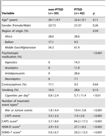
Table 1.Sociodemographic and clinical characteristics.
Variable
non-PTSD (n= 35)
PTSD (n= 42) p
Agea(years) 29.1
69.7 32.669.1 0.11
Gender (Female/Male) 22/13 21/21 0.26
Region of origin (%) 0.59
Africa 28.6 28.6
Balkan 17.1 9.5
Middle East/Afghanistan 54.3 61.9
Psychotropic medication (%)
,0.001
Hypnotics 0 14.3
Anxiolytics 0 11.9
Antidepressants 0 28.6
Neuroleptics 0 7.1
Contraceptives (%) 17.1 9.5 0.64
Smoking (%) 14.3 28.6 0.13
Cigarettes per daya 0.862.4 5.7611.4 ,0.01
Number of traumatic event typesa
War or torture events 1.864.4 10.465.8 ,0.001
CAPS events 3.562.3 7.462.0 ,0.001
CAPS scorea 3.7
68.0 84.2617.3 ,0.001
HAM-D scorea 2.9
63.5 27.168.3 ,0.001
SOMS-7 scorea 5.0
66.7 29.2612.5 ,0.001
aData are presented as mean
6SD. Group differences were analyzed using chi-square tests (categorical data) andt-tests (continuous data). CAPS, Clinician Administered PTSD Scale; HAM-D, Hamilton Depression Rating Scale; SOMS-7, Screening for Somatoform Symptoms-7.
Clinical Interviews
All participants underwent an extensive standardized clinical interview administered by experienced clinical psychologists and trained translators. PTSD symptoms and the number of traumatic event types experienced were assessed with the CAPS [23]. The vivo Checklist of War, Detention and Torture events [24], which assesses common traumatic experiences in conflict regions and during torture, allowed for a detailed evaluation of the number of traumatic event types experienced. The Mini International Neuropsychiatric Interview (M.I.N.I.) was used to screen for potential comorbid mental disorders [25]. In addition, the severity of depressive symptoms was assessed with the Hamilton Depres-sion Rating Scale (HAM-D; [26]). The Screening for Somatoform Symptoms-7 (SOMS-7) was used to check for potential somato-form disorders [27]. After complete description of the study to the subjects, written informed consent was obtained.
Specimen Collection
Blood samples were obtained from antecubital veins between 10 a.m. and 11 a.m., before starting with the clinical interviews. Blood for total RNA extraction was collected in PAXgene Blood RNA Tubes (PreAnalytiX GmbH, Hombrechtikon, Switzerland) and was, after an initial freezing step at –20uC for 24 h, stored at – 80uC until further processing. Blood for plasma cortisol determi-nation and complete blood cell (CBC) analysis was drawn into EDTA-treated tubes (BD Vacutainer, Franklin Lakes, NY, USA). Plasma was separated by centrifugation (30006g, 10 min, 4uC) and stored at –28uC.
GR Isoform mRNA Expression
PAXgene Blood RNA Tubes were thawed at room temperature and were incubated for 2 h after reaching ambient temperature. Total RNA was isolated from whole blood samples using the PAXgene Blood RNA Kit (Qiagen, Hilden, Germany) according to the manufacturer’s instructions. RNA concentration and purity were determined photometrically using a LabelGuard Microliter Cell (Implen, Munich, Germany) together with a BioPhotometer (Eppendorf, Hamburg, Germany). The A260/A280 ratios of all samples were within the recommended range between 1.8 and 2.1. First-strand cDNA was synthesized from 1.5mg total RNA using
the High Capacity cDNA Reverse Transcription Kit (Applied Biosystems, Darmstadt, Germany). Real-time quantitative PCR was performed on a 7500 Fast Real-Time PCR System (Applied Biosystems) using FAST qPCR MasterMix Plus Low ROX (Eurogentec, Seraing, Belgium) and the following cycling condi-tions: 5 min at 95uC followed by 50 cycles of 15 s at 95uC, 30 s at 60uC and 30 s at 72uC. Primers and probes were adapted from [28] and purchased from Microsynth (Balgach, Switzerland). A common forward primer (59 -TGTTTTGCTCCTGATCTGA-39), located in exon 6, as well as a common probe (59 -6-FAM-TGACTCTACCCTGCATGTACGAC-BHQ1–39), located in exon 7, were used for both GR splice variants. To discriminate among GR isoforms, specific reverse primers for GRa (59 -TCGGGGAATTCAATACTCA-39), located in exon 9a, and GRb (59- TGAGCGCC-AAGATTGT-39), located in exon 9b, were used. Hypoxanthine phosphoribosyl transferase (HPRT) served as endogenous reference gene (forward primer: 59 -CACTGGCAAAACAATGCAGACT-39; reverse primer: 59 -GTCTGGCTTATATCCAACACTTCGT-39; probe: 59 -6-FAM-CAAGCTTGCGACCTTGACCATCT-TTGGA-BHQ1– 39). For quantification of gene expression, serially diluted cDNA samples generated from purified specific PCR products (High Pure PCR Product Purification Kit, Roche Diagnostics, Mannheim,
Germany) were used as external standards in each run. Results are expressed as fg/mg total RNA.
Complete Blood Count
A complete blood count (CBC) including white blood cell differential was obtained using an automated hematology analyzer (XT-2000i, Sysmex, Horgen, Switzerland).
Cortisol Analysis
Total plasma cortisol concentrations were measured using a commercial enzyme-linked immunosorbent assay (Cortisol ELISA, IBL International, Hamburg, Germany) according to the manu-facturer’s instructions. Intra- and interassay variances were 5.6% and 6.9%, respectively.
Statistical Analysis
Data analysis was performed using SPSS 17.0 (SPSS Inc., Chicago, IL, USA). The level of significance was set atp,0.05. Normality of residuals was examined using the Kolmogorov-Smirnov test and data were log-transformed when necessary. Non-transformed data are presented in figures and tables. Group comparisons between non-PTSD subjects and PTSD patients were performed applying chi-square tests (categorical data) and t-tests (continuous data). For multiple group comparisons, data were analyzed by analysis of variance (ANOVA). Tukey’s HSD test was used for post hoc comparisons. Spearman’s rank correlation coefficient was applied to measure the strength of association between GR mRNA expression and clinical variables.
Results
Sociodemographic and Clinical Characteristics
PTSD (n =42) and non-PTSD groups (n =35) did not signifi-cantly differ in age, gender, region of origin, and intake of contraceptives (Table 1). However, PTSD patients smoked significantly more cigarettes per day than non-PTSD subjects. With respect to clinical variables, the PTSD group had experienced significantly more traumatic event types than the comparison group and had significantly higher CAPS, HAM-D, and SOMS-7 scores.
Cellular Composition of the Blood
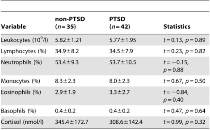
Since GRa and GRb expression levels were shown to vary among leukocyte subpopulations [29,30], we examined whether the cellular composition of the blood samples was comparable between PTSD patients and non-PTSD subjects. As shown in Table 2, total leukocyte numbers and percentages of leukocyte subsets did not significantly differ between study groups.
Expression of GRaand GRbIsoforms
GRaand GRbmRNA was detectable in blood samples from all study participants. Independent of PTSD diagnosis, GRamRNA levels were about 2,000-fold higher than GRb mRNA levels. PTSD patients exhibited significantly lower GRamRNA expres-sion levels compared to the non-PTSD comparison group [F(1,75) = 6.89,p= 0.01] whereas expression of the GRb isoform did not differ between groups [F(1,75) = 0.85,p= 0.36] (Fig. 1). In addition, no significant group difference was observed for the GRa/b ratio [F(1,75) = 2.56, p= 0.11]. Expression of HPRT, which served as reference gene, did not differ between PTSD patients and non-PTSD individuals [F(1,75) = 0.58,p= 0.45].
[F(1,74) = 6.70, p,0.05] was introduced as covariate into the model. Moreover, the effect became even stronger when all participants with psychotropic medication were excluded from the statistical analysis [F(1,59) = 12.53, p,0.01]. In a next step, we repeated the analyses with ‘gender’ or ‘major depression’ as additional between-subject factors to account for the possible influence of these variables on the effect reported here. For the expression of both GRaand GRbmRNA neither significant main effects of ‘gender’ nor significant ‘group6gender’ interactions
could be identified. After introducing ‘gender’ as additional covariate, the significant group difference for the GRaremained statistically significant [F(1,73) = 8.33, p,0.01]. Regarding the influence of major depression on GR expression, we found a main effect of ‘depression’ on the expression of GRa [F(1,73) = 5.58, p,0.05]. Again, the effect of PTSD diagnosis on GRaexpression remained statistically significant [F(1,73) = 13.34,p,0.001].
GR Isoform Expression and Plasma Cortisol
To determine whether the observed group difference in GRa
mRNA expression was related to individual differences in basal glucocorticoid secretion, we additionally measured blood cortisol levels in samples that were obtained at the same time as the
mRNA samples. Plasma cortisol concentrations did not signifi-cantly differ between PTSD patients and non-PTSD subjects (Table 2). Furthermore, no significant correlations between plasma cortisol and GRa (r =0.09, p= 0.46) or GRb (r= 0.09, p= 0.42) mRNA expression levels could be found.
GR Isoform Expression and Clinical Variables
To elucidate whether the observed reduction in GRa mRNA expression is a specific feature of PTSD or rather constitutes a general consequence of trauma exposure, we repeated the above reported analyses after subdividing the non-PTSD group into a group with no or few traumatic experiences (low trauma-exposed; n= 20) and a group with substantial exposure to traumatic stressors (high trauma-exposed;n= 15). With respect to the reduction in GRamRNA expression, the high trauma-exposed non-PTSD group displayed an intermediate phenotype, positioned between PTSD patients and the low trauma-exposed non-PTSD group, indicating a cumulative effect of trauma exposure on GRamRNA expression (Fig. 2). Furthermore, the number of traumatic event types assessed with the CAPS event list was significantly negatively correlated with GRamRNA expression level (r= –0.29,p,0.01). Within the PTSD group, GRa mRNA expression was again negatively correlated with the number of traumatic event types experienced (r= –0.32, p,0.05). In addition, GRa mRNA expression was negatively correlated with the hyperarousal subscale of the CAPS (r= –0.37,p,0.05) but was neither related to the total CAPS score (r= –0.02, p= 0.88), nor to intrusions (r =0.02, p= 0.90) or avoidance symptoms (r =0.08, p= 0.62). Furthermore, GRa mRNA expression showed no significant correlations with the duration of PTSD symptoms (r =0.13, p= 0.40), HAM-D (r= –0.04,p= 0.80) or SOMS-7 scores (r =0.06, p= 0.71).
Discussion
This study provides evidence for an altered GR isoform expression in individuals suffering from chronic and severe forms of PTSD. Peripheral blood mRNA expression of the predominant and functionally active GRasplice variant was about 20% lower in patients with war- and torture-related PTSD compared to non-PTSD control subjects without psychiatric diagnosis. In contrast, expression of the less abundant GRbisoform, which does not bind
Table 2.Blood cell parameters and plasma cortisol concentration.
Variable
non-PTSD (n= 35)
PTSD
(n= 42) Statistics
Leukocytes (109/l) 5.82
61.21 5.7761.95 t= 0.13,p= 0.89
Lymphocytes (%) 34.968.2 34.567.9 t= 0.23,p= 0.82
Neutrophils (%) 53.469.3 53.7610.5 t=20.15, p= 0.88
Monocytes (%) 8.362.3 8.062.3 t= 0.67,p= 0.50
Eosinophils (%) 2.961.9 3.362.7 t=20.84, p= 0.40
Basophils (%) 0.460.2 0.460.2 t= 0.47,p= 0.64
Cortisol (nmol/l) 345.46172.7 308.66142.4 t= 0.99,p= 0.32
Data are presented as mean6SD. doi:10.1371/journal.pone.0086333.t002
Figure 1. Glucocorticoid receptor isoform expression.Expression levels of glucocorticoid receptor (GR)aandbisoforms as well as GRa/GRb
ratio in the peripheral blood of non-PTSD subjects (n= 35) and PTSD patients (n= 42) measured by real-time quantitative PCR. Means and SEM are
shown. **p= 0.01.
GCs, was comparable between PTSD and non-PTSD groups. The results were neither influenced by the cellular composition of the blood samples nor by gender, smoking or psychotropic medica-tion. In addition, GRamRNA expression levels were not related to individual plasma cortisol concentrations, which did not significantly differ between PTSD and non-PTSD groups.
The observation of a diminished expression of the ligand-binding GRa isoform in severely traumatized PTSD patients is consistent with the findings of various studies that reported reduced GR numbers and/or GC binding capacity in blood immune cells of individuals with war-related PTSD [17–19,21]. However, other reports found that GR numbers may actually be higher [31,32] or similar [33,34] in PTSD compared to reference subjects. This inconsistency in the findings may be related to variations in sample characteristics (e.g., type of trauma, time elapsed since the traumatic event, PTSD symptom severity, use of reference groups with or without history of trauma) and differences in the techniques used to estimate GR numbers (e.g., mRNA/ protein expression, radioligand binding, flow cytometry).
A major risk factor for the development and persistence of PTSD constitutes the number of different traumatic event types experienced, with studies showing that higher trauma exposure is associated with higher prevalence of PTSD [35,36] as well as with lower probability of spontaneous remission from PTSD [37]. Our data suggests that such a dose-response effect of trauma load exists not only at the level of psychiatric symptoms but also at the physiological level, as non-PTSD subjects with substantial trauma experience (4–8 traumatic event types) show an intermediate GRa
expression level between low trauma-exposed non-PTSD subjects (0–3 traumatic event types) and PTSD patients. This assumption is further supported by the observation that the number of traumatic event types was negatively correlated with GRaexpression levels in the whole sample as well as within the PTSD patient group.
PTSD is not the only psychiatric condition that can develop following exposure to traumatic events. More than three-quarter of individuals suffering from PTSD meet the criteria for at least one other psychiatric disorder, with major depression being the most prevalent condition occurring concurrently with PTSD [38]. The present study included a sample of severely traumatized
subjects (victims of war and torture) of which 78.6% fulfilled the criteria for a current major depressive episode. Since reduced GRa mRNA expression as well as unchanged GRb mRNA expression in peripheral blood cells were also found in individuals with major depressive disorder, both during the depressive episode and in remission [16], it is plausible to argue that comorbid depression might have contributed to the observed GR isoform expression pattern in PTSD patients. However, various etiological routes may lead to depression symptoms, trauma exposure being one of them [38,39]. Since the inclusion of comorbid depression as an additional factor in our analysis did not eliminate the reported effect and GRa mRNA expression levels did not correlate with HAM-D scores, we assume that the observed group difference in GRa expression cannot be attributed to forms of depression unrelated to trauma exposure.
The reduced expression of the functionally active GRaisoform in the peripheral blood of PTSD subjects might be linked to the heightened inflammatory activity and immune dysregulation that has previously been found in subjects suffering from PTSD [22,40–43]. For example, PTSD patients were reported to exhibit increased peripheral NF-kB pathway activity [44] as well as increased circulating levels of inflammatory markers such as interleukin (IL)-6 and C-reactive protein (CRP) [45,46]. More-over, these changes were reported to correlate with PTSD symptom severity [43,44]. Since endogenous GCs restrain inflammatory responses, e.g., via repression of NF-kB-mediated activation of pro-inflammatory genes, it can be speculated whether the observed reduction in peripheral GRaexpression contributes to low-grade inflammation in PTSD.
The current study has notable strengths such as the assessment of PTSD diagnosis by applying the CAPS as clinical interview by experienced experts and the inclusion of low and high trauma-exposed non-PTSD reference groups. Nevertheless some limita-tions should be considered as well. For instance, we did not assess GR isoform expression beyond the transcriptional level to determine whether the observed group differences in mRNA expression are also reflected at the protein level, even though earlier studies have shown that, in most human tissues including peripheral blood cells, GRa protein expression matches up quite Figure 2. Relationship between trauma load and GRaisoform expression.(A) Number of different traumatic event types, (B) PTSD symptom
severity, and (C) GRamRNA expression levels in non-PTSD subjects with no or few traumatic experiences (0–3 traumatic event types;low
trauma-exposed non-PTSD;n= 20), in non-PTSD subjects with substantial exposure to traumatic stressors (4–8 traumatic event types;high trauma-exposed non-PTSD;n= 15), and in PTSD patients (n= 42). Means and SEM are shown. *p,0.05, **p,0.01, ***p,0.001.
well with GRamRNA levels [28,30]. Furthermore, the design of our study does not allow disentangling the effects of PTSDper se from the effects of trauma load, a problem which is, however, not easily solved since trauma exposure itself constitutes a well-described risk factor for the development of PTSD [35,36]. The approach to collect a reference group matching the PTSD group with respect to the number of traumatic events experienced, sometimes considered as gold standard, is problematic itself (e.g., by recruiting an especially resilient comparison group), and was therefore not adopted in our study.
Taken together, this study provides novel evidence for reduced expression of the ligand-binding and functionally active GRa
isoform in the blood of severely traumatized individuals, suggest-ing that the responsiveness of peripheral target cells towards GCs might be diminished in patients with chronic and severe forms of PTSD. However, GC sensitivity is not only determined by GR expression but also influenced by several other factors such as post-translational modifications, the composition of the GR complex,
and the efficiency of the ligand-induced nuclear translocation of the GR [47,48]. Thus, future studies should aim at elucidating whether important steps in the GR signaling cascade are additionally affected in severely traumatized subjects.
Acknowledgments
We thank Claudia Catani, Gilava Hamuni, Frank Neuner, Martina Ruf-Leuschner, and Maggie Schauer for clinical supervision and treatment of patients, Stephan Kolassa for support in the statistical analysis of the data, and Alexandra Kornowski, Heike Riedke, and Christiane Wolf for technical assistance.
Author Contributions
Conceived and designed the experiments: HG AE TE IK HE. Performed the experiments: HG AE JM HA HE. Analyzed the data: HG AE HE. Contributed reagents/materials/analysis tools: AE TE IK HE. Wrote the paper: HG AE HE.
References
1. American Psychiatric Association (1994) Diagnostic and Statistical Manual of Mental Disorders (DSM-IV). American Psychiatric Press, Washington, DC. 2. de Kloet CS, Vermetten E, Geuze E, Kavelaars A, Heijnen CJ, et al. (2006)
Assessment of HPA-axis function in posttraumatic stress disorder: pharmaco-logical and non-pharmacopharmaco-logical challenge tests, a review. J Psychiatr Res 40: 550–567.
3. Gola H, Engler H, Schauer M, Adenauer H, Riether C, et al. (2012) Victims of rape show increased cortisol responses to trauma reminders: a study in individuals with war- and torture-related PTSD. Psychoneuroendocrinology 37: 213–220.
4. Rohleder N, Wolf JM, Wolf OT (2010) Glucocorticoid sensitivity of cognitive and inflammatory processes in depression and posttraumatic stress disorder. Neurosci Biobehav Rev 35: 104–114.
5. Yehuda R (2009) Status of glucocorticoid alterations in post-traumatic stress disorder. Ann NY Acad Sci 1179: 56–69.
6. De Bosscher K, Vanden Berghe W, Haegeman G (2003) The interplay between the glucocorticoid receptor and nuclear factor-kB or activator protein-1: molecular mechanisms for gene repression. Endocr Rev 24: 488–522. 7. Nicolaides NC, Galata Z, Kino T, Chrousos GP, Charmandari E (2010) The
human glucocorticoid receptor: molecular basis of biologic function. Steroids 75: 1–12.
8. Hollenberg SM, Weinberger C, Ong ES, Cerelli G, Oro A, et al. (1985) Primary structure and expression of a functional human glucocorticoid receptor cDNA. Nature 318: 635–641.
9. Encio IJ, Detera-Wadleigh SD (1991) The genomic structure of the human glucocorticoid receptor. J Biol Chem 266: 7182–7188.
10. Revollo JR, Cidlowski JA (2009) Mechanisms generating diversity in glucocorticoid receptor signaling. Ann NY Acad Sci 1179: 167–178. 11. Bamberger CM, Bamberger AM, de Castro M, Chrousos GP (1995)
Glucocorticoid receptorb, a potential endogenous inhibitor of glucocorticoid action in humans. J Clin Invest 95: 2435–2441.
12. Oakley RH, Sar M, Cidlowski JA (1996) The human glucocorticoid receptorb
isoform. Expression, biochemical properties, and putative function. J Biol Chem 271: 9550–9559.
13. de Castro M, Elliot S, Kino T, Bamberger C, Karl M, et al. (1996) The non-ligand bindingb-isoform of the human glucocorticoid receptor (hGRb): tissue levels, mechanism of action, and potential physiologic role. Mol Med 2: 597– 607.
14. Yudt MR, Jewell CM, Bienstock RJ, Cidlowski JA (2003) Molecular origins for the dominant negative function of human glucocorticoid receptorb. Mol Cell Biol 23: 4319–4330.
15. Alt SR, Turner JD, Klok MD, Meijer OC, Lakke EA, et al. (2010) Differential expression of glucocorticoid receptor transcripts in major depressive disorder is not epigenetically programmed. Psychoneuroendocrinology 35: 544–556. 16. Matsubara T, Funato H, Kobayashi A, Nobumoto M, Watanabe Y (2006)
Reduced glucocorticoid receptoraexpression in mood disorder patients and first-degree relatives. Biol Psychiatry 59: 689–695.
17. de Kloet CS, Vermetten E, Bikker A, Meulman E, Geuze E, et al. (2007) Leukocyte glucocorticoid receptor expression and immunoregulation in veterans with and without post-traumatic stress disorder. Mol Psychiatry 12: 443–453. 18. Gotovac K, Sabioncello A, Rabatic S, Berki T, Dekaris D (2003) Flow
cytometric determination of glucocorticoid receptor (GCR) expression in lymphocyte subpopulations: lower quantity of GCR in patients with post-traumatic stress disorder (PTSD). Clin Exp Immunol 131: 335–339.
19. Matic G, Milutinovic DV, Nestorov J, Elakovic I, Jovanovic SM, et al. (2013) Lymphocyte glucocorticoid receptor expression level and hormone-binding properties differ between war trauma-exposed men with and without PTSD. Prog Neuropsychopharmacol Biol Psychiatry 43: 238–245.
20. Su TP, Zhang L, Chung MY, Chen YS, Bi YM, et al. (2009) Levels of the potential biomarker p11 in peripheral blood cells distinguish patients with PTSD from those with other major psychiatric disorders. J Psychiatr Res 43: 1078– 1085.
21. Vidovic A, Gotovac K, Vilibic M, Sabioncello A, Jovanovic T, et al. (2011) Repeated assessments of endocrine- and immune-related changes in posttrau-matic stress disorder. Neuroimmunomodulation 18: 199–211.
22. Gola H, Engler H, Sommershof A, Adenauer H, Kolassa S, et al. (2013) Posttraumatic stress disorder is associated with an enhanced spontaneous production of pro-inflammatory cytokines by peripheral blood mononuclear cells. BMC Psychiatry 13: 40.
23. Blake DD, Weathers FW, Nagy LM, Kaloupek DG, Gusman FD, et al. (1995) The development of a Clinician-Administered PTSD Scale. J Trauma Stress 8: 75–90.
24. Schauer M, Neuner F, Elbert T (2011) Narrative exposure therapy. In: A short-term treatment for traumatic stress disorders. Go¨ttingen, Hogrefe & Huber, 77– 79.
25. Sheehan DV, Lecrubier Y, Sheehan KH, Amorim P, Janavs J, et al. (1998) The Mini-International Neuropsychiatric Interview (M.I.N.I.): the development and validation of a structured diagnostic psychiatric interview for DSM-IV and ICD-10. J Clin Psychiatry 59 Suppl 20: 22–33.
26. Hamilton M (1960) A rating scale for depression. J Neurol Neurosurg Psychiatry 23: 56–62.
27. Rief W, Hiller W (2003) A new approach to the assessment of the treatment effects of somatoform disorders. Psychosomatics 44: 492–498.
28. Hagendorf A, Koper JW, de Jong FH, Brinkmann AO, Lamberts SW, et al. (2005) Expression of the human glucocorticoid receptor splice variantsa,b, and P in peripheral blood mononuclear leukocytes in healthy controls and in patients with hyper- and hypocortisolism. J Clin Endocrinol Metab 90: 6237–6243. 29. Li LB, Leung DY, Hall CF, Goleva E (2006) Divergent expression and function
of glucocorticoid receptorbin human monocytes and T cells. J Leukoc Biol 79: 818–827.
30. Pujols L, Mullol J, Roca-Ferrer J, Torrego A, Xaubet A, et al. (2002) Expression of glucocorticoid receptor a- and b-isoforms in human cells and tissues. Am J Physiol Cell Physiol 283: C1324–1331.
31. van Zuiden M, Geuze E, Willemen HL, Vermetten E, Maas M, et al. (2011) Pre-existing high glucocorticoid receptor number predicting development of posttraumatic stress symptoms after military deployment. Am J Psychiatry 168: 89–96.
32. Yehuda R, Lowy MT, Southwick SM, Shaffer D, Giller EL, Jr. (1991) Lymphocyte glucocorticoid receptor number in posttraumatic stress disorder. Am J Psychiatry 148: 499–504.
33. Golier JA, Schmeidler J, Legge J, Yehuda R (2006) Enhanced cortisol suppression to dexamethasone associated with Gulf War deployment. Psycho-neuroendocrinology 31: 1181–1189.
34. Yehuda R, Golier JA, Yang RK, Tischler L (2004) Enhanced sensitivity to glucocorticoids in peripheral mononuclear leukocytes in posttraumatic stress disorder. Biol Psychiatry 55: 1110–1116.
36. Neuner F, Schauer M, Karunakara U, Klaschik C, Robert C, et al. (2004) Psychological trauma and evidence for enhanced vulnerability for posttraumatic stress disorder through previous trauma among West Nile refugees. BMC Psychiatry 4: 34.
37. Kolassa IT, Ertl V, Eckart C, Onyut LP, Kolassa S, et al. (2010) Spontaneous remission from PTSD depends on the number of traumatic event types experienced. Psychol Trauma 2: 169–174.
38. O’Donnell ML, Creamer M, Pattison P (2004) Posttraumatic stress disorder and depression following trauma: understanding comorbidity. Am J Psychiatry 161: 1390–1396.
39. Rosen GM, Lilienfeld SO (2008) Posttraumatic stress disorder: an empirical evaluation of core assumptions. Clin Psychol Rev 28: 837–868.
40. Baker DG, Nievergelt CM, O’Connor DT (2012) Biomarkers of PTSD: neuropeptides and immune signaling. Neuropharmacology 62: 663–673. 41. Pace TW, Heim CM (2011) A short review on the psychoneuroimmunology of
posttraumatic stress disorder: from risk factors to medical comorbidities. Brain Behav Immun 25: 6–13.
42. Sommershof A, Aichinger H, Engler H, Adenauer H, Catani C, et al. (2009) Substantial reduction of naive and regulatory T cells following traumatic stress. Brain Behav Immun 23: 1117–1124.
43. von Kanel R, Hepp U, Kraemer B, Traber R, Keel M, et al. (2007) Evidence for low-grade systemic proinflammatory activity in patients with posttraumatic stress disorder. J Psychiatr Res 41: 744–752.
44. Pace TW, Wingenfeld K, Schmidt I, Meinlschmidt G, Hellhammer DH, et al. (2012) Increased peripheral NF-kB pathway activity in women with childhood abuse-related posttraumatic stress disorder. Brain Behav Immun 26: 13–17. 45. Maes M, Lin AH, Delmeire L, Van Gastel A, Kenis G, et al. (1999) Elevated
serum interleukin-6 (IL-6) and IL-6 receptor concentrations in posttraumatic stress disorder following accidental man-made traumatic events. Biol Psychiatry 45: 833–839.
46. Spitzer C, Barnow S, Volzke H, Wallaschofski H, John U, et al. (2010) Association of posttraumatic stress disorder with low-grade elevation of C-reactive protein: evidence from the general population. J Psychiatr Res 44: 15– 21.
47. Bamberger CM, Schulte HM, Chrousos GP (1996) Molecular determinants of glucocorticoid receptor function and tissue sensitivity to glucocorticoids. Endocr Reviews 17: 245–261.