Are GM-CSF-Independent and Are Not Required for Th
Cell Priming after Subcutaneous Immunization
Brian T. Edelson , Tara R. Bradstreet , Wumesh KC , Kai Hildner1 1 1 1,2¤ 1 3 John H. Russell3, Theresa L. Murphy1, Emil R. Unanue1, Kenneth M. Murphy1,2*
1Department of Pathology and Immunology, Washington University School of Medicine, St. Louis, Missouri, United States of America,2Howard Hughes Medical Institute, Washington University School of Medicine, St. Louis, Missouri, United States of America,3Department of Developmental Biology, Washington University School of Medicine, St. Louis, Missouri, United States of America
Abstract
Dendritic cells (DCs) subsets differ in precursor cell of origin, functional properties, requirements for growth factors, and dependence on transcription factors. Lymphoid-tissue resident CD8a+conventional DCs (cDCs) and CD11blow/2CD103+
non-lymphoid DCs are developmentally related, each being dependent on FMS-like tyrosine kinase 3 ligand (Flt3L), and requiring the transcription factors Batf3, Irf8, and Id2 for development. It was recently suggested that granulocyte/ macrophage colony stimulating factor (GM-CSF) was required for the development of dermal CD11blow/2Langerin+CD103+
DCs, and that this dermal DC subset was required for priming autoreactive T cells in experimental autoimmune encephalitis (EAE). Here, we compared development of peripheral tissue DCs and susceptibility to EAE in GM-CSF receptor deficient (Csf2rb2/2) andBatf32/2mice. We find thatBatf3-dependent dermal CD11blow/2Langerin+DCs do develop inCsf2rb2/2
mice, but that they express reduced, but not absent, levels of CD103. Further,Batf32/2mice lacking all peripheral CD11blow/2
DCs show robust Th cell priming after subcutaneous immunization and are susceptible to EAE. Our results suggest that defective T effector priming and resistance to EAE exhibited byCsf2rb2/2mice does not result from the absence of dermal
CD11blow/2Langerin+CD103+DCs.
Citation:Edelson BT, Bradstreet TR, KC W, Hildner K, Herzog JW, et al. (2011)Batf3-Dependent CD11blow/2Peripheral Dendritic Cells Are GM-CSF-Independent and Are Not Required for Th Cell Priming after Subcutaneous Immunization. PLoS ONE 6(10): e25660. doi:10.1371/journal.pone.0025660
Editor:Charalampos Babis Spilianakis, University of Crete, Greece
ReceivedAugust 9, 2011;AcceptedSeptember 8, 2011;PublishedOctober 17, 2011
Copyright:ß2011 Edelson et al. This is an open-access article distributed under the terms of the Creative Commons Attribution License, which permits unrestricted use, distribution, and reproduction in any medium, provided the original author and source are credited.
Funding:KMM was supported by the Howard Hughes Medical Institute (www.hhmi.org), BTE by a Burroughs Wellcome Fund Career Award for Medical Scientists (www.bwfund.org), a Jack H. Ladenson Fellowship in Experimental Clinical Pathology at Washington University School of Medicine, and an American Society of Hematology Scholar Award (www.hematology.org), KH by the Emmy Noether Program of the German Research Foundation (www.dfg.de/index.jsp), ERU by a grant from the National Institutes of Health (www.nih.gov, AI024742), and JHR by a grant from the National Multiple Sclerosis Society (www.nationalmssociety. org, RG 4352). The funders had no role in study design, data collection and analysis, decision to publish, or preparation of the manuscript.
Competing Interests:The authors have declared that no competing interests exist.
* E-mail: [email protected]
¤ Current address: Department of Internal Medicine, Medical Clinic 1, University of Erlangen-Nu¨rnberg, Erlangen, Germany
Introduction
GM-CSF regulates the development and activity of several myeloid cell types and influences both the initiation and maintenance of adaptive immune responses [1]. Mice lacking GM-CSF or its receptor show decreased antigen specific T cell priming against the encephalitogenic peptide of myelin oligoden-drocyte glycoprotein (MOG35–55) and are resistant to EAE [2]. Beyond this role in priming adaptive immune responses, GM-CSF acts to sustain ongoing effector responses, both in EAE initiated by MOG35–55 peptide [2] and in collagen-induced arthritis [3]. Similarly, in a murine model of autoimmune myocarditis, GM-CSF acts during priming of Th17 responses by promoting IL-6 and IL-23 production from DCs and during ongoing autoimmune responses by promoting survival of autoreactive CD4+
T cells [4]. Several recent studies indicate that it is GM-CSF rather than IL-17 that is the primary Th17-derived cytokine responsible for the development of EAE [5–7]. Initially, it was thought that IL-17 production would explain why Th17 cells and RORct were required for EAE. However,in vivo neutralization of IL-17 only
reduced EAE severity but did not eliminate disease, and non-IL-17 and non-IFN-cpathways have been implicated [8–10]. IL-23, but not IL-6 or TGF-b, was found to be critical for regulating the pathogenicitiy of Th17 cells [11,12]. GM-CSF derived from T cells, but not other CNS-resident or peripheral immune cells, was required for development of EAE [5], and acted during the effector phase to augment pathogenicity of Th17 cells [6,7]. IL-23 induced Th17 cells to increase production of GM-CSF, which further enhanced IL-23 production by antigen-presenting cells [6]. RORct was shown to induce GM-CSF, which acted on infiltrating myeloid cells entering the CNS, rather than resident microglia, during the development of EAE [7]. These studies suggest a model of EAE in which pathogenic T cells secrete GM-CSF that activates myeloid cells infiltrating the CNS to produce pathogenic lesions and further amplifies IL-23 production by DCs [13].
A recent report has suggested that the role for GM-CSF in EAE was based on its requirement for the development of dermal CD11blow/2Langerin+CD103+ DCs [14]. Dermal CD11blow/2
Langerin+
CD103+
DCs represent one anatomic subtype of peripheral tissue CD11blow/2CD103+
lymphoid tissue CD8a+
cDCs developmental dependence onBatf3,
Irf8, and Id2 [15,16]. CD8a+
cDCs are cross-presenting DCs, characterized by consistent expression of DEC205, and variable expression of CD103 which can be regulated by GM-CSF, IL-3, and TGF-b1 [17–19]. Mice lacking GM-CSF or the GM-CSF receptor were reported to lack dermal CD11blow/2
Langer-in+CD103+ DCs in the skin and peripheral lymph nodes under
steady state and inflammatory conditions, and were resistant to EAE [14]. In addition, radiation chimeras expressing the human diphtheria toxin receptor (DTR) controlled by the Langerin promoter [20] and depleted of peripheral CD11blow/2
Langer-in+CD103+
DCs were resistant to EAE and had reduced priming of MOG35–55-specific T cells [14]. This study concluded that GM-CSF was required for the initiation of EAE because it was required for the development of dermal CD11blow/2Langerin+
CD103+
DCs, which were required to prime pathogenic T cells [14]. To test whether loss of GM-CSF receptor protects from EAE by eliminating dermal CD11blow/2Langerin+
CD103+DCs, we asked ifBatf32/2mice, which also lack this DC subset, are also protected
from EAE. We find that Batf32/2 mice develop EAE after
immunization and have increased numbers of MOG35–55 peptide-specific T cells, excluding a requirement for either peripheral CD11blow/2CD103+
DCs or lymphoid tissue-resident CD8a+
DCs in EAE development. Direct comparison of Batf32/2 and
GM-CSF receptor deficient mice revealed that GM-CSF signaling was not required for the development of dermal CD11blow/2
Langerin+
CD103+
DCs, but rather regulated the expression level of CD103 on all peripheral DCs.
Results
Batf32/2mice develop EAE following MOG immunization
To compare Batf32/2 mice with mice lacking the GM-CSF
receptor (Csf2rb2/2) on the same genetic background, we
backcrossed Batf32/2 mice onto the C57BL/6 background for
10 generations. Csf2rb2/2 mice lack the common b-chain for
signaling through receptors for GM-CSF, IL-5, and IL-3. However, mice have a secondb-chain,Csf2rb2, which selectively pairs with the IL-3 receptor a-chain. Thus,Csf2rb2/2mice lose
responsiveness to GM-CSF and IL-5, but not to IL-3 [21,22]. Wild type (C57BL/6),Batf32/2, andCsf2rb2/2mice were
subcutane-ously immunized with MOG35–55peptide as previously described and followed for clinical disease [14]. Csf2rb2/2 mice remained
disease free as reported [14], butBatf32/2
mice developed EAE with the same severity and kinetics as wild type C57BL/6 mice (Fig. 1A).
In draining lymph nodes, examination by ELISPOT assay showed that immunized Batf32/2 mice had an increased
frequency of MOG35–55-specific T cells producing IL-17 and IFN-c(Fig. 1B), whileCsf2rb2/2mice had a significantly reduced
frequency of such cells, as previously reported [14]. Thus,Batf32/2
mice, which lack all peripheral CD11blow/2CD103+
DCs [16], are able to prime pathogenic CD4+T cells, and remain susceptible to
EAE. These findings would disagree with the interpretation that resistance to EAE in Csf2rb2/2mice results from the absence of
dermal CD11blow/2
Langerin+
CD103+
DCs as was recently suggested [14].
Csf2rb2/2 mice retain development ofBatf3-dependent
dermal CD11blow/2Langerin+DCs
GM-CSF was recently reported to directly regulate CD103 expression on splenic CD8a+ DCs [17–19], so conceivably Csf2rb2/2mice might retain dermal CD11blow/2Langerin+
DCs that simply lack CD103. To test this, we examined DCs from
C57BL/6, Batf32/2, and Csf2rb2/2 mice for expression of
markers that distinguish between Batf3-dependent and Batf3 -independent DC subsets (Fig. 2A).
In skin draining lymph nodes (SDLNs), migratory DCs are evident as DEC205+
CD8a2 cells, which can be divided into at
least three subsets based on CD11b, Langerin, and CD103 expression [23–25]. C57BL/6, Batf32/2, and Csf2rb2/2 mice
each contained normal populations of dermal Langerin2DCs and
Langerhans cells of the epidermis (CD11b+
Langerin+
CD1032
Ep-CAMhigh) (Fig. 2A). However, differences were evident in the dermal CD11blow/2Langerin+
CD103+
DCs between these strains. C57BL/6 mice contained the normal population of dermal CD11blow/2Langerin+CD103+EpCAMmidDCs, which were miss-ing inBatf32/2mice [16]. Notably,Csf2rb2/2mice retained this
population of dermal DCs, but in this strain of mice, these DCs expressed slightly lower levels of CD103 relative to C57BL/6 mice. CD24 can also been used to distinguish between populations of dermal DCs [26], and similarly C57BL/6 mice contained a population of dermal Langerin+
CD24+
EpCAMmidDCs that were completely missing inBatf32/2 mice, but which were present in Csf2rb2/2 mice (Fig. 2B). Csf2rb2/2 mice showed reduced total Figure 1.Batf32/2mice are susceptible to EAE.(A) Clinical course
of EAE in C57BL/6 mice,Batf32/2 mice (C57BL/6 background), and Csf2rb2/2mice. Data are combined from two experiments (n = 10 mice
per group). Points represent the mean clinical score. For clarity, error bars are not displayed. (B) ELISPOT assays on popliteal lymph node cells at day 7 following footpad immunization (MOG35–55peptide in CFA)
and restimulation with MOG35–55peptide. Data are from one of two
similar experiments (n = 3 per group). Mean 6 SEM, p values by unpaired student’s t test.
doi:10.1371/journal.pone.0025660.g001
CD11blow/-Peripheral DCs Are GM-CSF-Independent
lymph node cellularity and a reduced number of overall lymph node DCs, but normal numbers of Langerhans cells and dermal CD11blow/2Langerin+
DCs (Fig. 2C). Batf32/2 mice showed a
selective,100-fold reduction in the number of dermal CD11blow/2
Langerin+
DCs. These results indicate that Csf2rb2/2 mice do
contain dermal CD11blow/2Langerin+
DCs, but that in the absence of GM-CSF signaling, CD103 expression is reduced.
C57BL/6Batf32/2mice contain CD8a+cDCs in SDLNs but
not spleen
In analyzing SDLNs of C57BL/6Batf32/2mice, we noticed an
unexpected population of DEC205+CD8a+ cDCs that were
eliminated in the spleen and SDLNs of 129S6/SvEv Batf32/2
mice [16,27] (Fig. 2A, 2C). Therefore, we directly compared DCs from SDLNs ofBatf32/2mice on both genetic backgrounds (Fig.
S1A). C57BL/6Batf32/2mice lacked dermal CD11blow/2CD103+
DCs (Fig. S1A) but had a persistence of DEC205+CD8a+cDCs in
peripheral lymph nodes (Fig. S1A, S1B). In spleens,Batf32/2mice
showed a significant decrease in DEC205+
CD8a+
cDCs on both C57BL/6 and 129S6/SvEv backgrounds compared to controls (Fig. S1C, S1D). On the 129S6/SvEv background, DEC205+
CD8a+
cDCs were reduced from 4.8% of splenic cDCs in control mice to only 0.11% of splenic cDCs inBatf32/2 mice. On the C57BL/6
background, DEC205+
CD8a+
cDCs were reduced from 14% of splenic cDCs in control mice to only 2.5% of splenic cDCs in
Batf32/2 mice (Fig. S1C, S1D). Thus, for splenic cDCs,Batf3is
non-redundant in the development of DEC205+
CD8a+
cDCs. While loss of Batf3 does not eliminate the population of DEC205+
CD8a+
cDCs in SDLNs, these cells did not express CD103 normally (Fig. S1E). 9% of the DEC205+
CD8a+
cDCs in SDLNs of control mice expressed CD103, while only 1.8% expressed CD103 from Batf32/2 mice. Notably, CD103 expres
sion was also reduced on this population from SDLNs ofCsf2rb2/2
mice. Conceivably, the persistence of DEC205+
CD8a+
cDCs in SDLNs on the C57BL/6 background could result from genetic polymorphisms that impact the redundancy of Batf3 in the development of these cells. Alternately, variable inflammation between strains could be involved, since infection withToxoplasma gondii can regulate the size of splenic CD11blow/2CD103+
DC populations [28].
129S6/SvEvBatf32/2are susceptible to EAE
Conceivably, persistence of DEC205+
CD8a+
cDCs in SDLNs of C57BL/6Batf32/2mice was the basis for their susceptibility to
EAE, based on potentially overlapping functions for DEC205+
CD8a+
cDCs and dermal CD11blow/2CD103+
DCs [29]. To test this, we examinedBatf32/2mice on the 129S6/SvEv
background, which lack both DEC205+
CD8a+
and dermal CD11blow/2CD103+
DCs in both spleen and SDLNs (Fig. 2D).
As with C57BL/6Batf32/2 mice,Batf32/2 mice on the129S6/
SvEv background were equally as susceptible to EAE as wild type controls. Thus, loss of dermal CD11blow/2CD103+
DCs could not have explained the resistance to EAE that was observed in
Csf2rb2/2mice.
Csf2rb2/2mice retainBatf3-dependent CD11blow/2
peripheral tissue DCs with reduced CD103 expression We next compared C57BL/6,Batf32/2, andCsf2rb2/2mice for
the presence of DC subsets in peripheral tissues, specifically the lung and kidney. In the lung, DCs exist as two populations, characterized as Batf3-dependent CD11blow/2LangerinmidCD103+
DCs and
Batf3-independent CD11b+
Langerin2CD1032 DCs. Lungs from Batf32/2mice completely lacked CD11blow/2LangerinmidCD103+
DCs (Figure 3A) as expected [16]. Importantly, lungs from
Csf2rb2/2 mice retained a normal population of pulmonary
CD11blow/2
DCs, which expressed reduced levels of CD103 and undetectable levels of Langerin (Fig. 3A).Csf2rb2/2 mice develop
alveolar proteinosis due to defective surfactant handling by lung macrophages in the absence of GM-CSF signaling [21]. Conceiv-ably, this inflammatory process could have influenced CD103 expression by CD11blow/2 lung DCs. To test this, we examined
CD103 expression by DCs of the kidney, since this organ shows no pathology inCsf2rb2/2mice. Again, whileBatf32/2mice completely
lacked CD11blow/2CD103+
kidney DCs, Csf2rb2/2 mice had
normal numbers of CD11blow/2kidney DCs, but which expressed
moderately reduced levels of CD103 (Fig. 3B). In summary, it appears that Csf2rb2/2 mice retain normal development of
peripheral tissue CD11blow/2 which express reduced levels of
CD103.
Basal CD103 expression by CD8a+-equivalent cDCs
requiresBatf3but not GM-CSF signaling
While CD103 has been used as a marker for the dermal CD11blow/2Langerin+
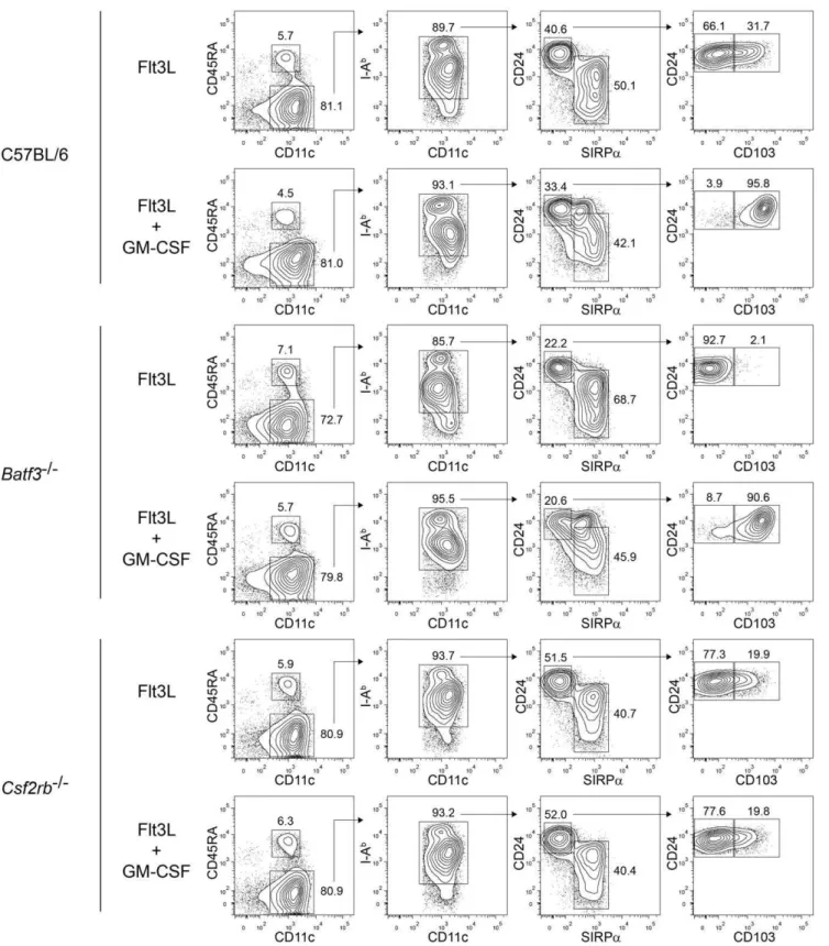
DC lineage [14], recent work shows that CD103 expression can be dynamically regulated by cytokines [17,18]. Therefore, we wished to re-examine the relationships between CD103 expression,Batf3, and GM-CSF signaling. We selected the Flt3L-induced DC differentiation from bone marrow cultures for this purpose since it has allowed the analysis of CD103 expression as dependent on both Batf3 and GM-CSF [17,18] (Fig. 4).
Previously, CD24high SIRPalow DCs were considered an ‘‘equivalent’’ of CD8a+ cDCs in Flt3L-treated bone marrow
cultures [30], despite the fact that only a subpopulation of these cells express CD103 (Fig. 4). The CD103+
fraction of CD24high SIRPalow
DCs was more potent than the CD1032 fraction for
cross-presentation [17], suggesting a correlation between CD103 expression and CD8a+
cDC function. We find that addition of
Figure 2.Batf3-dependent dermal CD11blow/2Langerin+DCs develop in
Csf2rb2/2 mice, but lack expression of CD103.(A) FACS
analysis of SDLN (inguinal) DCs from C57BL/6,Batf32/2 (C57BL/6 background), andCsf2rb2/2mice. Left plots are gated on CD11chighcells. The
second column is gated on migratory (DEC205+
CD8a2) DCs. The third, fourth, and fifth columns are gated on Langerin+
migratory DCs. Numbers represent the percentage of cells within the indicated gates. MFI are shown for the indicated gates. Data are representative of eight to ten individual inguinal lymph nodes from four to five mice per genotype over two experiments. (B) FACS analysis of SDLN (inguinal) DCs from C57BL/6,Batf32/2,
andCsf2rb2/2mice. Left plots are gated on CD11chighcells. Middle plots are gated on migratory (DEC205+
CD8a2) DCs. Right plots are gated on all
Langerin+migratory DCs, independent of expression of CD11b. Numbers represent the percentage of cells within the indicated gates. Data are representative of inguinal lymph nodes from at least five mice per genotype for all stains except CD24, which has been performed on one mouse per genotype. (C) Absolute cell numbers per individual inguinal lymph nodes calculated from the FACS analysis in (A). Langerhans cells were gated as Langerin+
CD11b+
EpCAMhigh DCs, and dermal Langerin+
DCs were gated as Langerin+
CD11blow/2EpCAMmidDCs. Horizontal bars represent the
geometric mean; p values by unpaired student’s t test. (D) Clinical course of EAE in 129S6/SvEv mice andBatf32/2mice (129S6/SvEv background).
Data are from one of three similar experiments (n = 5 mice per group). Points represent the mean clinical score. For clarity, error bars are not displayed.
doi:10.1371/journal.pone.0025660.g002
CD11blow/-Peripheral DCs Are GM-CSF-Independent
GM-CSF during day 7 through 9 of Flt3L culture induces CD103 expression on all CD24highSIRPalow
DCs (Fig. 4), in agreement with recent reports which showed that such GM-CSF-induced cells exhibit robust cross-presentation [17,18]. However, while addition of IL-3 and TGF-b1 to Flt3L cultures can both induce expression of CD103, only IL-3 induced the capacity for presentation [17], dissociating CD103 expression from cross-presentation. Likewise, we found that GM-CSF treatment induced CD103 expression on all CD24highSIRPalow
DCs, even in bone marrow cultures from Batf32/2 mice (Fig. 4), which completely
lack CD103+ DCs in vivo [16], further dissociating CD103
expression from the properties of true CD8a+cDC equivalents.
In agreement, a recent report found that GM-CSF-induced CD103+
CD24highSIRPalow
Flt3L-derived DCs from Batf32/2
mice lacked the capacity for cross-presentation [18]. Finally,
Csf2rb2/2 bone marrow generated a subpopulation of
CD103+
CD24highSIRPalow
DCs, indicating development that is independent of GM-CSF signaling, although whether these arise from the actions of cytokines such as IL-3, or arise independently of soluble factors present in the culture remains unknown. In summary, these results indicate that CD103 expression can be regulated by GM-CSF, independently of Batf3, and may not represent a static marker ofBatf3-dependent DC lineages.
Discussion
Our study makes two new observations. First, we show that CD11blow/2 non-lymphoid DCs develop in the absence of
GM-CSF signaling. Second, we show thatBatf3-dependent CD11blow/2
non-lymphoid DCs are not required for T effector cell priming or clinical EAE after subcutaneous immunization. King et al. recently concluded that mice deficient for GM-CSF or GM-CSF receptor were resistant to EAE because they lacked dermal CD11blow/2
Langerin+
CD103+
DCs, which they proposed were required for priming effector CD4 T cells with MOG35–55 [14]. Dermal CD11blow/2Langerin+
CD103+
DCs are a conventional DC subset that requires the transcription factor Batf3for their development [16]. Here, we directly comparedCsf2rb2/2withBatf32/2mice for
their susceptibility to EAE and for their development of various DC subsets using a broad panel of DC markers, including Langerin, CD11b, EpCAM, CD24, and CD103. We confirm that the dermal CD11blow/2Langerin+
CD103+
DC subset is absent in Batf32/2
mice, but surprisingly find that Csf2rb2/2 mice harbor normal
numbers of this DC subset in SDLNs as a CD11blow/2
Langerin+
EpCAMmidCD24high cell that expresses reduced, but not absent, levels of CD103. Moreover, in other peripheral tissues,
Csf2rb2/2mice have similar CD11blow/2CD103+DCs that express
reduced, but not absent, levels of CD103.
Our data agree with recent reports that GM-CSF directly regulates CD103 expression on Flt3L-derived CD8a+equivalent
cDCs and splenic DEC205+
CD8a+
cDCs [17–19]. Beyond this, we show that GM-CSF also regulates levels of CD103 expression on peripheral tissue-resident CD11blow/2 DCs, which were not
examined in the previous studies. Notably, a separate report has suggested that GM-CSF signaling is required for the development of small intestinal lamina propria CD11b+
CD103+
DCs [31], although their data could be reinterpreted as simply representing reduced CD103 expression by these DCs. In that report,Csf2rb2/2
mice had half as many lamina propria CD11b+
CD103+
DCs as wild type mice, but 1.7-fold more CD11b+
CD1032DCs than wild type
mice, consistent with a reduction of CD103 expression as an explanation for the ‘‘missing’’ subset.
King et al. also found decreased T cell priming to subcutaneous immunization with MOG35–55peptide and partial EAE resistance afterin vivo depletion of peripheral Langerin+
CD103+
DCs using radiation chimeras reconstituted with bone marrow from transgenic mice expressing DTR under the control of the Langerin promoter [20]. The use of radiation chimeras allowed the selective depletion of dermal CD11blow/2Langerin+
DCs, avoiding depletion of epidermal CD11b+
Langerin+
Langerhans cells, because the latter are radiation-resistant, and were therefore of host origin and did not express the DTR. Our results indicate thatCsf2rb2/2mice are not
deficient in dermal CD11blow/2Langerin+
DCs, but rather that these cells have reduced CD103 expression. However, Batf32/2
mice lack this dermal DC subset, and yet are susceptible to EAE induction and display increased, not decreased, T cell priming to MOG35–55peptide immunization. It is unclear why DTR-mediated depletion of donor-derived, radiation-sensitive Langerin+
DCs reduces priming and delays EAE, when genetic ablation of these cells inBatf32/2mice leaves priming and EAE intact. Their use of
DTR-mediated depletion involved irradiation of mice prior to immunization, which may have had unintended consequences. Furthermore, a study by Henri et al. has identified additional dermal DC subsets [26], including one that is Langerin+ but
CD1032, although it’s Batf3-dependence was not characterized.
Conceivably, the depletion of this subset by diphtheria toxin treatment could contribute to differences between our results and those of King et al. Notably, DTR-mediated depletion reduced the severity of EAE, but with a profound decrease only in MOG35–55 -specific IFN-c-secreting T cells, with minimal change in the number of IL-17-secreting T cells. In contrast, MOG35–55-immunized
Csf2rb2/2mice had profound reductions in antigen-specific
IFN-c-secreting and in IL-17-secreting T cells, suggesting thatCsf2rb2/2
mice differ from mice in which Langerin+
DCs are depleted using DTR.
Our results suggest that the absence of dermal CD11blow/2
Langerin+
CD103+
DCs leads to augmented T cell priming of both Th17 and Th1 responses. Igya´rto´ et al. have recently shown that skin infection ofBatf32/2mice with recombinantCandida albicans
led to reduced Th1 but increased Th17 responses compared to control mice [32]. These authors demonstrated that dermal Langerin+
CD103+
DCs expressed IL-12, explaining their ability to promote Th1 cell responses, and IL-27, a cytokine known to inhibit Th17 cell responses [33,34]. While we found increased Th17 responses inBatf32/2mice immunized with MOG
35–55and CFA, we also observed increased Th1 responses compared to control
Figure 3. GM-CSF controls the level of CD103 expression onBatf3-dependent CD11blow/2peripheral DCs.(A) FACS analysis of lung DCs
from C57BL/6,Batf32/2(C57BL/6 background), andCsf2rb2/2mice. Left plots are gated on live, single cells. The second column of plots is gated on
CD11c+ CD45.2+
cells, consisting of a mixture of lung macrophages and DCs. Note that inCsf2rb2/2mice, lung macrophages lack CD11c expression
[38], and therefore this gate includes only DCs. The third and fourth columns of plots are gated on I-AbhighautofluorescencelowDCs. Numbers
represent the percentage of cells within the indicated gates. Data are representative of six individual lungs from three mice per genotype over two experiments. (B) FACS analysis of kidney DCs from C57BL/6,Batf32/2(C57BL/6 background), andCsf2rb2/2mice. Left plots are gated on live, single
cells. The second column of plots is gated on CD11chighCD45.2+cells, consisting of DCs. The third column of plots is gated on CD11chighI-Ab+DCs. Numbers represent the percentage of cells within the indicated gates. Data are representative of six individual kidneys from three mice per genotype over two experiments.
doi:10.1371/journal.pone.0025660.g003
CD11blow/-Peripheral DCs Are GM-CSF-Independent
Figure 4. GM-CSF induces CD103 expression on Flt3L-derived CD8a-equivalent DCs.FACS analysis of developing DCs in bone marrow
cells from C57BL/6,Batf32/2(C57BL/6 background), andCsf2rb2/2mice cultured with Flt3L for 9 days, with or without the addition of GM-CSF to the
culture on day 7. The left column is gated on live single cells, the second on CD11c+
CD45RA2cDCs, the third on I-Ab+
cDCs, and the fourth on CD24highSirpalowcells. Numbers represent the percentage of cells within the indicated gates. Data are representative of bone marrow cells from two
mice. Conceivably, the different Th1 responses between our study and Igya´rto´ et al. could arise from differences in the form of immunization, relying on CFA versus fungal infection. Notably, blood-derived inflammatory monocytes have been reported to serve as a critical source of IL-12 after CFA-based subcutaneous immunization [35]. While we observed between a 2 to 3-fold higher frequency of IL-17-producing T cells in MOG35–55 -immunized Batf32/2 mice relative to controls, the clinical EAE
scores were similar between these groups. Conceivably, the frequency of IL-17-producing T cells is unrelated to the severity of EAE when Th17 cells are above some threshold frequency. Alternatively, IL-17 itself may not be the limiting factor in regulating the intensity of EAE, since GM-CSF also recently was implicated as the major Th17-derived cytokine responsible for driving pathogenic lesions in this model [5–7]. In fact, however, we do observe a slightly more rapid onset of EAE inBatf32/2mice,
which was more pronounced in the 129S6/SvEv genetic back-ground (Fig. 2D), which could relate to increased Th17 develop-ment.
If a lack of peripheral CD11blow/2 DCs does not abrogate
priming of T effector cells after subcutaneous immunization, why then does this priming defect occur in Csf2rb2/2 mice? One
possibility is that other DC subsets besides dermal CD11blow/2
DCs can prime CD4 T cells [35], but that the ability of these other DCs to prime requires that they receive a GM-CSF signal. Indeed, GM-CSF is essential for IL-6 and IL-23 production by DCs during the priming phase of autoimmune myocarditis [4] and is required for inflammatory DC development from monocytes in a model of repeated immunization with methylated BSA and CFA [36]. T cells do not express GM-CSF receptor, so an intrinsic T cell defect seems unlikely. While the source of GM-CSF after subcutaneaous immunization is still unclear, antigen-specific T cells can produce GM-CSF [5–7] and may augment responses by DCs during initial priming of T effector cells.
Materials and Methods
Ethics statement
This study was carried out in strict accordance with the recommendations in the Guide for the Care and Use of Laboratory Animals of the National Institutes of Health. The protocol was approved by the Animal Studies Committee of Washington University (#20090320).
Mice
Wild-type 129S6/SvEv and C57BL/6 mice were purchased from Taconic.Batf32/2mice on a 129S6/SvEv background were
previously generated in our laboratory [27], and were backcrossed for 10 generations to the C57BL/6 background.Csf2rb2/2
mice on the C57BL/6 background were purchased from Jackson Laboratory. Experiments were performed with sex-matched mice at 8–20 weeks of age. Mice were bred and maintained in our specific pathogen-free animal facility according to institutional guidelines.
Induction of EAE
EAE was induced as previously described [14]. Briefly, mice were immunized subcutaneously with 100 micrograms MOG35–55 peptide (Sigma Genosys) emulsified in complete Freund’s adjuvant (CFA) (made with 5 mg/ml heat-killed Mycobacterium tuberculosis
H37Ra (BD Difco) in incomplete Freund’s adjuvant (BD Difco)). Pertussis toxin (List Biological Laboratories) was injected intra-peritoneally (300 ng) on days zero and two. Mice were observed
for signs of EAE and graded on a standard 0–5 scale as described [37].
T cell priming and ELISPOT assays
Mice were immunized subcutaneously in the hind footpads with 10 nanomoles MOG35–55peptide (Sigma Genosys) emulsified in complete Freund’s adjuvant (CFA) (BD Difco). Popliteal lymph nodes were collected at day 7, and single cell suspensions were used in ELISPOT assays with the IFNcELISPOT antibody pair from BD Biosciences on Multiscreen Filter Plates from Millipore. An IL-17 ELISPOT was designed using anti-IL17A capture (clone TC11-18H10) and detection (clone TC11-8H4.1) antibodies (BD Biosciences). Cells were plated at 16106cells/well, in duplicate, and stimulated with either no antigen, or 10 micromolar MOG35–55 peptide. Plates were developed after 16 hours of stimulation at 37uC, and spots were counted on an Immunospot counter (Cellular Technology Ltd.).
Antibodies
The following antibodies were purchased from BD Biosciences: PE-Cy7 anti-CD11b (M1/70), PerCP-Cy5.5 anti-CD8a(53-6.7), V500 anti-CD8a (53-6.7), PE CD45RA (14.8), biotin anti-CD24 (30F1), FITC anti-CD24 (M1/69), APC anti-CD172a/SIRPa
(P84). FITC anti-CD11b (M1/70), APC eFluor 780-anti-CD11c (N418), PE anti-CD103 (2E7), biotin anti-CD103 (2E7), eFluor450 anti-CD317/BST2 (eBio927), eFluor450 anti-MHCII (I-A/I-E) (M5/114.15.2), APC anti-CD45.2 (104), and streptavidin-eFluor450 were purchased from eBioscience. PE anti-CD205/ DEC205 145) and APC anti-CD205/DEC205 (NLDC-145) were purchased from Miltenyi. Alexa Fluor 488 anti-CD207/ Langerin (929F3.01) was purchased from Imgenex. PE anti-CD326/EpCAM (G8.8) was purchased from Biolegend.
Flow cytometry
Inguinal lymph nodes and spleens were minced and digested in 5 ml Iscove’s modified Dulbecco’s media +10% FCS (cIMDM) with 250mg/ml collagenase B (Roche) and 30 U/ml DNase I (Sigma-Aldrich) for 1 h at 37uC with stirring. Lungs and kidneys were perfused with 10 ml DPBS via injection into the right ventricle after transection of the lower aorta. Dissected lungs and kidneys were minced and digested in 5 ml of cIMDM with 4 mg/ ml collagenase D (Roche) for 1 h at 37uC with stirring. EDTA (5 mM final concentration) was added to cell suspensions, and cells were incubated on ice for 5 min. Cells were passed through an 80mm strainer before red blood cells lysis with ACK lysis buffer. Cells were counted on a Vi-CELL analyzer, and 2–56106 cells were used per antibody staining reaction.
Staining was performed at 4uC in the presence of Fc Block (clone 2.4G2, BD Biosciences or BioXcell) in FACS buffer (DPBS+0.5% BSA+2 mm EDTA+0.02% sodium azide). For experiments involv-ing intracellular anti-Langerin staininvolv-ing, cells were surface antibody stained prior to fixation in 4% formaldehyde (Thermo Scientific) for 15 minutes at room temperature. Cells were subsequently permea-bilized (DPBS+0.1% BSA+0.5% saponin) for 5 min at 4uC and stained in the same permeabilization buffer with anti-Langerin antibody. Cells were washed twice in permeabilization buffer before returning them to FACS buffer. Flow cytometry was performed on a FACSCantoII (BD Biosciences), and data was analyzed with FlowJo software (Tree Star, Inc.).
Bone marrow cultures
Bone marrow cells from femurs and tibias were collected and red blood cells were lysed in ACK lysis buffer. Cells were cultured
CD11blow/-Peripheral DCs Are GM-CSF-Independent
in four ml cIMDM in six-well plates at 26106cells/ml containing 160 ng/ml murine Flt3L (Peprotech). At day seven of culture, murine GM-CSF (Peprotech) was added to some wells at 20 ng/ ml. Non-adherent cells were collected at day nine for flow cytometry.
Supporting Information
Figure S1 Flow cytometric analyses of DCs inBatf32/2
mice.(A) FACS analysis of SDLN (inguinal) DCs fromBatf3+/+
and Batf32/2 mice on both the C57BL/6 and 129S6/SvEv
backgrounds. Left plots from mice of each background are gated on CD11c+
BST22 cDCs. Right plots from mice of each
background are gated on migratory (DEC205+
CD8a2) DCs.
Numbers represent the percentage of cells within the indicated gates. Data are representative of at least six mice per genotype and background obtained in several independent experiments. (B) Absolute cell numbers of DEC205+
CD8a+
DCs per individual inguinal lymph nodes from C57BL/6 and Batf32/2 mice
(C57BL/6 background) calculated from the FACS analysis performed in Figure 2A. Horizontal bars represent the geometric mean, p value by unpaired student’s t test. (C) and (D) FACS analysis of splenic DCs fromBatf3+/+
andBatf32/2mice on both
the (C) C57BL/6 and (D) 129S6/SvEv backgrounds. Plots are gated on CD11c+
I-Ab+
cDCs. Numbers represent the percentage of cells within the indicated gates. Data are representative of at least six mice per genotype and background obtained in several experiments. (E) FACS analysis of SDLN (inguinal) DCs from C57BL/6,Batf32/2(C57BL/6 background), andCsf2rb2/2mice.
Left plots are gated on CD11chighcells. Right plots are gated on DEC205+
CD8a+
cDCs. Numbers represent the percentage of cells within the indicated gates. Data are representative of eight to ten individual inguinal lymph nodes from four to five mice per genotype obtained in two experiments. Data is depicted in these plots are derived from analysis of the same mice used in Figure 2A, but gated to analyze CD103 expression on DEC205+
CD8a+
cDCs. (TIF)
Author Contributions
Conceived and designed the experiments: KMM BTE ERU TLM JHR. Performed the experiments: BTE TRB WKC KH JWH JS. Analyzed the data: BTE KMM TLM ERU JHR. Contributed reagents/materials/ analysis tools: BTE KMM. Wrote the paper: BTE KMM.
References
1. Hamilton JA (2008) Colony-stimulating factors in inflammation and autoim-munity. Nat Rev Immunol 8: 533–544.
2. McQualter JL, Darwiche R, Ewing C, Onuki M, Kay TW, et al. (2001) Granulocyte macrophage colony-stimulating factor: a new putative therapeutic target in multiple sclerosis. J Exp Med 194: 873–882.
3. Cook AD, Braine EL, Campbell IK, Rich MJ, Hamilton JA (2001) Blockade of collagen-induced arthritis post-onset by antibody to granulocyte-macrophage colony-stimulating factor (GM-CSF): requirement for GM-CSF in the effector phase of disease. Arthritis Res 3: 293–298.
4. Sonderegger I, Iezzi G, Maier R, Schmitz N, Kurrer M, et al. (2008) GM-CSF mediates autoimmunity by enhancing IL-6-dependent Th17 cell development and survival. J Exp Med 205: 2281–2294.
5. Ponomarev ED, Shriver LP, Maresz K, Pedras-Vasconcelos J, Verthelyi D, et al. (2007) GM-CSF production by autoreactive T cells is required for the activation of microglial cells and the onset of experimental autoimmune encephalomyelitis. J Immunol 178: 39–48.
6. El Behi M, Ciric B, Dai H, Yan Y, Cullimore M, et al. (2011) The encephalitogenicity of T(H)17 cells is dependent on IL-1- and IL-23-induced production of the cytokine GM-CSF. Nat Immunol 12: 568–575.
7. Codarri L, Gyulveszi G, Tosevski V, Hesske L, Fontana A, et al. (2011) RORgammat drives production of the cytokine GM-CSF in helper T cells, which is essential for the effector phase of autoimmune neuroinflammation. Nat Immunol 12: 560–567.
8. Hofstetter HH, Ibrahim SM, Koczan D, Kruse N, Weishaupt A, et al. (2005) Therapeutic efficacy of IL-17 neutralization in murine experimental autoim-mune encephalomyelitis. Cell Immunol 237: 123–130.
9. Lovett-Racke AE, Yang Y, Racke MK (2011) Th1 versus Th17: are T cell cytokines relevant in multiple sclerosis? Biochim Biophys Acta 1812: 246–251. 10. Kroenke MA, Chensue SW, Segal BM (2010) EAE mediated by a
non-IFN-gamma/non-IL-17 pathway. Eur J Immunol 40: 2340–2348.
11. McGeachy MJ, Bak-Jensen KS, Chen Y, Tato CM, Blumenschein W, et al. (2007) TGF-beta and IL-6 drive the production of IL-17 and IL-10 by T cells and restrain T(H)-17 cell-mediated pathology. Nat Immunol 8: 1390– 1397.
12. Ghoreschi K, Laurence A, Yang XP, Tato CM, McGeachy MJ, et al. (2010) Generation of pathogenic T(H)17 cells in the absence of TGF-beta signalling. Nature 467: 967–971.
13. McGeachy MJ (2011) GM-CSF: the secret weapon in the T(H)17 arsenal. Nat Immunol 12: 521–522.
14. King IL, Kroenke MA, Segal BM (2010) GM-CSF-dependent, CD103+dermal dendritic cells play a critical role in Th effector cell differentiation after subcutaneous immunization. J Exp Med 207: 953–961.
15. Ginhoux F, Liu K, Helft J, Bogunovic M, Greter M, et al. (2009) The origin and development of nonlymphoid tissue CD103+DCs. J Exp Med 206: 3115–3130. 16. Edelson BT, KC W, Juang R, Kohyama M, Benoit LA, et al. (2010) Peripheral CD103+ dendritic cells form a unified subset developmentally related to CD8alpha+conventional dendritic cells. J Exp Med 207: 823–836.
17. Sathe P, Pooley J, Vremec D, Mintern J, Jin JO, et al. (2011) The acquisition of antigen cross-presentation function by newly formed dendritic cells. J Immunol 186: 5184–5192.
18. Jackson JT, Hu Y, Liu R, Masson F, D’Amico A, et al. (2011) Id2 expression delineates differential checkpoints in the genetic program of CD8alpha(+) and CD103(+) dendritic cell lineages. EMBO J 30: 2690–2704.
19. Zhan Y, Carrington EM, van Nieuwenhuijze A, Bedoui S, Seah S, et al. (2011) GM-CSF increases cross presentation and CD103 expression by mouse CD8(+) spleen dendritic cells. Eur J Immunol;In press.
20. Kissenpfennig A, Henri S, Dubois B, Laplace-Builhe C, Perrin P, et al. (2005) Dynamics and function of Langerhans cells in vivo: dermal dendritic cells colonize lymph node areas distinct from slower migrating Langerhans cells. Immunity 22: 643–654.
21. Robb L, Drinkwater CC, Metcalf D, Li RL, Kontgen F, et al. (1995) Hematopoietic and Lung Abnormalities in Mice with A Null Mutation of the Common Beta-Subunit of the Receptors for Granulocyte-Macrophage Colony-Stimulating Factor and Interleukin-3 and Interleukin-5. Proc Natl Acad Sci U S A 92: 9565–9569.
22. Nicola NA, Robb L, Metcalf D, Cary D, Drinkwater CC, et al. (1996) Functional inactivation in mice of the gene for the interleukin-3 (IL-3)-specific receptor beta-chain: implications for IL-3 function and the mechanism of receptor transmodulation in hematopoietic cells. Blood 87: 2665–2674. 23. Bursch LS, Wang L, Igyarto B, Kissenpfennig A, Malissen B, et al. (2007)
Identification of a novel population of Langerin+dendritic cells. J Exp Med 204: 3147–3156.
24. Ginhoux F, Collin MP, Bogunovic M, Abel M, Leboeuf M, et al. (2007) Blood-derived dermal langerin+dendritic cells survey the skin in the steady state. J Exp Med 204: 3133–3146.
25. Poulin LF, Henri S, de Bovis B, Devilard E, Kissenpfennig A, et al. (2007) The dermis contains langerin+dendritic cells that develop and function indepen-dently of epidermal Langerhans cells. J Exp Med 204: 3119–3131.
26. Henri S, Poulin LF, Tamoutounour S, Ardouin L, Guilliams M, et al. (2010) CD207+ CD103+ dermal dendritic cells cross-present keratinocyte-derived antigens irrespective of the presence of Langerhans cells. J Exp Med 207: 189–206.
27. Hildner K, Edelson BT, Purtha WE, Diamond M, Matsushita H, et al. (2008) Batf3 deficiency reveals a critical role for CD8alpha+dendritic cells in cytotoxic T cell immunity. Science 322: 1097–1100.
28. Mashayekhi M, Sandau MM, Dunay IR, Frickel EM, Khan A, et al. (2011) CD8a+Dendritic Cells Are the Critical Source of Interleukin-12 that Controls Acute Infection by Toxoplasma gondii Tachyzoites. Immunity 35: 249–259. 29. Bedoui S, Whitney PG, Waithman J, Eidsmo L, Wakim L, et al. (2009)
Cross-presentation of viral and self antigens by skin-derived CD103(+) dendritic cells. Nat Immunol 10: 488–495.
30. Naik SH, O’Keeffe M, Proietto A, Hochrein H, Shortman K, et al. (2010) CD8+, CD82, and plasmacytoid dendritic cell generation in vitro using flt3
ligand. Methods Mol Biol 595: 167–176.
31. Bogunovic M, Ginhoux F, Helft J, Shang L, Hashimoto D, et al. (2009) Origin of the lamina propria dendritic cell network. Immunity 31: 513–525.
32. Igya´rto´ BZ, Haley K, Ortner D, Bobr A, Gerami-Nejad M, et al. (2011) Skin-Resident Murine Dendritic Cell Subsets Promote Distinct and Opposing Antigen-Specific T Helper Cell Responses. Immunity 35: 260–272.
T helper cells during chronic inflammation of the central nervous system. Nat Immunol 7: 937–945.
34. Diveu C, McGeachy MJ, Boniface K, Stumhofer JS, Sathe M, et al. (2009) IL-27 blocks RORc expression to inhibit lineage commitment of Th17 cells. J Immunol 182: 5748–5756.
35. Nakano H, Lin KL, Yanagita M, Charbonneau C, Cook DN, et al. (2009) Blood-derived inflammatory dendritic cells in lymph nodes stimulate acute T helper type 1 immune responses. Nat Immunol 10: 394–402.
36. Kamphorst AO, Guermonprez P, Dudziak D, Nussenzweig MC (2010) Route of antigen uptake differentially impacts presentation by dendritic cells and activated monocytes. J Immunol 185: 3426–3435.
37. Lees JR, Sim J, Russell JH (2010) Encephalitogenic T-cells increase numbers of CNS T-cells regardless of antigen specificity by both increasing T-cell entry and preventing egress. J Neuroimmunol 220: 10–16.
38. Guth AM, Janssen WJ, Bosio CM, Crouch EC, Henson PM, et al. (2009) Lung environment determines unique phenotype of alveolar macrophages. Am J Physiol Lung Cell Mol Physiol 296: L936–L946.
CD11blow/-Peripheral DCs Are GM-CSF-Independent