125
P akistan V eterinary J ournal
ISSN: 0253-8318 (PRINT), 2074-7764 (ONLINE)Accessible at: www.pvj.com.pk
Renal-Adenocarcinoma-Associated Erythrocytosis in a Cat
Sungjun Noh, Ji-Houn Kang*, Gonhyung Kim, Dongwoo Chang, Byeongwoo Ahn, Ki-Jeong Na and Mhan-Pyo Yang College of Veterinary Medicine, Chungbuk National University, Cheongju, Chungbuk 361-763, Republic of Korea *Corresponding author: jhkang@chungbuk.ac.kr
A R T I C L E H I S T O R Y A B S T R A C T Received:
Revised: Accepted:
July 05, 2012 July 22, 2012 July 24, 2012
A 9-year-old spayed female domestic shorthair cat was referred for erythrocytosis. Even after the correction of dehydration, blood analyses showed that there had been no improvement. An abdominal ultrasonography and computed tomography identified the presence of a mass on the left kidney. Measurement of serum erythropoietin (EPO) showed higher concentration than the reference interval. These findings suggested a direct association of the erythrocytosis with excessive EPO production. The cat underwent nephrectomy of the affected (left) kidney. Subsequent histopathology was consistent with a diagnosis of renal adenocarcinoma. Following the nephrectomy, serum EPO concentrations decreased gradually, and the erythrocytosis resolved 15 days postoperatively. This case describes the diagnosis and treatment of secondary inappropriate erythrocytosis in a cat with renal adenocarcinoma.
©2012 PVJ. All rights reserved Key words:
Erythropoietin FELINE Renal tumor
To Cite This Article: Noh S, JH Kang, G Kim, D Chang, B Ahn, KJ Na and MP Yang, 2013. Renal-adenocarcinoma-associated erythrocytosis in a cat. Pak Vet J, 33(1): 125-127.
INTRODUCTION
Erythrocytosis is characterized by increases in packed cell volume (PCV), red blood cell (RBC) count, and hemoglobin (Hb) concentration (Randolph et al., 2010).
Based on its etiology, two forms of the disease are recognized, relative and absolute (Nitsche, 2004). Relative erythrocytosis is caused by severe dehydration and body-fluid shifts and resolves after appropriate body-fluid therapy (Weiss and Tvedten, 2012). Absolute erythrocytosis is due to an increased total RBC mass and may be either primary (polycythemia vera) or secondary. The later can be further defined as physiologically appropriate or inappropriate (Weiss and Tvedten, 2012).
Appropriate secondary erythrocytosis occurs as a response to conditions associated with poor systemic tissue oxygenation, such as cardiovascular disease, chronic pulmonary disease, and hemoglobinopathies (Nitsche, 2004; Weiss and Tvedten, 2012), whereas inappropriate secondary erythrocytosis is characterized by increased EPO levels without evidence of a hypoxic stimulus. In companion animals, inappropriate secondary erythrocytosis may follow from the presence of a space-occupying renal lesions, such as renal cysts, hydro-nephrotic lesions, chronic pyelonephritis, or renal neoplasms (Kessler, 2008; Durno et al., 2011). In cats,
renal-tumor-related inappropriate secondary
erythro-cytosis is very rare, with only two cases fully reported in the English veterinary literature (Klainbart et al., 2008).
Herein we describe the diagnosis and treatment of a renal adenocarcinoma in a domestic shorthair cat with erythrocytosis.
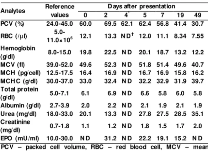
Case Description: A 9-year-old spayed female domestic shorthair cat was referred for the evaluation of severely elevated PCV. The cat had been previously brought to the referring hospital for anorexia and vomiting of 3 days duration, with fluid therapy administered by the referring veterinarian to correct the dehydration. However, after treatment, the PCV remained high. On presentation, the cat had brick-red mucous membranes. A routine hematological assessment revealed severe erythrocytosis, characterized by increases in the PCV, RBC count, and Hb (Table 1). The remainder of the complete blood count (CBC) was within normal limits, and serum biochemistry panels were without remarkable findings. Thoracic radiographs and the results of the arterial blood gas analysis were also normal. However, abdominal ultrasound revealed a round mass of mixed echogenicity originating from the caudal pole of the left kidney. The cat was hospitalized and administered fluid therapy to correct the erythrocytosis. A re-evaluation on the following day, including a CBC, showed the persistence of marked erythrocytosis (Table 1).
Pak Vet J, 2013, 33(1): 125-127. 126
Fig. 1: A computed tomography image of the left kidney in a cat
diagnosed with inappropriate polycythemia secondary to a renal adenocarcinoma. N ote the contrast-enhanced mass in the left kidney (arrows).
Fig. 2: T issues obtained from the affected left kidney. N ote the
well-differentiated neoplastic cells, with an eosinophilic cytoplasm, uniform nuclei, and no mitosis. Tubules contain eosinophilic homogeneous fluid. H&E stain. ×200
On day 3, abdominal computed tomography (CT) confirmed a 3.0 × 3.0 cm heterogeneous round mass within the left kidney (Fig. 1). The lesion enhanced following the administration of intravenous contrast medium (Iohexol; Omnipaque®, GE Healthcare Korea, Seoul, Korea). No remarkable findings were seen on the thoracic CT. The serum EPO concentration in the sample collected at presentation was markedly elevated (31.2 mU/mL) compared to the reference interval (10.0–30.0 mU/mL), as determined in a radioimmunoassay (Table 1). On the basis of these findings, the erythrocytosis was considered to be due to inappropriate EPO production, which in turn was probably directly related to the mass in the left kidney. Nephrectomy was subsequently performed, which revealed a well-defined encapsulated mass on the caudal pole of the left kidney. Histopathology showed that the tumor contained highly infiltrative tumor cells with areas of solid and tubular differentiation, consistent with a primary renal adenocarcinoma (Fig. 2).
An improvement of the cat’s erythrocytosis was observed 3 days after surgical removal of the left kidney (Table 1). At 19 days follow-up, the cat was in good condition, without abnormalities on the physical examination and no further clinical signs, as reported by
T able 1: Serial laboratory parameters in a domestic shorthair cat with
secondary inappropriate polycythemia caused by a renal
adenocarcinoma
Analytes Reference
values
D ays after presentation
0 2 4* 5 7 19 49
PCV (%) 24.0-45.0 60.0 69.5 62.1 62.4 56.8 41.4 30.7
RBC (/µl)
5.0-11.0×10⁶ 12.1 13.3 N D
† 12.0 11.1 8.34 7.55
Hemoglobin
(g/dl) 8.0-15.0 19.8 22.5 N D 20.1 18.7 13.2 12.2
MCV (fl) 39.0-52.0 49.6 52.3 N D 51.8 51.4 49.6 40.7
MCH (pg/cell) 12.5-17.5 16.4 16.9 N D 16.7 16.9 15.8 16.2
MCH C (g/dl) 30.0-37.0 33.0 32.4 N D 32.2 32.9 31.9 39.7
Total protein
(g/dl) 5.0-7.1 6.1 6.9 N D 6.6 5.8 6.0 5.8
Albumin (g/dl) 2.7-3.9 2.0 2.2 N D 2.1 1.9 2.1 1.9
Urea (mg/dl) 18.0-33.0 20.1 13.3 N D 27.8 27.5 28.5 35.1
Creatinine
(mg/dl) 0.7-1.8 1.1 1.2 N D 1.8 1.5 1.7 2.0
EPO (mU/ml) 10.0-30.0 N D 31.2 N D 22.2 19.1 15.2 N D
PCV – packed cell volume, RBC – red blood cell, MCV – mean corpuscular volume, MCH – mean corpuscular hemoglobin, MCH C – mean corpuscular hemoglobin concentration, EPO – erythropoietin;
*The day of the nephrectomy; †N D – N ot done.
the owner. Serial blood analyses confirmed that the levels of all the analytes had returned to within the reference interval. The cat remains healthy 1 year after the nephrectomy.
DISCUSSION
In this case of feline secondary erythrocytosis, the abnormal blood values were directly related to the high serum EPO concentration, which resulted from a primary renal adenocarcinoma. Measurement of serum EPO levels in animals can be useful to differentiate among the various forms of erythrocytosis (Hasler and Giger, 1996; Nitsche, 2004). Generally, primary erythrocytosis is characterized by low to normal EPO levels, whereas secondary erythrocytosis is associated with increased serum EPO levels (Hasler and Giger, 1996). In this case, the EPO level was relatively high, almost certainly the consequence of renal adenocarcinoma; accordingly, the diagnosis was inappropriate secondary erythrocytosis. However, in contrast to our case, serum EPO levels were normal in the previously reported two cats with secondary erythrocytosis due to a primary renal neoplasia (Klainbart
et al., 2008). In both human and veterinary medicine, the
reasons for the abnormal EPO production in secondary renal-neoplasia-related erythrocytosis are not completely understood. It was recently suggested that EPO production by renal tumors is triggered by one of two mechanisms: 1) autonomous excretion of EPO or EPO-like substances from renal tumors, documented to directly cause erythrocytosis (Kessler, 2008; McMullin, 2008) and 2) as a consequence of local renal hypoxia, due to compression of the normal tissue or vasculature by the infiltrative neoplasm, which induces the secretion of EPO by the remaining, normal kidney tissue (Kessler, 2008; McMullin, 2008). Additionally, in a report on two dogs with renal T-cell lymphoma immunohistochemical staining showed that EPO was produced directly by the tumor tissues but also by the normal renal cells and vasculature, in response to local hypoxia (Durno et al.,
Pak Vet J, 2013, 33(1): 125-127. 127
In cats, renal neoplasia generally manifests as non-specific clinical signs, including loss of appetite, weight loss, lethargy, vomiting, abdominal pain, and/or hematuria (Henry et al., 1999). The chief complaints in the two
polycythemic cats mentioned above were central nervous system disturbances, i.e., changes in mentation, ataxia, blindness, and seizures, which are caused by the hyperviscosity of the blood (Klainbart et al., 2008). In the
cat described herein, anorexia and vomiting were not accompanied by neurologic signs. Presumably, the decreased blood flow due to the hyperviscosity was not enough to induce neurologic abnormalities, although blood viscosity was not measured directly. Feline primary renal neoplasia is often an aggressive disease, with a high metastatic rate and thus a grave prognosis (Henry et al.,
1999; Meuten, 2002). The prognosis of secondary erythrocytosis depends on the ability to control the primary disease. In this case, the erythrocytosis resolved after nephrectomy and the cat has remained healthy, without a recurrence of clinical abnormalities. Thus, despite the above-mentioned grave prognosis of feline renal neoplasia, nephrectomy seems to be warranted in some cases.
In conclusion, our report describes the diagnosis and treatment of secondary inappropriate erythrocytosis associated with renal adenocarcinoma in a cat with high EPO concentration. Further study will be needed to
clarify the mechanism on the EPO production by renal tumor in cats.
REFERENCES
D urno AS, JA W ebb, MJ G autheier and D Bienzle, 2011. Polycythemia and inappropriate erythropoietin concentrations in two dogs with renal T-cell lymphoma. J Am Anim H osp Assoc, 47: 122-128. H asler AH and U Giger, 1996. Serum erythropoietin values in
polycythemic cats. J Am Anim H osp Assoc, 32: 294-301.
H enry CJ, SE Turnquist, A Smith, JC Graham, D H Thamm, M O ’Brien and CA Clifford, 1999. Primary renal tumors in cats: 19 cases (1992-1998). J Feline Med Surg, 1: 165-170.
Kessler M, 2008. Secondary polycythemia associated with high plasma erythropoietin concentrations in a dog with a necrotizing
pyelonephritis. J Small Anim Pract, 49: 363-366.
Klainbart S, G Segev, E Loeb, D Melamed and I Arocho, 2008.
Resolution of renal adenocarcinoma-induced secondary
inappropriate polycythaemia after nephrectomy in two cats. J Feline Med Surg, 10: 264-268.
McMullin MF, 2008. The classification and diagnosis of erythrocytosis. Int J Lab Hematol, 30: 447-459.
Meuten DJ, 2002. Tumors of the urinary system. In: Tumors in Domestic Animals (Meuten DJ, ed): 4th Ed, Blackwell, Ames, USA, pp: 509-546.
N itsche EK, 2004. Erythrocytosis in dogs and cats: diagnosis and management. Compend Contin Educ Vet, 26: 104-118.
Randolph JF, ME Peterson, T Stokol and D J W eiss, 2010. Erythrocytosis and Polycythemia. In: Schalm’s Veterinary Haematology (W eiss DJ and W ardrop KJ, eds): 6th Ed, Blackwell, Ames, USA, pp: 162-165. W eiss DJ and H Tvedten, 2012. Erythrocyte disorder. In: Small Animal