Immunoadjuvant Properties of the Rho
Activating Factor CNF1 in Prophylactic and
Curative Vaccination against
Leishmania
infantum
Grégory Michel1,2,3
*, Bernard Ferrua1,2, Patrick Munro1,2, Laurent Boyer1,2,
Nassim Mathal4, Daniel Gillet4, Pierre Marty1,2,3, Emmanuel Lemichez1,2*
1Inserm U1065, Centre Méditerranéen de Médecine Moléculaire, Team“Microbial toxins in host pathogen interactions”, Equipe labellisée ligue contre le cancer, Nice, France,2Université de Nice-Sophia Antipolis, Faculté de Médecine, Nice, France,3Centre Hospitalier Universitaire de Nice, Laboratoire de Parasitologie-Mycologie, Nice, France,4CEA, iBiTecS, SIMOPRO, Paris Saclay University, LabEx LERMIT, Gif sur Yvette, France
*[email protected](GM);[email protected](EL)
Abstract
There is a need to develop new effective immunoadjuvants for prophylactic or therapeutic vaccines against intracellular pathogens. The activation of Rho GTPases by bacterial cyto-toxic necrotizing factor 1 (CNF1) elicits humoral protective responses against protein anti-gens. Here, we set out to investigate whether CNF1 activity initiates humoral immunity against co-administered parasite antigens and anti-microbial immune signaling. We report that co-administration of wild-type (WT) CNF1 withLeishmania(L.) promastigote antigens at the nasal mucosa triggered prophylactic and curative vaccine responses against this par-asite. Vaccination of the mucosa with promastigote lysate antigens combined with WT CNF1 conferred protection against high inoculumL.infantuminfection, which reached 82% in the spleen. Immune parameter analysis by antigen recall indicated robust T-helper (Th)1 polarization of immune memory cells, with high IL-2 and IFN-γproduction combined with
decreased IL-4 production. Additionally, we explored the curative effect of WT CNF1 on pre-viously infected animals. We observed that PL combined with WT CNF1, but not the inac-tive C866S mutant CNF1 (mCNF1), induced a 58% decrease in the parasite burden in the spleen.
Introduction
The discovery of the molecular basis of innate immunity has boosted the development of vac-cine adjuvants on the basis of their capacity to stimulate innate immune receptors [1]. The family of bacterial effectors catalyzing the activation of Rho proteins has attracted growing
attention because of their capacity to stimulate the immune system [2–6]. It has now been
established in different model systems that cells perceive robust activation of Rho GTPases by a11111
OPEN ACCESS
Citation:Michel G, Ferrua B, Munro P, Boyer L, Mathal N, Gillet D, et al. (2016) Immunoadjuvant Properties of the Rho Activating Factor CNF1 in Prophylactic and Curative Vaccination against
Leishmania infantum. PLoS ONE 11(6): e0156363. doi:10.1371/journal.pone.0156363
Editor:Michel R. Popoff, Institute Pasteur, FRANCE
Received:March 30, 2016
Accepted:May 12, 2016
Published:June 3, 2016
Copyright:© 2016 Michel et al. This is an open access article distributed under the terms of the
Creative Commons Attribution License, which permits unrestricted use, distribution, and reproduction in any medium, provided the original author and source are credited.
Data Availability Statement:All relevant data are within the paper and its Supporting Information files.
virulence factors as a danger signal that is translated into effective anti-bacterial immune
responses [5–7]. Here, we address whether a microbial effector targeting Rho GTPases can be
translated into an adjuvant for vaccination againstLeishmania infantum.
Host Rho GTPases are essential elements in host-pathogen interactions. The cytotoxic
nec-rotizing factor-1 (CNF1) is an A-B toxin produced by uropathogenic strains ofEscherichia coli.
Wild-type (WT) CNF1 specifically deamidates the glutamine 61 in Rac1/Cdc42 (Q63 in RhoA)
into a glutamic acid [8–10]. WT CNF1, but not the catalytically inactive mutant C866S of
CNF1 (mCNF1), catalyzes the activation of small Rho GTPases [9,10]. Consequently, WT CNF1 can be used to increase the flux of activated Rho GTPases in host cells and the down-stream Rho GTPase signaling pathways [11]. Although the small GTPases of the Rho protein family are frequent targets of post-translational modifications that are catalyzed by bacterial toxins, they contribute to sensing bacterial virulence [6,12]. Genome-wide gene expression
analysis in cells treated with WT CNF1 has revealed the induction of a large panel of NF-κ
B-driven pro-inflammatory cytokines and chemokines [2]. More recent studies have begun to
underscore the importance of the RIPK/NF-κB and ASC/Caspase-1 signaling axis in host
anti-bacterial responses modulated by WT CNF1 and the Rac GTPases [5–7,13]. It is important to
determine the spectrum of pathogens for which the CNF1 activity can be exploited to develop vaccines.
Leishmania infantum/chagasiis the causative agent of visceral leishmaniasis (VL), which is endemic in numerous southern countries, notably in the Mediterranean basin [14,15]. VL is fatal if left untreated and represents the second most challenging infectious disease worldwide [15]. Hence, part of the human population is chronically affected by poorly understood health consequences. Apart from humans, dogs are the main victims and reservoir. The current treat-ments are based on antibiotherapy and have serious limitations, such as high costs and toxicity [16]. For these reasons, and on the basis of the robust immunity to reinfection observed in
cured patients, several vaccine trials against VL have been performed [17].Leishmania
para-sites harness phagocytic cells, notably monocytes, in order to survive and replicate. Clinical studies of VL have suggested that decreased T-helper (Th)1 and increased Th2 responses are the hallmarks of the disease [15]. Hence, treatments that actively increase Th1 immune responses can promote the clearance of the parasites [18].
CNF1 activity and the downstream activation of Rac are sufficient to promote efficient host
immune responses against bacteria [7,13]. We have begun to divert this toxin’s Rho activating
property in the development of an immunoadjuvant for mucosal vaccination. We established that CNF1 activity stimulates the systemic and mucosal production of IgG and IgA antibodies against ovalbumin and tetanus toxoid [2,3]. Mice immunized against tetanus toxoid together with WT CNF1 show specific and long-lasting protection against a challenge by 10-fold of the
LD50of tetanus toxin [4]. It is now of interest to determine whether WT CNF1 can also
stimu-late Th-1 cellular immunity against an intracellular pathogen.
Materials and Methods
Mice and ethics statement
The protocol was approved by the Committee on the Ethics of Animal Experiments of the
School of Medicine of the University of Nice, France (Permit Number: 2010–45). Groups of
BALB/c female mice were purchased from Charles River at 6 weeks of age (Le Genest St. Isle, France). The mice were maintained and handled according to the regulations of the European Union and the French Ministry of Agriculture as well as to the FELASA (the Federation of Lab-oratory Animal Science Associations) recommendations. All efforts were made to minimize or avoid suffering.
supported by the grant from the Agence Nationale de la Recherche (ANR-10-LABEX-33).
L
.
infantum
parasites, antigens and CNF1
L.infantumMON-1 (MHOM/FR/94/LPN101) was isolated from a patient with Mediterranean
visceralLeishmaniathat was contracted in Nice, France.L.infantumpromastigotes were
rou-tinely grown at 26°C in Schneider’s medium, as previously described [19].L.infantumclones
encoding firefly luciferase were generated as previously described [20].
For the promastigote lysate (PL) preparation, stationary phaseL.infantumpromastigotes
were washed and suspended at 109/ml in distilled water [19]. The suspension was submitted to
5 freeze/thaw cycles to generate PL. Typically, 5 mg ofLeishmaniaprotein was obtained from
109parasites.
Recombinant wild-type CNF-1 (WT CNF1) and its catalytically inactive form
(CNF1-C866S; mCNF1) were produced and purified as previously reported [21]. Both recom-binant proteins were passed through a polymixin B column (Affinity pack TM-detoxy gel TM, Pierce), and the lack of endotoxin content was verified using a colorimetric LAL assay (LAL QCL-1000, Cambrex). Each CNF1 preparation stock (2 mg/ml) was shown to contain less than 0.5 endotoxin units/ml.
Endonasal immunization and challenge in BALB/c mice
Groups of 7 mice were immunized 3 times at 2-weeks intervals with 15μg of PL together with
1μg WT CNF1 or 1μg catalytically inactive CNF1 C866S (mCNF1). PL preparations were
delivered into the nasal mucosa with a micropipette in 10μl volumes of Dulbecco’s
phosphate-buffered saline (PBS, from Gibco life technologies) (5μl per nostril). Fourteen days after the
last boost, mice were challenged via the intraperitoneal route with 108stationary phaseL.
infantummetacyclic parasites. One month later, the mice were sacrificed, and spleen section were collected and analyzed for parasite content by ELISA sandwich technique [22]. Briefly, parasite antigens in infected tissues were extracted with Nonidet-P40 detergent and were
cap-tured by anti-L.infantumhuman IgG that were insolubilized onto microtiter plate and were
subsequently revealed using anti-L.infantumF(ab)' fragments labelled with peroxidase.
Analysis of vaccine-induced immune responses
To assess total IgG titers, blood samples were recovered from the tail vein after vaccination (one day before infection) and before mouse dissection (one month after infection). IgG anti-body responses were assessed at a 1/100 dilution by ELISA using PL-coated plates, as reported
[19]. Vaccine-induced cellular immunity was measured post-vaccination usingin vitroantigen
recall experiments on spleen homogenates as follows: the spleens from each individual mouse (5 per group) were homogenized in sterile PBS, and erythrocytes were lysed at room
tempera-ture using 10 mM NaHCO3containing 155 mM NH4Cl and 0.1 mM EDTA. Splenocytes were
then washed twice with PBS, counted and suspended at 5×10⁶cells/ml in DMEM containing 2
mM glutamine, 1 mM sodium pyruvate, 100 U/ml penicillin, 100μg/ml streptomycin, 50μM
2-mercaptoethanol and 10% fetal calf serum. Cell suspensions were cultured for 48 h in the
presence or absence of 50μg/ml of PL. Supernatants were harvested and assayed for IL-2, IL-4
and IFN-γcontent by indirect sandwich ELISA (Pharmingen, Clinisciences). The threshold
sensitivities of the techniques were in the range of 20–30 pg/ml.
Statistical analysis
Non-parametric Mann-Whitney tests were performed using GraphPad Prism version 5.0d for
Results
CNF1 activity stimulates humoral IgG responses against
L
.
infantum
antigens
In this study, we first sought to determine the efficacy of WT CNF1 as a specific immunoadju-vant for the induction of protective responses against an intracellular pathogen. Additionally, we sought to evaluate the efficacy of this adjuvant for needle-free vaccination by topical delivery through the nasal mucosa. The immuno-modulatory effects of WT CNF1 rely on its catalytic activity, with mCNF1 catalytically inactive mutant having no effect on humoral responses [3,4]. Therefore, we directly compared the effect of WT CNF1, to that of mCNF1. As infectious model,
we choose mice infection withL.infantum. Mice were immunized 3 times at 2-week intervals
with promastigote lysate (PL), which was supplemented with either WT CNF1 (PL + WT CNF1) or the catalytically inactive mutant CNF1-C866S (PL + mCNF1) as a control. At first, we moni-tored the adjuvant effect of WT CNF1 by measuring the IgG antibody titers against PL in the sera. Under these conditions, we observed a modest but reproducible 4-fold increase in the serum IgG-titer of PL + WT CNF1 immunized mice (Fig 1). We concluded that CNF1 activity
enhances immune responses againstL.infantumantigens.
Fig 1. Antibody responses toL.infantumantigens post-vaccination.The anti-PL IgG antibody responses were measured post-vaccination by ELISA (one day before infection). Mice were immunized intranasally with 3x15μg promastigote lysate (PL) plus either wild-type CNF1 (PL + WT CNF1) or catalytically
inactive CNF1 (PL + mCNF1). The controls represent infected but non-immunized animals. Serum samples were tested at a 1/100 dilution and evaluated using HRP-labeled anti-mouse IgG. The interquartile ranges as well as the 10–90% percentiles are presented for each group.***: p<0.001. The results are representative of
2 independent experiments. n = 7.
We then established the extent of protection againstL.infantumin animals under different immunization conditions. Groups of 7 mice were immunized with PL supplemented with either WT CNF1 (PL + WT CNF1) or mCNF1 (PL + mCNF1), prior to infection with high
loads of 108infective metacyclic parasites. Mice were sacrificed one month later to analyze the
parasite content in the spleen (Fig 2). In the naïve group, we measured a typical parasite burden
ranging from 6-15x106parasites/spleen; mean = 8.5x10⁶(Fig 2). The live parasite level
dramati-cally decreased by 23-fold in mice that were immunized with PL together with WT CNF1 com-pared with controls. To evaluate the effects of CNF1 activity, we also quantified the parasite levels in a group of mice immunized with PL + mCNF1. The results revealed that the WT CNF1 catalytic activity produced a marked 6-fold increase of protection compared with mCNF1. Together, these experiments suggest that mice immunized against PL together with active WT CNF1 develop a strong resistance to infection.
In parallel, we assessed the IgG-titer against PL in infected mice (Fig 3). The results showed a 3-fold increase in the IgG level in PL + WT CNF1 immunized mice compared with naïve and PL + mCNF1 conditions (Fig 3). Our data are in good agreement with our findings that active CNF1 together with promastigote lysate conferred a high resistance to infection in vaccinated mice.
Fig 2. Protective effects of nasal immunizations againstL.infantuminfection.BALB/c mice were immunized with promastigote lysate plus either wild-type CNF1 (PL + WT CNF1) or catalytically inactive CNF1 (PL + mCNF1). Fourteen days after the last boost, the mice were intraperitoneally challenged with 108 stationary phaseL.infantummetacyclic parasites. The controls represent infected but non-immunized animals. Spleen parasite burdens were quantified 1 month later by ELISA. The bars indicate the mean parasite loads±SEM.**: p<0.01. The results are representative of 2 independent experiments. n = 7.
WT CNF1 primes T-cell stimulatory responses against
L
.
infantum
Th1 cellular immune responses confer animals and humans with a capacity to control
Leish-maniamultiplication and dissemination [23,24]. We investigated whether WT CNF1 might stimulate T-helper Th1 responses. This was assessed in isolated splenocytes by means of
anti-gen recall.Fig 4shows IL-2, IFN-γand IL-4 production levels recorded after PL-antigen recall.
No cytokine production was recorded afterin vitroPL-antigen stimulation in naïve mice spleen
cells. In contrast, robust cytokine responses were recorded in mice immunized with PL. Inter-estingly, these responses differed among the different immunization conditions with
catalyti-cally active or inactive CNF1. The highest IL-2 and IFN-γmemory responses to PL recall were
measured in mice immunized with PL + WT CNF1 compared with PL + mCNF1 (Fig 4A and 4B). Additionally, we measured a 2-fold decrease in IL-4 production in mice immunized with a
catalytic form of CNF1 (Fig 4C), which produced an IFN-γ/IL-4 ratio approximately 4-fold
higher in the PL + WT CNF1 vaccinated mouse group compared with the group immunized with PL + mCNF1. This profile of immune cell responses against PL, which included an
increase in IL-2 and IFN-γcombined with a decrease in IL-4, indicates that CNF1 activity
Fig 3. Antibody responses toL.infantumantigens post-infection.Anti-PL IgG antibody responses measured by ELISA post-infection in vaccinated mice. Mice were immunized intranasally with 3x15μg
promastigote lysate plus either wild-type CNF1 (PL + WT CNF1) or catalytically inactive CNF1 (PL + mCNF1). Serum samples were collected one month after infection and tested at a 1/100 dilution and were evaluated using HRP-labeled anti mouse IgG. The interquartile ranges as well as the 10–90% percentiles are presented for each group.***: p<0.001. The results are representative of 2 independent experiments. n = 7.
Fig 4.In vitroantigen recall experiments.Spleen homogenates from mice immunized via the nasal route with promastigote lysate (PL) plus either wild-type CNF1 (PL + WT CNF1) or catalytically inactive CNF1 (PL + mCNF1) and infected with 108stationary phaseL.infantummetacyclic parasites were challenged with 50
μg/ml
PL for 48 hours. The supernatants were collected and assayed for IL-2 (A), IFN-γ(B) and IL-4 (C) by ELISA.
The bars represent the mean cytokine production±SEM.*: p<0.05,**: p<0,01,***: p<0,001. n = 7.
stimulates pro-T-helper Th1 cellular responses. This result is in agreement with the capacity of
WT CNF1 to promote protection againstL.infantuminfection in mice.
WT CNF1 shows curative activity against
L
.
infantum
The above data revealed a previously unknown property of WT CNF1 in stimulating a cytokine response, thus demonstrating its strong capacity to stimulate pro T-helper Th1 immune responses. This prompted us to assess whether the CNF1 activity might also be endowed with adjuvant curative properties. Mice were first infected and later treated with or without PL in the presence or absence of either WT CNF1 or the catalytically inactive mutant, mCNF1, as a control. Therapeutic vaccination was repeated twice at one-week intervals, and the infection
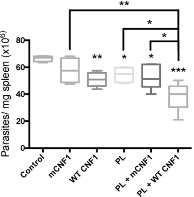
was monitored at day 42 post-immunization.Fig 5depicts the parasite burden in the spleen.
The infected mice treated with WT CNF1 had a reduced parasite burden compared with the mCNF1 and control mice. Second, the infected mice treated with PL alone had a significantly reduced parasite burden. Third, the PL + WT CNF1 treatment produced a maximal parasite clearance effect. Altogether, our results show a robust reduction in parasite burden triggered by PL in combination with WT CNF1. Our previous observations indicated a higher IL-2 and
IFN-γresponse after PL-antigen recall when mice were vaccinated with PL + WT CNF1
com-pared with PL + mCNF1. We tested whether the curative properties of WT CNF1, PL and PL
+ WT CNF1 correlated with an increase in IL-2 and IFN-γlevels after PL-antigen recall. We
measured a significant increase in IL-2 and IFN-γcytokine responses with the PL treatment
Fig 5. CNF1 activity confers curative immunoadjuvant properties.BALB/c mice were first infected with 3x10⁶of stationary phase parasites. Fourteen days post-infection, mice were immunized via the nasal route with promastigote lysate (PL), wild-type CNF1 (WT CNF1), catalytically inactive CNF1 (mCNF1), PL plus either WT CNF1 (WT CNF1 + PL) or catalytically inactive CNF1 (mCNF1 + PL). The controls represent infected but non-immunized animals. Twenty-one and twenty-eight days post infection, the mice were immunized again. At day 42, the mice were sacrificed, and parasite numbers were determined by quantitative PCR using mouse spleen DNA extracts. The bars represent the mean cytokine production±SEM.*: p<0.05,
**: p<0,01,***: p<0,001. n = 5.
but not with the WT CNF1 treatment alone, thus suggesting a role for WT CNF1 in
stimulat-ing the Th1 response initiated by PL (Fig 6A and 6B). The highest IFN-γcytokine response
level was found when PL + WT CNF1 were combined, a result consistent with maximal
para-site clearance in this treatment condition (Figs5and6B). Additionally, in these experiments,
we measured a decreased IL-4 production of approximately 2.1-fold (Fig 6C). The ratio of
IFN-γ/IL-4 was approximately 3.8-fold higher for the mice treated with PL + WT CNF1
com-pared with the mice immunized with PL + mCNF1 (Fig 6). Both of our curative and prophylac-tic vaccination settings suggest a similar mode of action for CNF1 activity, which exacerbates the Th1 cellular responses induced by PL.
Discussion
Here, we report the use of WT CNF1 as an immunoadjuvant in a prophylactic and curative
vaccination againstL.infantumInfection. We linked this property of WT CNF1 to its
enzy-matic activity, and we provide evidence that CNF1 activity induces an immunostimulatory cytokine profile that is biased toward a Th1 response. This study suggests that Rho GTPases
are targets of great value to stimulate cellular immunity againstL.infantumintracellular
parasite.
A limited number of vaccine trials against the visceral species,L.infantum/chagasi, have
been reported to date [15,17,25]. Second- and third-generation vaccine candidates are based
on the use of variousLeishmaniaantigen preparations combined with different adjuvants [15].
Second- and third-generation vaccines using purified or recombinantL.infantumsubfractions
represent a feasible option for mass vaccination campaigns; however, their efficacy generally requires the co-administration of an adjuvant [15,17]. Several compounds with adjuvant
prop-erties, including cytokines, monophosphoryl lipid A, saponins,Cryptosporidium parvum,
Pro-pionibacterium acnesand Complete Freund Adjuvant have been described in vaccination trials
againstL.infantum[26]. However, to our knowledge, the adjuvant effect of WT CNF1, a Rho
GTPase activating protein, during vaccination againstLeishmaniaspecies has not yet been
reported. In this study, we found that catalytically active CNF1 exerted a protective effect
againstL.infantuminfection when mice were immunized in the nasal mucosa with a
promasti-gote lysate. Notably, WT CNF1 significantly increased the resistance toLeishmaniainfection
in animals despite the use of high doses of metacyclic parasites. Extending previous reports
showing that vaccination withLeishmaniaantigens confers some protection in animals [27],
here, we established that this protection was dramatically improved by using WT CNF1 reach-ing 82% protection in the spleen and 94% in liver tissues (S1 Fig).
WT CNF1 confers protection againstL.infantuminfection through molecular mechanisms
that remain to be fully elucidated; however, they involve the toxin catalytic activity toward Rho GTPases. Here, we showed that treatment with WT CNF1 elicited specific cellular responses
characterized by increased secretion of IFN-γand 2 cytokines and decreased secretion of
IL-4. WT CNF1 had no effect on the levels of IL-10 production after antigen recall, a down
regula-tor of Th1 responses (not shown). IFN-γproduction has a major role in eliciting anti-parasite
macrophage responses, notably it induces production of H2O2and induction of NO synthase,
which are required for intracellular parasite killing. Additionally, IL-2 production and lympho-proliferation contribute to conferring cellular immunoprotection. These protective immune responses are balanced by the immunosuppressive responses triggered by the parasite. Immu-nosuppressive cytokines, notably IL-4, are involved in the exacerbation of infection. Although WT CNF1 had no effect on IL-10 production, we found that catalytically active CNF1 was able
to decrease IL-4 production. Thus, decreased IL-4 production combined with increased IFN-γ
polarization toward the Th1 phenotype. The mechanistic behind this Th1 polarization trig-gered by WT CNF1 requires further investigation. One possibility is that WT CNF1 directly targets the T cell compartment. A second possibility is that WT CNF1 targets other immune cells allowing them to produce signals triggering the polarization of T cells toward the Th1 phe-notype. We also reveal a significant curative effect triggered by WT CNF1 during the treatment ofLeishmaniainfected animals. This curative effect is greatly enhanced in the presence of
Leishmaniaantigen. While the exact mechanism by which CNF1 activity confers protection
againstLeishmaniaremains to be uncovered, interestingly we show here that this curative
effect of WT CNF1 is specifically promoted by the co-administered antigens.
Collectively, our data provide the first indication that active WT CNF1 has vaccinal properties
in promoting prophylactic and curative protection againstL.infantumintracellular parasites.
Supporting Information
S1 Fig. Liver Protective effects of nasal immunizations againstL.infantuminfection. BALB/c mice were immunized with promastigote lysate plus either wild-type CNF1 (PL + WT CNF1) or catalytically inactive CNF1 (PL + mCNF1). Fourteen days after the last boost, the mice
were intraperitoneally challenged with 108stationary phaseL.infantummetacyclic parasites. The
controls represent infected but non-immunized animals. Liver parasite burdens were quantified
1 month later by ELISA. The bars indicate the mean parasite loads ± SEM.: p<0.01. The
results are representative of 2 independent experiments. n = 7. (TIFF)
Acknowledgments
We are grateful to Dr Mery Tulic and Carmelo Luci for critical reading of the manuscript. This
work was supported by the Groupe d’Action Contre la Leishmaniose (GACL), by institutional
funding from INSERM, grants from the Fondation Infectiopôle Sud, the Agence Nationale de la Recherche (ANR 11BSV3 004 01), the "Investments for the Future" LABEX SIGNALIFE ANR-11-LABX-0028-01 and the C3M animal facility. NM was supported by DGA and the joint ministerial program of R&D against CBRNE threats. SIMOPRO is a member of the labo-ratory of Excellence LERMIT supported by the grant from the Agence Nationale de la Recher-che (ANR-10-LABEX-33). We thank Veronique Corcelle and the C3M animal facility team (Yannick Michelle, Sandrine Verger and Alexandre Ipekdjian) for their help and valuable assis-tance with animal care.
Author Contributions
Conceived and designed the experiments: GM BF EL. Performed the experiments: GM BF P. Munro. Analyzed the data: GM BF LB EL. Contributed reagents/materials/analysis tools: GM BF P. Munro LB EL. Wrote the paper: GM BF P. Munro DG NM LB P. Marty EL.
References
1. Mbow ML, De Gregorio E, Valiante NM, Rappuoli R (2010) New adjuvants for human vaccines. Curr Opin Immunol 22: 411–416. doi:10.1016/j.coi.2010.04.004PMID:20466528
animals. At day 42, the mice were sacrificed, and spleen homogenates were challenged with 50μg/ml PL for
48 hours. Supernatants were collected and assayed for IL-2 (A), IFN-γ(note, on graph it is INF) (B) and IL-4
(C) by ELISA. The bars represent the mean cytokine production±SEM.*: p<0.05,*: p<0.05,**: p<0,01, ***: p<0,001. n = 5.
2. Munro P, Flatau G, Doye A, Boyer L, Oregioni O, Mege JL, et al. (2004) Activation and proteasomal degradation of rho GTPases by cytotoxic necrotizing factor-1 elicit a controlled inflammatory response. J Biol Chem 279: 35849–35857. PMID:15152002
3. Munro P, Flatau G, Anjuere F, Hofman V, Czerkinsky C, Lemichez E (2005) The Rho GTPase activa-tors CNF1 and DNT bacterial toxins have mucosal adjuvant properties. Vaccine 23: 2551–2556. PMID:15780436
4. Munro P, Flatau G, Lemichez E (2007) Intranasal immunization with tetanus toxoid and CNF1 as a new mucosal adjuvant protects BALB/c mice against lethal challenge. Vaccine 25: 8702–8706. PMID:
18035455
5. Boyer L, Magoc L, Dejardin S, Cappillino M, Paquette N, Hinault C, et al. (2011) Pathogen-Derived Effectors Trigger Protective Immunity via Activation of the Rac2 Enzyme and the IMD or Rip Kinase Signaling Pathway. Immunity 35: 536–549. doi:10.1016/j.immuni.2011.08.015PMID:22018470 6. Stuart LM, Paquette N, Boyer L (2013) Effector-triggered versus pattern-triggered immunity: how
ani-mals sense pathogens. Nat Rev Immunol 13: 199–206. doi:10.1038/nri3398PMID:23411798 7. Diabate M, Munro P, Garcia E, Jacquel A, Michel G, Obba S, et al. (2015)Escherichia coli
alpha-Hemo-lysin Counteracts the Anti-Virulence Innate Immune Response Triggered by the Rho GTPase Activat-ing Toxin CNF1 durActivat-ing Bacteremia. PLoS Pathog 11: e1004732. doi:10.1371/journal.ppat.1004732
PMID:25781937
8. Lemonnier M, Landraud L, Lemichez E (2007) Rho GTPase-activating bacterial toxins: from bacterial virulence regulation to eukaryotic cell biology. FEMS Microbiol Rev 31: 515–534. PMID:17680807 9. Schmidt G, Sehr P, Wilm M, Selzer J, Mann M, Aktories K (1997) Gln 63 of Rho is deamidated by
Escherichia colicytotoxic necrotizing factor-1. Nature 387: 725–729. PMID:9192900
10. Flatau G, Lemichez E, Gauthier M, Chardin P, Paris S, Fiorentini C, et al. (1997) Toxin-induced activa-tion of the G protein p21 Rho by deamidaactiva-tion of glutamine. Nature 387: 729–733. PMID:9192901 11. Mettouchi A, Lemichez E (2012) Ubiquitylation of active Rac1 by the E3 Ubiquitin-Ligase HACE1.
Small GTPases 3: 102–106. doi:10.4161/sgtp.19221PMID:22790197
12. Lemichez E, Aktories K (2013) Hijacking of Rho GTPases during bacterial infection. Exp Cell Res 319: 2329–2336. doi:10.1016/j.yexcr.2013.04.021PMID:23648569
13. Keestra AM, Winter MG, Auburger JJ, Frassle SP, Xavier MN, Winter SE, et al. (2013) Manipulation of small Rho GTPases is a pathogen-induced process detected by NOD1. Nature 496: 233–237. doi:10. 1038/nature12025PMID:23542589
14. Marty P, Izri A, Ozon C, Haas P, Rosenthal E, Del Giudice P, et al. (2007) A century of leishmaniasis in Alpes-Maritimes, France. Ann Trop Med Parasitol 101: 563–574. PMID:17877875
15. Das A, Ali N (2012) Vaccine Development Against Leishmania donovani. Front Immunol 3: 99. doi:10. 3389/fimmu.2012.00099PMID:22615707
16. Sundar S, Chakravarty J (2015) An update on pharmacotherapy for leishmaniasis. Expert Opin Phar-macother 16: 237–252. doi:10.1517/14656566.2015.973850PMID:25346016
17. Nagill R, Kaur S (2011) Vaccine candidates for leishmaniasis: a review. Int Immunopharmacol 11: 1464–1488. doi:10.1016/j.intimp.2011.05.008PMID:21616175
18. Fernandes AP, Coelho EA, Machado-Coelho GL, Grimaldi GJ, Gazzinelli RT (2012) Making an anti-amastigote vaccine for visceral leishmaniasis: rational, update and perspectives. Curr Opin Microbiol 15: 476–485. PMID:22698479
19. Ferrua B, Luci C, Le Fichoux Y, Paul A, Marty P (2006) Imprinting of BALB/c mice with lowLeishmania infantumparasite dose markedly protects spleen against high-dose challenge. Vaccine 24: 589–596. PMID:16157427
20. Michel G, Ferrua B, Lang T, Maddugoda MP, Munro P, Pomares C, et al. (2011) Luciferase-expressing
Leishmania infantumallows the monitoring of amastigote population size,in vivo,ex vivoandin vitro. PLoS Negl Trop Dis 5: e1323. doi:10.1371/journal.pntd.0001323PMID:21931877
21. Doye A, Boyer L, Mettouchi A, Lemichez E (2006) Ubiquitin-mediated proteasomal degradation of Rho proteins by the CNF1 toxin. Methods Enzymol 406: 447–456. PMID:16472677
22. Ferrua B, Le Fichoux Y, Suffia I, Rousseau D, Roptin C, Kubar J (2001) Quantitation ofLeishmania infantumin tissues of infected BALB/c mouse by sandwich ELISA. J Immunoassay Immunochem 22: 165–181. PMID:11486813
23. Reis AB, Giunchetti RC, Carrillo E, Martins-Filho OA, Moreno J (2010) Immunity toLeishmaniaand the rational search for vaccines against canine leishmaniasis. Trends Parasitol 26: 341–349. doi:10.1016/ j.pt.2010.04.005PMID:20488751
25. Lemesre JL, Holzmuller P, Cavaleyra M, Goncalves RB, Hottin G, Papierok G (2005) Protection against experimental visceral leishmaniasis infection in dogs immunized with purified excreted secreted anti-gens ofLeishmania infantumpromastigotes. Vaccine 23: 2825–2840. PMID:15780731
26. Holmgren J, Adamsson J, Anjuere F, Clemens J, Czerkinsky C, Eriksson K, et al. (2005) Mucosal adju-vants and anti-infection and anti-immunopathology vaccines based on cholera toxin, cholera toxin B subunit and CpG DNA. Immunol Lett 97: 181–188. PMID:15752556