v. 45 – no.2 – abr./jun. 2008 Arq Gastroenterol
152
GASTROENTEROLOGIA PEDIÁ
TRICA / PEDIA
TRIC GASTROENTEROLOG
ARQGA/1357
CLINICAL AND LABORATORY EVALUATION
OF 101 PATIENTS WITH
INTRAHEPATIC NEONATAL CHOLESTASIS
Maria Angela
BELLOMO-BRANDÃO
1, Gilda
PORTA
2and Gabriel
HESSEL
1ABSTRACT – Background - Intrahepaticneonatal cholestasis can be the initial manifestation of a very heterogeneous group of illnesses
of different etiologies. Aim - To evaluate and compare clinical and laboratory data among intrahepaticneonatal cholestasis groups of infectious, genetic-endocrine-metabolic and idiopathic etiologies. Methods - The study evaluated retrospectively clinical and laboratory data of 101 infants, from March 1982 to December 2005, 84 from the State University of Campinas Teaching Hospital, Campinas, SP, Brazil, and 17 from the Child’s Institute of the University of São Paulo, SP, Brazil. The inclusion criteria consisted of: jaundice beginning at up to 3 months of age and hepatic biopsy during the 1st year of life. It had been evaluated: clinical findings (gender, age, birth weight, weight during the first medical visit, stature at birth, jaundice, acholia/hipocholia, choluria, hepatomegaly and splenomegaly) and laboratorial (ALT, AST, FA, GGT, INR). Results - According to diagnosis, patients were classified into three groups: group 1 (infectious) n = 24, group 2 (genetic-endocrine-metabolic) n = 21 and group 3 (idiopathic) n = 56. There were no significant differences in relation to the variables: age, gender, stature at birth, jaundice, acholia/hipocholia, choluria, hepatomegaly, splenomegaly, AST, ALT, ALP, GGT, DB and albumin. Significant differences were observed in relation to the following variables: birth weight and weight during the first medical visit. Birth weight of group 1 was lower in relation group 2 and 3. Weight during the first medical visit followed the same pattern. There was a statistically significant difference in relation to the INR, as the patients of the group 2 presented higher values in relation to groups 2 and 3, despite the median was still pointing out normal values. Conclusions - There were no significant differences in relation to age, gender, stature at birth, jaundice, acholia/hipocholia, choluria, hepatomegaly, splenomegaly, AST, ALT, ALP, GGT, BD and albumin. Birth weight and the weight during the first medical visit were lower in the group with infectious etiology. In addition, a significant difference in INR reflected impaired coagulation of patients of the group of the genetic-endocrine-metabolic disease.
HEADINGS – Cholestasis, intrahepatic. Jaundice, neonatal.
INTRODUCTION
The frequency of the cholestatic jaundice is difficult to evaluate, varying from 1:2500 to 1:5000 newborns(3, 6, 9). The initial approach aims to distinguish the diagnosis between intrahepatic causes and extrahepatic, as the latter require precocious surgical intervention(19). In general, intrahepatic neonatal cholestasis (IHNC) represents 2/3 of the cases of neonatal cholestasis(8, 12, 15, 18, 28). The most common causes of the disease are
of infectious origin(25, 21, 29). In septicemia, signal and symptom manifestations of hepatic origin represent only one component of the involvement of multiple organs, to which the adequate treatment offers the best chance of recovery(4). Any serious bacterial infection during the neonatal period can result in jaundice(26); however, there seems to be a more frequent association with urinary tract infections, especially when the bacterium is Escherichia coli(14). In addition to these infections, others have been
observed, such as syphilis, toxoplasmosis, rubella and cytomegalovirus (CMV)(1, 11, 20, 21, 22, 23).
Despite the many possible etiologies for IHNC(8), 13%-78% of the cases have been reported to be related to idiopathic etiology(7, 10, 16, 17, 18, 27). Idiopathic IHNC implies that the liver suffers inflammatory alterations of unknown cause, with no evidence of blockage of the biliary tree, and those infectious agents or metabolic errors for the praised methods have been discarded(2, 3). Concerning the evolution, there are cases of idiopathic IHNC considered spontaneous, in which there is family recurrence; therefore, sporadic cases could possibly consist in a viral injury or another environmental factor that affected the transitory form of the immature liver of the newborn, however characteristics are similar in both cases(4, 5).
The objectives of the present study were to evaluate and compare clinical and laboratory data among etiologic groups, searching for clinical and laboratories findings that can help in the diagnostic process.
1Department of Pediatrics, Campinas State University (UNICAMP), Campinas, SP, Brazil; 2Department of Pediatrics, Children’s Institute of the University of São Paulo
Medical School (USP), Sao Paulo, SP, Brazil
Bellomo-Brandão MA, Porta G, Hessel G. Clinical and laboratory evaluation of 101 patients with intrahepatic neonatal cholestasis
Arq Gastroenterol 153
v. 45 – no.2 – abr./jun. 2008
METHODS
This study evaluated retrospectively clinical and laboratory data of patients submitted to hepatic biopsy during IHNC investigation, from March 1982 to December 2005. The inclusion criteria consisted of: jaundice beginning at up to 3 months of age and hepatic biopsy during the 1st year of life.
There were evaluated 101 patients (84 from the State University of Campinas Teaching Hospital (UNICAMP), Campinas, SP, and 17 from the Children’s Institute of the University of São Paulo (USP). The approach to the diagnosis of NIHC was standardized and a uniform diagnostic approach was taken throughout the observation period.
The information comprised in: neonate’s identification, antecedents, clinical findings, physical examination and results of the following laboratorial tests: alanine aminotransferase (ALT), aspartate aminotransferase (AST), alkaline phosphatase (ALP), gamma glutamyltransferase (GGT) international normalized ratio (INR), direct bilirubin (DB) and albumin; carried out in the first medical visit. Hepatic enzymes (AST, ALT, ALP and GGT) are presented in number of times in which the superior limit of normality was reached. Albumin is presented in gram per deciliter (g/dL) and DB in milligram per deciliter (mg/dL).
In order to establish the IHNC etiology, the following examinations were reviewed: liver biopsy, alpha-1-antitrypsin serum, sweat sodium and chloride, innate metabolism errors in urine, polimerase chain reaction (PCR) and CMV antigenemia, serology of: CMV, HIV, EBV, rubella, toxoplasmosis and syphilis.
Ethical aspects
The present research study was approved by the Medical Research Ethics Committees of both institutions.
Statistical analysis
In order to verify if there was association with the categorical variable, the Qui-square test was used. When the expected values were under 5, the Fisher’s exact test was used(24).
For comparing the continuous variable among the groups, the analysis of variance (ANOVA) and Tukey’s tests was performed to identify the differences(13). Significance was established as
P<0.05 in all tests.
RESULTS
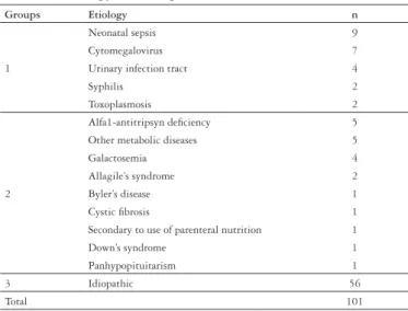
According to diagnosis, patients were classified into three groups: group 1 (infectious) n = 24, group 2 (genetic-endocrine -metabolic) n = 21 and group 3 (idiopathic) n = 56. The diagnosis of NIHC is presented in Table 1.
The median age of patients at initial presentation was 2 months and 7 days. There was no statistical difference among the groups (P = 0.595). Patients were predominating boys in all groups (P = 0.770).
Table 2 shows clinical characteristics during the first medical visit (age, gender, birth weight, weight during the first medical visit, stature at birth, jaundice, acholia/hypocholia, choluria,
hepatomegaly and splenomegaly) of the groups. There were no significant differences among the groups according the variables: age, gender, jaundice, acholia, choluria, hepatomegaly and splenomegaly. However, a significant difference was observed according the variables: birth weight and weight during the first medical visit. Birth weight of group 1 was lower in relation to groups 2 and 3 (P = 0.002). The weight during the first medical visit followed the same pattern (P = 0.047).
TABLE 1. Etiology of intrahepatic neonatal cholestasis (NIHC)
Groups Etiology n
1
Neonatal sepsis 9
Cytomegalovirus 7
Urinary infection tract 4
Syphilis 2
Toxoplasmosis 2
2
Alfa1-antitripsyn deficiency 5
Other metabolic diseases 5
Galactosemia 4
Allagile’s syndrome 2
Byler’s disease 1
Cystic fibrosis 1
Secondary to use of parenteral nutrition 1
Down’s syndrome 1
Panhypopituitarism 1
3 Idiopathic 56
Total 101
TABLE 2. Clinical characteristics of the patients during the first evaluation, in accordance with the groups. Numbers in parentheses correspond to the numbers of patients evaluated in the different groups. n = total number of patients, M = male, F = female. Median values: Age (months), Weight expressed in grams (g) and stature in centimeters (cm). SD = Standard deviations
*ANOVA - P-value = 0.021 Tukey’s test: (1 ≠ 2; 1 ≠ 3; 2 = 3) **ANOVA - P-value = 0.047 Tukey’s test :(1 ≠ 2; 1 ≠ 3; 2 = 3)
n Infectious NIHC (24) Genetic-endocrine-metabolic NIHC (21) Idiopathic NIHC (56) P
Age (months) 101 2.40
(SD = 1.15)
2.04 (SD = 1.24)
2.43 (SD = 1.68)
0.595
Gender M/F 101 17/7 13/8 39/17 0.770
Birth weight (g) 96 (21)
2160g (SD = 843)
(20) 2820g (SD = 612)
(55) 2750g (SD = 763)
0.021 *
Weight during the first medical visit (g)
81 (20)
3105g (SD = 931)
(18) 3725g (SD = 1122)
(43) 3970g (SD = 1074)
0.047 **
Stature at birth (cm)
69 (13)
44,5 (SD = 4.69)
(12) 47 (SD = 4.22)
(44) 48 (SD = 5,22)
0.683
Jaundice 98 (23)
21 (21) 19 (54) 51 0.657 Acholia/ hiphocolia 94 (20) 12 (19) 10 (55) 38 0.403
Choluria 80 (19)
10 (17) 8 (44) 30 0.241
Hepatomegaly 94 (22)
20 (20) 15 (52) 44 0.439
Splenomegaly 93 (22)
Bellomo-Brandão MA, Porta G, Hessel G. Clinical and laboratory evaluation of 101 patients with intrahepatic neonatal cholestasis
Arq Gastroenterol
154 v. 45 – no.2 – abr./jun. 2008
TABLE 3. Median of results of the laboratory tests: alanine aminotransferase (ALT), aspartate aminotransferase (AST), alkaline phosphatase (ALP), gama glutamyltransferase (GGT), albumin, international normalized ratio (INR) and direct bilirubin (BD) carried out in the first medical visit. Hepatic enzymes (AST, ALT, ALP and GGT) are presented in number of times in which the superior limit of normality was reached. Albumin is presented in gram per deciliter (g/dL) and DB in milligram per deciliter (mg/dL). The numbers in parentheses represent the number of patients whose data were obtained from each group
* ANOVA - P-value = 0.033 Tukey’s test (1 ≠ 2; 1 = 3; 2 ≠ 3)
Variables n
Infectious NIHC
(24)
Genetic-endocrine-metabolic NIHC
(21)
Idiopathic NIHC
(56) P
ALT 92 (20)
3.62
(21) 3.80
(51) 3.48
0.397
AST 96 (20)
5.28
(21) 6.69
(55) 5.4
0.462
AlkPhos 80 (18)
1.67
(18) 1.39
(44) 1.50
0.893
GGT 76 (17)
3.91
(18) 5.57
(41) 4.31
0.676
Albumin (g/dL) 62 (17)
3.70
(12) 3.27
(33) 3.91
0.074
INR 70 (15)
1.04
(15) 1.24
(40) 1.10
0.033*
DB (mg/dL) 78 (22)
5.73
(20) 6.55
(56) 6.91
0.683
Table 3 shows the laboratorial tests carried out in the beginning of the inquiry (ALT, AST, ALP, GGT, BD, albumin and INR), using comparison between groups. There was significant difference in relation to the INR (P = 0.033). The group 2 presented a higher value in relation to group 1 and 3; however, the median one still was pointing out normality values. Four patients evolved to death: three of the group 2 (two with metabolic disease and one with Allagile’s syndrome) and one patient of the group 3 (idiopathic). The autopsy of this last patient showed intense hepatocelular necrosis, positive serology for the virus of the
acquired immunodeficiency (HIV-AIDS) and negativity for HIV in the liver biopsy using PCR.
DISCUSSION
There was accordance with the literature concerning the clinical findings, in which jaundice was the main manifestation, followed by acholia and choluria(21). Birth weight values and the weight during the first medical visit were lower in the group with infectious etiology, possibly reflecting the impaired growth expected in cases of neonatal diseases and congenital infections. Differences of clinical presentation were not observed either among other clinical findings (gender, age, stature at birth, jaundice, acholia/hypocholia, choluria, hepatomegaly, splenomegaly) or among the following laboratory tests: aminotransferases, AlkPhos, DB and GGT. This last test was evaluated by WANG et al.(27), who concluded that normal or low levels of GGT could indicate greater severity of idiopathic IHNC, although reversible. INR statistical significant difference reflected impaired coagulation of patients of the group of the genetic-endocrine-metabolic disease. This data may reflect severity of these cases, since three of the four deaths occurred in the group of the genetic-endocrine-metabolic disease.
CONCLUSION
There were no differences between the groups of infectious, genetic-endocrine-metabolic and idiopathic etiologies regarding to: age, gender, stature at birth, jaundice, acholia/hypocholia, choluria, hepatomegaly, splenomegaly, ALT, AST, AlkPhos, GGT and DB.
Bellomo-Brandão MA, Porta G, Hessel G. Clinical and laboratory evaluation of 101 patients with intrahepatic neonatal cholestasis
Arq Gastroenterol 155
v. 45 – no.2 – abr./jun. 2008
REFERENCES
1. American Academy of Pediatrics. Cytomegalovirus infection. In: Pickering LK, editor. Redbook 2003: report of the Committee on Infectious Diseases. 26th ed. Elk Grove Village: American Academy of Pediatrics; 2003. p.259–62.
2. Balistreri WF, Heubi JE, Suchy FJ. Immaturity of the enterohepatic circulation in early life: factors predisposing to “physiologic” maldigestion and cholestasis. J Pediatr Gastroentrol Nutr. 1983;18:346-54.
3. Balistreri WF. Neonatal cholestasis. J Pediatr. 1985;106:171-84.
4. Bezerra JA. Colestase neonatal. In: Ferreira CT, Carvalho E, Silva LR, editors. Gastroenterologia e hepatologia em pediatria. Rio de Janeiro: Medsi; 2003. p.582-97.
5. Bezerra JA, Balistreri W. Whatever happened to ‘‘neonatal hepatitis’’? Clin Liver Dis. 2006;10:27–53.
6. Danks DM, Campbell PE, Jack I, Rogers J, Smith AL. Studies of the etiology of neonatal hepatitis and biliary atresia. Arch Dis Child. 1977;52:360-7.
7. Dehghani SM, Haghighat M, Imanieh MH, Geramizadeh B. Comparison of different diagnostic methods in infants with cholestasis. World J Gastroenterol. 2006;12:5893-6.
8. Dellert SF, Balistreri WF. Neonatal cholestasis. In: Walker WA, Durie PR, Hamilton JR, Walker-Smith JA, Watkins JB, editors. Pediatric gastrointestinal disease: pathophysiology, diagnosis, management. 3rd ed. Hamilton, Ont.: BC Decker; 2000. p.880-94.
9. Dick MC, Mowat AP. Hepatitis syndrome in infancy: an epidemiology survey with 10-year follow up. Arch Dis Child. 1985;60:512-6.
10. Eliot N. Analyze statistique des donnees cliniques, biologiques et histologiques dans 288 observations de cholestase neonatale. Arch Franç Pédiatr. 1977;34(2 Suppl):213-20.
11. Felber S, Sinatra M. Systemic disorders associated with neonatal cholestasis. Semin Liver Dis. 1987;7:108-18.
12. Fischler B, Papadogiannakis N, Nemeth A. Aetiological factors in neonatal cholestasis. Acta Paediatr. 2001;90:88-92.
13. Fleiss JL. Statistical methods for rates and proportions. 2nd ed. New York: John Wiley; 1981. p.321.
14. Garcia FJ, Nager AL. Jaundice as an early diagnostic sign of urinary tract infection in infancy. Pediatrics. 2002;109:846-51.
15. Henriksen NT, Drablos PA, Aagenaes O. Cholestatic jaundice in infancy. The importance of familial and genetic factors in aetiology and prognosis. Arch Dis Child. 1981;56:622-7.
Bellomo-Brandão MA, Porta G, Hessel G. Avaliação clínica e laboratorial de 101 pacientes com colestase neonatal intra-hepática. Arq Gastroenterol. 2008;45(2): 152-5.
RESUMO – Racional - A colestase neonatal intra-hepática pode ser a manifestação inicial de um grupo muito heterogêneo de doenças de diferentes causas.
Objetivos - Avaliar e comparar características clínicas e laboratoriais entre os grupos de colestase neonatal intra-hepática de causa infecciosa, genético-endócrino-metabólica e idiopática. Métodos - Foram revistos os prontuários de 101 pacientes com diagnóstico de colestase neonatal intra-hepática no período de março de 1982 a dezembro de 2005, 84 avaliados no Hospital das Clínicas da Universidade Estadual de Campinas, SP, e 17 no Instituto da Criança da Universidade de São Paulo. Os critérios de inclusão foram: história de surgimento de icterícia até 3 meses de idade e realização da biopsia hepática durante o primeiro ano de vida. Foram avaliados: quadro clínico (gênero, idade, peso ao nascimento, peso à primeira consulta, estatura ao nascimento, icterícia, acolia ou hipocolia, colúria, hepatomegalia e esplenomegalia) e laboratorial (ALT, AST, FA, GGT, INR, BD). Resultados - Os pacientes foram divididos em grupos, de acordo com o diagnóstico etiológico: grupo 1 (infeccioso) n = 24; grupo 2 (genético-endócrino-metabólico) n = 21 e grupo 3 (idiopático) n = 56. Não houve diferença estatisticamente significante em relação às variáveis: gênero, idade, estatura ao nascimento, icterícia, acolia/hipocolia, colúria, hepatomegalia, esplenomegalia, AST, ALT, FA, GGT, BD e albumina. O peso ao nascimento e o peso na primeira consulta dos pacientes com colestase neonatal intra-hepática de etiologia infecciosa foi menor. Houve diferença estatisticamente significante em relação ao INR: os pacientes com causas genético-endócrino-metabólicas apresentaram valor mais prolongado, porém com a mediana se situando dentro dos valores de normalidade. Conclusão - Não houve diferença estatisticamente significativa entre os grupos em relação às variáveis: gênero, idade, estatura ao nascimento, icterícia, acolia/hipocolia, colúria, hepatomegalia, esplenomegalia, AST, ALT, FA, GGT, BD e albumina. Os pacientes com colestase neonatal intra-hepática de causa infecciosa apresentaram menores valores de peso em relação às demais causas e o INR foi mais prolongado nos pacientes com causas genético-endócrino-metabólicas, demonstrado alterações na função de coagulação.
DESCRITORES – Colestase intra-hepática. Icterícia neonatal.
16. Hessel G, Yamada RM, Escanhoela CA, Bustorff-Silva JM, Toledo RJ. Valor da ultra-sonografia abdominal e da biopsia hepática percutânea no diagnóstico diferencial da colestase neonatal. Arq Gastroenterol.1994;31:75-82.
17. Mieli-Vergani G, Howard ER, Mowat AP. Liver disease in infancy: a 20 year perspective. Gut. 1991;32:s123–s8.
18. Mowat AP. Hepatite e colestase em lactentes: afecções intra-hepáticas. In: Mowat AP, editor. Doenças hepáticas em pediatria. 2a ed. Rio de Janeiro: Revinter; 1991. p.41-80. 19. Moyer V, Freeser DK, Whitington PF, Olson AD, Brewer F, Colletti RR, Heyman MB,
North American Society for Pediatric, Gastroenterology, Hepatology and Nutrition. Guideline for the evaluation of cholestatic jaundice in infants: recommendations of the North American Society for Pediatric, Gastroenterology, Hepatology and Nutrition. J Pediatr Gastroenterol Nutr. 2004;39:115-28.
20. Munro C, Hall B, Whybin LR, Leader L, Robertson P, Maine GT, Rawlinson WD. Diagnosis of and screening for cytomegalovirus infection in pregnant women. J Clin Microbiol. 2005;43:4713–8.
21. Prado ET, Araujo MF, Campos JV. Colestase neonatal prolongada: estudo prospectivo. Arq Gastroenterol. 1999;36:185-94.
22. Roberts EA. A criança ictérica. In: Kelly DA, editor. Doenças hepáticas e do sistema biliar em crianças. São Paulo: Santos; 2001. p.11-45.
23. Shibata Y, Kitajima N, Kawada J, Sugaya N, Nishikawa K, Morishima T, Kimura H.Association of cytomegalovirus with infantile hepatitis. Microbiol Immunol. 2005;49:771-7.
24. Siegel S. Estatísticas não-paramétricas para as ciências do comportamento. São Paulo: McGraw-Hill. 1975. p.106-16.
25. Silveira TR, Pires ALG. Icterícia colestática neonatal. In: Fagundes Neto U, Wehba J, Penna FJ. Gastroenterologia pediátrica. 2ª edition. Rio de Janeiro: Medsi. 1991. p.465-87.
26. Tiker F, Tarcan A, Kilicdag H, Gurakan B. Early onset conjugated hiperbilirrubinemia in newborn infants. Indian J Pediatr. 2006;73:409-12.
27. Wang JS, Tan N, Dhawan A. Significance of low or normal serum gamma glutamyl transferase level in infants with idiopathic neonatal hepatitis. Eur J Pediatr. 2006;165:795–801.
28. Yachha SK, Sharma A. Neonatal cholestasis in India. Indian Pediatr. 2005;42:491-2. 29. Zerbini MC, Gallucci SD, Maezono R, Ueno Cm, Porta G, Maksoud JG, Gayotto
LC. Liver biopsy in neonatal cholestasis: a review on statistical ground. Mod Pathol. 1997;10:793-9.