Symposium
Spontaneous Clearance of
Mycobacterium ulcerans
in a
Case of Buruli Ulcer
Claire L. Gordon1, John A. Buntine2,3, John A. Hayman4, Caroline J. Lavender5, Janet A. Fyfe5, Patrick Hosking6, Paul D. R. Johnson1,5*
1Department of Infectious Diseases, Austin Health, Melbourne, Australia,2Department of Surgery, Box Hill Hospital, Melbourne, Australia,3Department of Surgery, Monash University, Melbourne, Australia,4Department of Anatomy and Developmental Biology, Monash University, Melbourne, Australia,5WHO Collaborating Centre for Mycobacterium ulcerans (Western Pacific Region) and Victorian Infectious Diseases Reference Laboratory, Melbourne, Australia,6Department of Pathology, Box Hill Hospital, Melbourne, Australia
Buruli ulcer (BU) is an infection of skin and soft tissue caused by Mycobacterium ulcerans, a toxin-producing environmental mycobacterium. Significant advances in the treatment of BU have been made over the past decade with the introduction of effective antibiotic therapy and there is a greater understanding of the pathogenesis and host immune response. Although it is generally held that early BU lesions may heal spontaneously, to our knowledge, there are no previously published cases that definitively document spontaneous resolution of culture-confirmed BU.
Presentation of Case
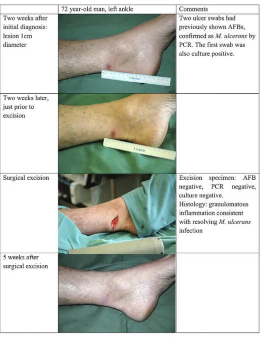
The patient was a 72-year-old man from Melbourne, Australia, who had visited his holiday house in Point Lons-dale, an endemic area of BU on the coast 70 km to the southwest [1,2], most weekends for the past 28 years. He had a past medical history of coronary artery disease but was otherwise fit and active. In October 2008 he noted a 5-week history of a slowly progressive painless papule with a punctate centre on his left ankle (Figure 1). His most recent visit to Point Lonsdale was 2 weeks before the lesion appeared.
The diagnosis of BU was made uncon-ventionally. The patient’s 12-year-old gran-dson was attending a clinic with his mother for treatment of IS2404 PCR-confirmed [3] BU on his lower back as reported elsewhere [4]. During the consultation she mentioned that her father also had a progressive lesion on his ankle. She was given a dry specimen swab to take home with instructions on how to sample her father’s ankle lesion. Material from the ankle swab tested positive for acid-fast bacilli (AFB) by Ziehl-Neelsen (Z-N) stain-ing andM. ulceransinfection was confirmed by PCR and later by culture. A repeat swab taken by the treating clinician (PDRJ) 2 weeks later was again positive for AFB, and PCR again confirmedM. ulceransinfection. Culture from this second specimen was ultimately negative, although the culture
result was not available at the time management decisions had to be taken. Antibiotics were considered but not pre-scribed [5], as there was a potential interaction between the patient’s cardiac medications and rifampicin. Instead, exci-sion with primary closure was performed without the use of antibiotics or other treatment modalities (Figure 1). Sections of the excised tissue showed a 3-mm area of skin ulceration (Figure 2, left panel) with mixed inflammatory cell infiltration and granulation tissue extending into subcuta-neous fat (Figure 2, right panel). AFB were not detected, but the appearances were considered consistent with M. ulcerans infection in a healing or resolving phase of the infection [6]. The excised tissue spec-imen was divided into five sub-sections and all portions screened by both PCR and culture according to standard operating procedures. Generally, this means that the entire sub-section of tissue is cut into smaller pieces and homogenised in a bottle containing glass beads and 2 ml of phos-phate buffered saline. If the sub-section is too large to fit into the bead bottle, smaller pieces of tissue are taken from throughout the specimen to maximise the likelihood of sampling organisms. One ml of tissue homogenate is then processed for PCR [3] and 1 ml used for culture in BACTEC 12B bottles and on Brown and Buckle medium [7] incubated at 31uC for up to 12 weeks.M. ulceranswas no longer detected by either method. The patient gave informed consent for publication.
Case Discussion
Buruli ulcer has an alarming potential for progressive tissue destruction and has the potential to leave patients permanently disabled due to widespread necrosis of subcutaneous fat, extensive fibrous scar tissue formation, and contractures [6,8,9]. However, even when no effective therapy is available, progression may cease [10,11]. The human immune system is therefore able to containM. ulcerans, albeit after some years and considerable tissue destruction. The details of how this final victory is won are of great importance to researchers working on BU vaccines.
Our patient first noted a lesion 5 weeks before the first diagnostic specimen was obtained. AFB were seen, IS2404 PCR was positive, andM. ulcerans was isolated by culture. The diagnosis was reconfirmed with a second swab. The lesion was excised a month later but no AFB were detectable by then, and both PCR and culture were negative. Of note, the patient’s grandson who visited his grand-father’s house at Point Lonsdale contem-poraneously developed a large progressive BU over his lower back [4].
M. ulcerans, like M. tuberculosis and M. leprae, does not cause clinical disease in all exposed individuals. Gooding et al. [12] showed that the prevalence of antibodies to M. ulcerans in exposed household controls in Queensland was similar to that in patients with proven BU using a whole cell antigen preparation. Similar findings
Citation:Gordon CL, Buntine JA, Hayman JA, Lavender CJ, Fyfe JA, et al. (2011) Spontaneous Clearance of Mycobacterium ulcerans in a Case of Buruli Ulcer. PLoS Negl Trop Dis 5(10): e1290. doi:10.1371/ journal.pntd.0001290
Editor:Pamela L. C. Small, University of Tennessee, United States of America PublishedOctober 25, 2011
Copyright: ß2011 Gordon et al. This is an open-access article distributed under the terms of the Creative Commons Attribution License, which permits unrestricted use, distribution, and reproduction in any medium, provided the original author and source are credited.
Funding:There were no sources of funding for this work.
Competing Interests:The authors have declared that no competing interests exist. * E-mail: [email protected]
Figure 1. Clinical appearance and laboratory characteristics. doi:10.1371/journal.pntd.0001290.g001
Figure 2. Histopathology of excised lesion.Left panel: Section of excised skin ulcer, showing one ulcer margin with fibrinous exudate in the base of the ulcer. Granulation tissue with a mixed inflammatory cell infiltrate extends into subcutaneous fat (H&E, orig mag640). Right panel: Section
from the base of the ulcer, showing granulation tissue with a mixed inflammatory cell infiltrate. Acid-fast bacilli were not seen in a Z-N stained section of the same area (H&E, orig mag6200).
doi:10.1371/journal.pntd.0001290.g002
with more specific antigens have confirmed this observation in West Africa [13,14]. However, antibodies toM. ulceransare not M. ulceransspecific and are encountered in persons from endemic and non-endemic regions, which makes it difficult to evaluate evidence for spontaneous remission based on the presence of antibodies. As with most infectious diseases, variation in host im-mune response genes is likely to influence susceptibility. In a study performed in Ghana, the SLC11A1 (NRAMP1) D543N polymorphism, which confers susceptibility to tuberculosis and leprosy, has been linked to increased susceptibility to BU, with an estimated 13% population attributable risk [15].
However, it also likely that inoculum size influences outcome. For example, our patient may have received a lower initial inoculum than his grandson, although the natural inoculum size has not been
definitively established. Fenner noted that the protective effect of BCG against M. ulcerans in a mouse model could be overcome by increasing the inoculum [16]. Unlike the grandson, it is also possible that our patient had a prior exposure to M. ulcerans that may have immunised him and enhanced his ability to control his infection, as he reported very frequent visits to Point Lonsdale where transmission of M. ulcerans has been common since at least 2002 [1,2].
This report of spontaneous clearance of M. ulcerans from a small but clinically apparent BU confirms previous anecdotal and some systematic field observations in patients likely to have BU, but from whom definitive laboratory confirmation was not available [17]. For example, Revill et al. reported that 29% of patients with small nodular lesions diagnosed clinically healed spontaneously while receiving placebo
during a randomised study of clofazamine therapy [18].
The key virulence factor ofM. ulcerans, mycolactone, has potent cytotoxic and immunosuppressive properties that act both locally and systemically. Initially, the histology of BU lesions shows bland necrosis with a remarkable absence of an acute inflammatory response (reviewed in Schu¨tte et al. [19]). However, natural halting of progressive infection, or treat-ment with antibiotics, appears to be associated with the development of a vigorous Th-1 response, the development of delayed type hypersensitivity to myco-bacterial antigens, and intense granuloma-tous inflammation on histology (reviewed by Demangel et al. [20] and Schu¨tte et al. [19]). At present, no satisfactory model exists to explain how the human host is able to sterilize activeM. ulceransinfections that are likely to be producing increasing local concentrations of mycolactone, par-ticularly as mycolactone itself does not appear to stimulate the production of neutralising antibodies [19]. The mecha-nism by which this occurs will be of great interest, as a vaccine able to induce this sterilizing response should be highly effec-tive for both primary prevention and as an adjuvant to therapy.
References
1. Johnson PD, Azuolas J, Lavender CJ, Wishart E, Stinear TP, et al. (2007)Mycobacterium ulceransin mosquitoes captured during outbreak of Buruli ulcer, southeastern Australia. Emerg Infect Dis 13: 1653–1660.
2. Johnson PD, Hayman JA, Quek TY, Fyfe JA, Jenkin GA, et al. (2007) Consensus recommen-dations for the diagnosis, treatment and control of
Mycobacterium ulcerans infection (Bairnsdale or Buruli ulcer) in Victoria, Australia. Med J Aust 186: 64–68.
3. Fyfe JA, Lavender CJ, Johnson PD, Globan M, Sievers A, et al. (2007) Development and application of two multiplex real-time PCR assays for the detection of Mycobacterium ulcerans in clinical and environmental samples. Appl Environ Microbiol 73: 4733–4740.
4. Gordon CL, Buntine JA, Hayman JA, Lavender CJ, Fyfe JA, et al. (2010) All-oral antibiotic treatment for buruli ulcer: a report of four patients. PLoS Negl Trop Dis 4: e770. doi:10.1371/journal.pntd.0000770.
5. Johnson PD (2010) Should antibiotics be given for Buruli ulcer? Lancet 375: 618–619.
6. Johnson PD, Stinear T, Small PL, Pluschke G, Merritt RW, et al. (2005) Buruli ulcer (M. ulcerans infection): new insights, new hope for disease control. PLoS Med 2: e108. doi:10.1371/ journal.pmed.0020108.
7. Mac CP, Tolhurst JC, et al. (1948) A new mycobacterial infection in man. J Pathol Bacteriol 60: 93–122.
8. Wansbrough-Jones M, Phillips R (2005) Buruli ulcer. BMJ 330: 1402–1403.
9. van der Werf TS, Stienstra Y, Johnson RC, Phillips R, Adjei O, et al. (2005)Mycobacterium ulceransdisease. Bull World Health Organ 83: 785–791.
10. Hayman J (1993) Out of Africa: observations on the histopathology ofMycobacterium ulcerans infec-tion. J Clin Pathol 46: 5–9.
11. Sizaire V, Nackers F, Comte E, Portaels F (2006)
Mycobacterium ulceransinfection: control, diagnosis, and treatment. Lancet Infect Dis 6: 288–296. 12. Gooding TM, Johnson PD, Smith M, Kemp AS,
Robins-Browne RM (2002) Cytokine profiles of patients infected withMycobacterium ulceransand unaffected household contacts. Infect Immun 70: 5562–5567.
13. Diaz D, Dobeli H, Yeboah-Manu D, Mensah-Quainoo E, Friedlein A, et al. (2006) Use of the immunodominant 18-kiloDalton small heat shock protein as a serological marker for exposure to
Mycobacterium ulcerans. Clin Vaccine Immunol 13: 1314–1321.
14. Pidot SJ, Porter JL, Marsollier L, Chauty A, Migot-Nabias F, et al. (2010) Serological evalua-tion ofMycobacterium ulceransantigens identified by
comparative genomics. PLoS Negl Trop Dis 4: e872. doi:10.1371/journal.pntd.0000872. 15. Stienstra Y, van der Werf TS, Oosterom E,
Nolte IM, van der Graaf WT, et al. (2006) Susceptibility to Buruli ulcer is associated with the SLC11A1 (NRAMP1) D543N polymorphism. Genes Immun 7: 185–189.
16. Fenner F (1957) Homologous and heterologous immunity in infections of mice withMycobacterium ulceransandMycobacterium balnei. Am Rev Tuberc 76: 76–89.
17. Thangaraj HS, Phillips RO, Evans MR, Wans-brough-Jones MH (2003) Emerging aspects of Buruli ulcer. Expert Rev Anti Infect Ther 1: 217–222.
18. Revill WD, Morrow RH, Pike MC, Ateng J (1973) A controlled trial of the treatment of
Mycobacterium ulceransinfection with clofazimine. Lancet 2: 873–877.
19. Schutte D, Pluschke G (2009) Immunosuppres-sion and treatment-associated inflammatory re-sponse in patients with Mycobacterium ulcerans
infection (Buruli ulcer). Expert Opin Biol Ther 9: 187–200.
20. Demangel C, Stinear TP, Cole ST (2009) Buruli ulcer: reductive evolution enhances pathogenicity ofMycobacterium ulcerans. Nat Rev Microbiol 7: 50–60.
Key Learning Points