Spatial Distribution of
Mycobacterium
ulcerans
in Buruli Ulcer Lesions: Implications
for Laboratory Diagnosis
Marie-Thérèse Ruf1,2, Miriam Bolz1,2¤, Moritz Vogel3, Pierre F. Bayi4, Martin W. Bratschi1,2, Ghislain Emmanuel Sopho5, Dorothy Yeboah-Manu6, Alphonse Um Boock4,
Thomas Junghanss3, Gerd Pluschke1,2*
1Swiss Tropical and Public Health Institute, Basel, Switzerland,2University of Basel, Basel, Switzerland, 3Section Clinical Tropical Medicine, Heidelberg University Hospital, Heidelberg, Germany,4Fairmed, Bureau Régional pour l’Afrique, B.P. 5807, Yaoundé, Cameroon,5Centre de Depistage et de Traitement de l'Ulcere de Buruli d'Allada, Allada, Benin,6Noguchi Memorial Institute for Medical Research, University of Ghana, Legon, Ghana
¤ Current address: Division of Infectious Diseases and Immunology, Department of Medicine, New York University School of Medicine, New York, New York, United States of America
Abstract
Background
Current laboratory diagnosis of Buruli ulcer (BU) is based on microscopic detection of acid fast bacilli, quantitative real-time PCR (qPCR), histopathology or cultivation. Insertion sequence (IS) 2404 qPCR, the most sensitive method, is usually only available at reference laboratories. The only currently available point-of-care test, microscopic detection of acid fast bacilli (AFB), has limited sensitivity and specificity.
Methodology/ Principal Findings
Here we analyzed AFB positive tissue samples (n = 83) for the presence, distribution and amount of AFB. AFB were nearly exclusively present in the subcutis with large extracellular clusters being most frequently (67%) found in plaque lesions. In ulcerative lesions small clusters and dispersed AFB were more common. Beside this, 151 swab samples from 37 BU patients were analyzed by IS2404 qPCR and ZN staining in parallel. The amount ofM. ulceransDNA in extracts from swabs correlated well with the probability of finding AFB in
direct smear microscopy, with 56.1% of the samples being positive in both methods and 43.9% being positive only in qPCR. By analyzing three swabs per patient instead of one, the probability to have at least one positive swab increased from 80.2% to 97.1% for qPCR and from 45% to 66.1% for AFB smear examination.
Conclusion / Significance
Our data show thatM.ulceransbacteria are primarily located in the subcutis of BU lesions, making the retrieval of the deep subcutis mandatory for examination of tissue samples for AFB. When laboratory diagnosis is based on the recommended less invasive collection of
a11111
OPEN ACCESS
Citation:Ruf M-T, Bolz M, Vogel M, Bayi PF, Bratschi MW, Sopho GE, et al. (2016) Spatial Distribution of Mycobacterium ulceransin Buruli Ulcer Lesions: Implications for Laboratory Diagnosis. PLoS Negl Trop Dis 10(6): e0004767. doi:10.1371/journal. pntd.0004767
Editor:Christian Johnson, Fondation Raoul Follereau, FRANCE
Received:March 4, 2016
Accepted:May 18, 2016
Published:June 2, 2016
Copyright:© 2016 Ruf et al. This is an open access article distributed under the terms of theCreative Commons Attribution License, which permits unrestricted use, distribution, and reproduction in any medium, provided the original author and source are credited.
Data Availability Statement:All relevant data are within the paper and its Supporting Information files.
Funding:This work was funded by the Volkswagen Foundation, Germany (grant I/82 232). The funders had no role in study design, data collection and analysis, decision to publish, or preparation of the manuscript.
swab samples, analysis of three swabs from different areas of ulcerative lesions instead of one increases the sensitivity of both qPCR and of smear microscopy substantially.
Author Summary
Currently, four laboratory methods are available to diagnose Buruli ulcer, a neglected trop-ical skin disease caused byMycobacterium ulceransaffecting mainly children in remote rural areas of West Africa. Only one of the four methods, direct microscopic examination of wound exudate for acid fast bacilli, is suitable as point-of-care test. The others, histopa-thology, culture and IS2404 quantitative PCR, require sophisticated laboratory infrastruc-ture. However, in comparison to the current gold standard, IS2404 quantitative PCR, microscopic smear examination has limited sensitivity. Our results on the distribution of
M.ulceransin Buruli ulcer lesions emphasize that the sensitivity of Buruli ulcer laboratory diagnosis is dependent on optimal sampling procedures. Accurate histopathology crucially depends on tissue samples containing all three skin layers, including the subcutis in which the majority of the bacteria are found. For IS2404 quantitative PCR, culture and direct smear detection, the margin of ulcerative lesions should be sampled at several positions, since bacteria and bacterial DNA are unevenly distributed. With optimized sampling, well-trained laboratory personnel and good microscopy infrastructure, direct smear exam-ination reached a sensitivity of 73%, as compared to IS2404 quantitative PCR.
Introduction
Buruli ulcer (BU) is a devastating disease of the skin and subcutis, resulting from infection with
Mycobacterium ulcerans[1–3]. BU lesions present as non-ulcerative (papules, nodules, plaques or oedema) or ulcerative forms. Extensive ulcerated wounds often lead to permanent disabili-ties. Especially in remote rural areas of Africa diagnosis is often clinical and its accuracy depends strongly on the experience of the local health staff [4]. The differential diagnosis of BU includes diseases like cutaneous tuberculosis, cutaneous leishmaniasis and squamous cell carci-noma [3]. Because of this and also because the current standard antibiotic treatment is associ-ated with relevant side effects it is important to reconfirm the initial clinical diagnosis by a laboratory test. The detection of theM.ulceransspecific insertion sequence 2404 (IS2404) in tissue samples, swabs or fine needle aspirates (FNA) by quantitative real-time PCR (qPCR) is the current gold standard to diagnose BU disease [3,4]. Other available diagnostic methods are based on the ability to culture the bacteria, the presence of typical histopathological features in skin biopsies or the direct detection of acid fast bacilli (AFB). Compared to qPCR, both micros-copy and culture have limited sensitivity and may give false negative results [5]. This is in part due to the uneven distribution of the bacteria in BU lesions. Already in 1948 Mac Callum described the presence of“grouped masses of acid fast bacilli”inside BU lesions [6]. In 2006 Rondiniet al. analyzed the horizontal distribution of AFB across entire lesions by correlating histopathology with semi-quantitative IS2404 qPCR results [7]. AFB were found to be focally clustered with some bacteria detectable in the peripheral, macroscopically healthy tissue [7].
instead of the lesion core because it is assumed that the bacterial load is higher there. The pres-ent study aimed at exploring the distribution and presence of AFB at the margin of ulcerative BU lesions and in plaques by qPCR, smear microscopy and histopathology in a large BU patient group.
Materials and Methods
Ethics statement
Ethical approval for analyzing patient specimens was obtained from the ethical review board of the Ministry of Health of Benin (N° IRB00006860), the Cameroon National Ethics Comitee (N°041/CNE/DNM/09,N°172/CNE/SE/2011 and ISRCTN72102977) and the Ghana Noguchi Memorial Institute for Medical research (FWA00001824). Written informed consent from the patients or their guardians was obtained before specimens were collected for reconfirmation of BU as well as for detailed histopathological analysis and all patient data have been
anonymized.
Tissue samples
Punch biopsies were taken before, during or after treatment, both for the initial diagnosis and subsequent monitoring of the response to treatment [8–11]. Excisions were only done for some patients, based on the judgment of the responsible clinician [8].
Altogether 378 tissue samples from 219 laboratory reconfirmed (at least one IS2404 qPCR positive swab) BU patients were histopathologically analyzed and AFB were found in 94 (24.9%) of these tissue samples coming from 72 patients. Eleven AFB positive skin samples had to be excluded from the analysis because of incomplete recovery of skin layers. The remaining 83 tissue specimens coming from 63 patients were analyzed for the abundance and distribution of AFB by histopathology. Lesions of 61/63 AFB positive patients were located at the extremi-ties. 68/83 (81.9%) tissue samples came from ulcerative lesions and 15/83 (18.1%) from pla-ques. From ulcerated BU lesions punch biopsies were taken at the outer margin of the lesion at the border of the indurated to normal tissue, well beyond the undercutting to not miss the sub-cutis. In contrast, from BU plaque lesions punch biopsies were collected from the non-ulcer-ated center of the lesion.
Histopathological analysis
Tissue samples (4 mm punch biopsies or surgical excisions) were aseptically removed and immediately fixed in 10% neutral-buffered formalin for 24 h to maintain tissue structures. Afterwards they were transferred to 70% ethanol for storage and transport. Tissue specimens were then dehydrated, embedded into paraffin, and cut into 5μm thin formalin fixed paraffin
embedded (FFPE) tissue sections. After deparaffinization and rehydration, sections were stained with Ziehl-Neelsen/Methylenblue (ZN, Sigma-Aldrich) according to WHO standard protocols [4].
Tissue sections were analyzed with a Leica DM2500 Microscope. Pictures were either taken with a Leica DFC 420C camera or with an Aperio ScanScope XT.
IS2404 quantitative PCR
Basel, Switzerland. DNA extraction of the whole swab and IS2404 qPCR was performed as described by Lavenderet al. [12]. CT values of samples taken at the same day from the same lesion were compared with each other and theΔCT value between the lowest and the highest CT value was determined. Based on theΔCT value, lesions were categorized into three different groups: minimal heterogeneity (ΔCT5), medium heterogeneity (5<ΔCT10) and maxi-mal heterogeneity (ΔCT>10). TheΔCT value reflects the differences in DNA amount and therefore the differences in bacterial load present at different positions of the lesions with a ΔCT of 3.32 correlating to 10 times more target DNA. For the qPCR runs 1/50 of the DNA extracted from the swabs was used.
Direct microscopic detection of AFB
Before storage of the swabs for qPCR analysis, part of the wound material on each swab was applied onto a glass slide. Smears were fixed by pulling the glass slide three times through a flame. Glass slides were stained with ZN [4] and embedded into Eukitt (Sigma-Aldrich) mounting medium. Entire slides were analyzed for the presence of AFB with a Leica DM2500 microscope using the 40x and the 100x objectives.
Statistical analysis
The proportion of positive qPCR or smear microscopy tests when different numbers of swabs are taken per patient was determined using a bootstrap with 1000 loops. 30 patients having had at least 4 swabs (134 swabs in total) were selected for the calculations. First, one swab per patient was chosen at random and the proportion of positive tests in the subsample was calcu-lated. This was repeated 1000 times and the probability of obtaining one positive test when only one swab is taken was computed by taking the mean of all proportions. A similar proce-dure was followed to determine the probability of obtaining at least one or more positive tests when two, three or four swabs are taken form a patient.
Results
Preferential localization of
M
.
ulcerans
in the subcutis
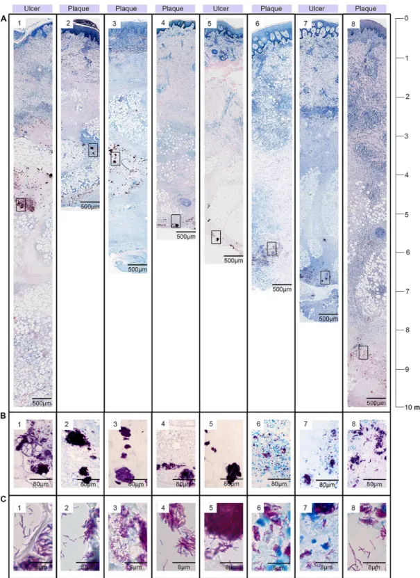
In histopathological sections of BU lesions AFB presented either as i. single bacteria (Fig 1B1), ii. small clusters of AFB (Fig 1B2) or iii. large clusters (Fig 2) located in a tissue depth of up to 10 mm in the FFPE sections (Fig 2). Since 10–30% shrinkage of the tissue is common in FFPE tissue sections the true skin depth of theM.ulceransbacteria was up to 1.3 cm (in excisions). Similar numbers of tissue specimens could be assigned to each category: in 29/83 (35%) of the microscopy AFB positive specimens only single bacteria were found, 28/83 (34%) samples con-tained small clusters of AFB and 26/83 (31%) large clusters (Fig 1A). When specimens were further stratified by lesion type, 67% (10/15) of the analyzed plaque specimens presented with large clusters of extracellular bacteria, 13% (2/15) contained only small clusters and 20% (3/15) displayed only single bacteria. In contrast, single bacteria (26/68; 38%) and small clusters (26/ 68; 38%) dominated in the specimens originating from ulcers; here large clusters of AFB were found in only 16/68 (24%) of the samples (Fig 1A).
Uneven distribution of
M
.
ulcerans
DNA at the margins of ulcerative
lesions
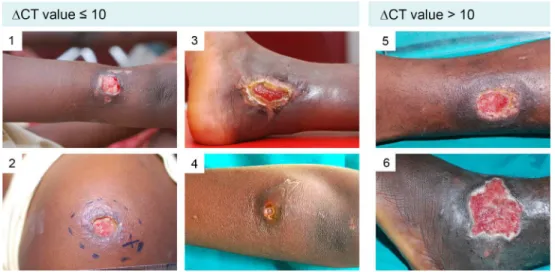
We investigated the distribution of bacterial DNA and of AFB in ulcerative lesions of 37 untreated qPCR reconfirmed (at least one positive IS2404 qPCR positive swab;S1andS2 Tables) BU patients by analyzing 151 swab samples taken from the lesion margins at different positions of the same lesions (Fig 3andS1andS2Figs). Altogether 18.5% (28/151) of the total number of analyzed swabs were qPCR negative. The 28 qPCR negative swabs came from the lesions of 15 patients (15/37, 40%). A total of 66 swabs from these 15 patients have been ana-lyzed (S1 Table) and 42.4% (28/66) of these swabs had a negative qPCR result. However, each of these 15 patients had at least one IS2404 qPCR positive swab. All 85 swabs taken from the other 22 patients were IS240 qPCR positive. In comparative qPCR analyses of different swabs taken from different areas of the same lesion, minimal heterogeneity (ΔCT5) was found in 9/37 (24%), medium heterogeneity (5<ΔCT10) in 17/37 (46%) and a maximal heterogene-ity (ΔCT>10) in 11/37 (30%) of the lesions (S1andS2Figs andS1andS2Tables), where a ΔCT>10 represents a more than 1000 fold difference in DNA quantity in different places of one lesion.
The 151 swab samples, obtained from the 37 BU patients were also analyzed by microscopy after direct ZN staining (Fig 4andS1andS2Tables). All 28 PCR negative swabs were also microscopy negative. Sixty-nine samples (56.1%) were positive in both tests and 54 samples (43.9%) were only qPCR positive (Fig 4). AFB were microscopically detected in smears of all 51 swabs (41.5%) yielding a qPCR CT value of27.8 (equaling about300 genomes in the total
Fig 1. Preferential location ofM.ulceransbacteria in the subcutis.A: In plaque lesions mainly large clusters of extracellular bacteria were found, whereas in ulcerated lesions single AFB and small clusters were more common. In both types of lesions AFB were primarily found in the subcutis. B: Examples for dispersed single bacteria (1) and small clusters of AFB (2) in histological sections stained with ZN (counterstain methylenblue).
Fig 2. Localisation of large clusters of AFB in the subcutis of BU lesions.Histological sections were stained with ZN (counterstain methylenblue). A: Cross sections through the excised tissue specimens from BU plaque lesions (A2-A4, A7-A8) or ulcerated BU lesions (A1, A5-A6) revealing large clusters of AFB located in the subcutis in different tissue depths (3 mm—10 mm). Boxed areas are shown in a higher magnification in (B). B: Large clusters of AFB. C: High magnification of AFB with typical rod shaped appearance.
extract of the swab [13]). In contrast, AFB were found in none of the 23 swabs (18.7%) yielding a CT value33.9 (equaling about2 genomes in the total extract of the swab) [14]. Of the 49 swabs (39.8%) with a CT between 27.8 and CT 33.9, 18 (36.7%) were also positive in ZN smear microscopy (Fig 4A), with a clear increase in the proportion of positive ZN smears with decreasing qPCR CT value (Fig 4B). In total, 27/37 (73%) of the patients with at least one qPCR positive swab had also at least one positive microscopy result.
Increase in test sensitivity by analyzing multiple swab samples
Results obtained with 134 swabs from those 30 patients who had at least four swabs taken from different lesion areas were statistically analyzed for the effect of testing multiple samples on assay sensitivity. Both for qPCR (Table 1) and for smear microscopy (Table 2) the probability of obtaining at least one positive test result per patient increased substantially, when analyzing two or three samples instead of one. In particular for qPCR the effect of analyzing a fourth swab was only marginal.
Discussion
To date, no systematic analysis of the distribution ofM.ulceransbacteria in BU lesions has been published. Here we have systematically evaluated the amount and distribution of AFB in 83 tissue specimens in which we could detect AFB, corroborating previous findings that extra-cellular clusters ofM.ulceransbacteria in the subcutis are a histopathological hallmark of BU [6–8,14,15]. In none of the 83 analyzed samples we found AFB in the epidermal layer, whereas AFB were present in the subcutis of all complete tissue specimens with occasional minor spread of bacteria into the lower layer of the dermis. As expected [8], large extracellular clusters of AFB were found in most (67%) of the plaque lesions analyzed, but only in 24% of the ulcerated lesions, where small clusters and dispersed AFB were more common. This is most likely related to sloughing of necrotic tissue including the bulk of bacteria during ulcer formation [16,17]. Consequently, improper sampling of tissue specimens for histopathological analysis (e.g. not including the subcutis) will often result in false negative laboratory results based on
Fig 3. Examples of lesions presenting with both IS2404 qPCR positive and negative swabs.Lesions of six laboratory confirmed BU patients are shown, whereof four (# 1–4) presented with aΔCT value10, indicating a minimal to medium heterogeneity and two (# 5 and 6) presented with aΔCT value>10, indicative for a maximum heterogeneity. From each lesion four swabs have been taken in a clockwise manner and IS2404 qPCR has been performed.
microscopic detection of AFB in tissue specimens. Since no samples from nodular or edema-tous lesions were available we are not able to draw any conclusions concerning these pre-ulcer-ative forms.
Fine-needle aspiration (FNA) rather than taking punch biopsies is recommended [4] to obtain samples from clinically-diagnosed non-ulcerative lesions (nodules, plaques and
Fig 4. Correlation between direct smear microscopy and IS2404 qPCR results.Of the 123 IS2404 qPCR positive swab samples analyzed by direct smear microscopic analysis after ZN staining, 54 samples (43.9%) were only positive by qPCR, whereas 69 samples (56.1%) were positive for both methods applied. Up to a qPCR CT of 27.8 all swabs were positive for both methods. Between CT 27.9 and CT 33.8 results were variable and above CT 33.8 all samples were microscopy negative. A: AFB positive and negative swabs in correlation to the qPCR values. Each dot represents one swab sample. B: Percentage of AFB positive and negative swabs in correlation to qPCR value ranges.
oedema). However, FNA samples were not available for this study. Our findings on the distri-bution of AFB may reflect a tropism ofM.ulceransfor the deeper subcutis, possibly related to special nutritional needs only provided by the destroyed fat tissue. However, it cannot be excluded that the location in the lower layers of the skin is rather the consequence of the inocu-lation route, which is still unclear, but may either be via skin trauma or by insect bites. Skin tro-pism is generally explained by the temperature preference ofM.ulcerans[4,18,19].
In ulcerative BU lesions disease activity is thought to be concentrated at the edges of ulcers stretching into the indurated surroundings. Bacterial yield is expected to be highest below the undermined edges, where laboratory specimen for direct smear microscopy, qPCR and cultiva-tion should be obtained. However, it needs to be emphasized that the textbook like presenta-tion with circularly undermined edges is the exceppresenta-tion rather than the rule in most confirmed BU lesions. In our patient cohort only a few patients had circular undermined edges, while all the others presented with only partial or even no undercutting (S1andS2Tables). Here we have addressed the question how evenly bacterial DNA is distributed in different areas of the lesion by taking several swabs from the same lesion but from different areas. Samples were ana-lyzed in parallel by IS2404 qPCR and direct smear microscopy for AFB. A high variability in the distribution ofM.ulceransDNA in different samples of the same lesions was observed and from 40% of the lesions both IS2404 qPCR positive and negative swabs were obtained. This supports the concept to repeat sampling if qPCR results are negative, but clinical signs and symptoms are highly indicative for BU.
We observed a good correlation between the amount ofM.ulceransDNA in extracts from swabs and the positivity of direct smear microscopy. All swabs yielding extracts
containing300M.ulceransgenomes had positive microscopy results, while swabs
yielding2 genomes were all negative. About 35% of the swabs with DNA amounts between
Table 1. Increase in the probability of obtaining one or more positive qPCR results with the number of swabs analyzed.
Number of swabs analyzed
Probability*of PCR positive tests (expressed as percentages)
% of one positive test (95% CI)
% of two positive tests (95% CI)
% of three positive test (95% CI)
% of four positive test (95% CI)
1 80.17 (79.81 to 80.52)
2 92.16 (91.96 to 92.41) 67.67 (67.37 to 67.98)
3 97.1 (96.25 to 97.24) 82.7 (82.47 to 82.95) 60.19 (59.95 to 60.43)
4 98.69 (98.57 to 98.80) 92.01 (91.90 to 92.10) 73.33 (73.28 to 73.38) 55.82 (55.67 to 55.97)
*A bootstrap with 1000 loops was used
doi:10.1371/journal.pntd.0004767.t001
Table 2. Increase in the probability of obtaining one or more positive smear microscopy results with the number of swabs analyzed.
Number of swabs analyzed
Probability*of ZN smear positive tests (expressed as percentages)
% of one positive test (95% CI)
% of two positive tests (95% CI)
% of three positive test (95% CI)
% of four positive test (95% CI)
1 45.00 (44.63 to 45.38)
2 59.01 (58.70 to 59.32) 30.67 (30.33 to 30.93)
3 66.11 (65.87 to 66.35) 44.58 (44.37 to 44.79) 23.38 (23.14 to 23.61)
4 71.26 (71.11 to 71.41) 51.30 (51.20 to 51.40) 38.01 (37.91 to 38.11) 18.52 (18.38 to 18.67)
*A bootstrap with 1000 loops was used
the above limits were microscopy positive. While the sensitivity of direct AFB detection in smears was thus lower than that of IS2404 qPCR, 73% of all IS2404 qPCR positive patients were also confirmed by microscopy by analyzing several swabs per patient. This value is high compared to previous studies [20–22] and might not only be due to the number of swabs ana-lyzed, but also to the covering of the slides with Eukitt mounting medium and a coverslip immediately after staining, which protects the slides from attachment of dust and dirt and good laboratory equipment. Our analyses also indicate that for qPCR analyzing three swab samples may be an optimal number. However we have to emphasize that for this study each swab was taken from a different area of the lesion and if the margin of the whole lesion is pled with one swab, results may be different. Overall, all diagnostic tests rely on a careful sam-pling and in case of a negative result but a high clinical suspicion repeated testing may be advisable.
Supporting Information
S1 Table. Patients from whom not all analyzed swabs were positive by IS2404 qPCR. (DOCX)
S2 Table. Patients from whom all analyzed swabs were positive by IS2404 qPCR. (DOCX)
S1 Fig. Lesions from which both IS2404 qPCR positive and negative swabs were obtained. Depicted are all analyzed lesions that presented with both positive and negative IS2404 qPCR results, sorted by theΔCT heterogeneity. Picture numbers correspond to the patient numbers inS1 Table.
(TIF)
S2 Fig. Lesions from which only positive IS2404 qPCR swabs were obtained.All analyzed lesions that presented with positive IS2404 qPCR results, sorted by theΔCT heterogeneity are shown. Picture numbers correspond to the patient numbers inS2 Table.
(TIF)
Acknowledgments
We thank Vincent Romanet, Caroline Stork, Ernesta Damassa, Patrizia Barzaghi-Rinaudo and Dr. Masato Murakami from Novartis Basel for excellent technical support and providing access to lab equipment for histopathology. Peter Schmid for advice and tissue scans and Leticia Grize for statistical support.
Author Contributions
Conceived and designed the experiments: MTR MB GP. Performed the experiments: MTR MB. Analyzed the data: MTR MB MWB TJ MV GP. Contributed reagents/materials/analysis tools: PFB GES DYM AUB. Wrote the paper: MTR MB GP TJ.
References
1. Walsh DS, Portaels F, Meyers WM. Buruli ulcer (Mycobacterium ulcerans infection). Trans R Soc Trop Med Hyg. 2008; 102: 969–978. doi:10.1016/j.trstmh.2008.06.006PMID:18657836
2. Van der Werf TS, Stienstra Y, Johnson RC, Phillips R, Adjei O, Fleischer B, et al. Mycobacterium ulcer-ans disease. Bull World Health Organ. 2005; 83: 785–791. PMID:16283056
3. Junghanss T, Johnson C, Pluschke G. Mycobacterium ulcerans disease. Manson’s tropical diseases. 23rd ed. Edinburgh: Saunders Ltd.; 2014. pp. 519–531.
5. Bratschi MW, Bolz M, Grize L, Kerber S, Minyem JC, Um Boock A, et al. Primary cultivation: factors affecting contamination and Mycobacterium ulcerans growth after long turnover time of clinical speci-mens. BMC Infect Dis. 2014; 14: 636. doi:10.1186/s12879-014-0636-7PMID:25433390
6. MacCallum P, Tolhurst JC. A new mycobacterial infection in man. J Pathol Bacteriol. 1948; 60: 93–122. 7. Rondini S, Horsfield C, Mensah-Quainoo E, Junghanss T, Lucas S, Pluschke G. Contiguous spread of
Mycobacterium ulcerans in Buruli ulcer lesions analysed by histopathology and real-time PCR quantifi-cation of mycobacterial DNA. The Journal of Pathology. 2006; 208: 119–128. PMID:16261539
8. Ruf M-T, Sopoh GE, Brun LV, Dossou AD, Barogui YT, Johnson RC, et al. Histopathological changes and clinical responses of Buruli ulcer plaque lesions during chemotherapy: a role for surgical removal of necrotic tissue? PLoS Negl Trop Dis. 2011; 5: e1334. doi:10.1371/journal.pntd.0001334PMID:
21980547
9. Chauty A, Ardant M-F, Marsollier L, Pluschke G, Landier J, Adeye A, et al. Oral treatment for Mycobac-terium ulcerans infection: results from a pilot study in Benin. Clin Infect Dis. 2011; 52: 94–96. doi:10. 1093/cid/ciq072PMID:21148526
10. Yeboah-Manu D, Kpeli GS, Ruf M-T, Asan-Ampah K, Quenin-Fosu K, Owusu-Mireku E, et al. Second-ary bacterial infections of buruli ulcer lesions before and after chemotherapy with streptomycin and rifampicin. PLoS Negl Trop Dis. 2013; 7: e2191. doi:10.1371/journal.pntd.0002191PMID:23658847
11. Vogel M, Bayi PF, Ruf M-T, Bratschi MW, Bolz M, Um Boock A, et al. Local heat application for the treat-ment of Buruli ulcer: results of a phase II open label single center non comparative clinical trial. Clin Infect Dis. 2015;
12. Lavender CJ, Fyfe JAM. Direct detection of Mycobacterium ulcerans in clinical specimens and environ-mental samples. Methods Mol Biol. 2013; 943: 201–216. doi:10.1007/978-1-60327-353-4_13PMID:
23104291
13. Fyfe JAM, Lavender CJ, Johnson PDR, Globan M, Sievers A, Azuolas J, et al. Development and appli-cation of two multiplex real-time PCR assays for the detection of Mycobacterium ulcerans in clinical and environmental samples. Appl Environ Microbiol. 2007; 73: 4733–4740. PMID:17526786
14. Hayman J. Out of Africa: observations on the histopathology of Mycobacterium ulcerans infection. J Clin Pathol. 1993; 46: 5–9. PMID:8432888
15. Portaels F, Silva MT, Meyers WM. Buruli ulcer. Clin Dermatol. 2009; 27: 291–305. doi:10.1016/j. clindermatol.2008.09.021PMID:19362692
16. Van der Werf TS, van der Graaf WT, Tappero JW, Asiedu K. Mycobacterium ulcerans infection. Lancet. 1999; 354: 1013–1018. PMID:10501380
17. Bolz M, Ruggli N, Borel N, Pluschke G, Ruf M-T. Local Cellular Immune Responses and Pathogenesis of Buruli Ulcer Lesions in the Experimental Mycobacterium Ulcerans Pig Infection Model. PLoS Negl Trop Dis. 2016; 10: e0004678. doi:10.1371/journal.pntd.0004678PMID:27128097
18. Eddyani M, Portaels F. Survival of Mycobacterium ulcerans at 37 degrees C. Clin Microbiol Infect. 2007; 13: 1033–1035. PMID:17697005
19. Eddyani M, Debacker M, Martin A, Aguiar J, Johnson CR, Uwizeye C, et al. Primary culture of Mycobac-terium ulcerans from human tissue specimens after storage in semisolid transport medium. J Clin Microbiol. 2008; 46: 69–72. PMID:17989199
20. Phillips R, Horsfield C, Kuijper S, Lartey A, Tetteh I, Etuaful S, et al. Sensitivity of PCR Targeting the IS2404 Insertion Sequence of Mycobacterium ulcerans in an Assay Using Punch Biopsy Specimens for Diagnosis of Buruli Ulcer. J Clin Microbiol. 2005; 43: 3650–3656. PMID:16081892
21. Yeboah-Manu D, Asante-Poku A, Asan-Ampah K, Ampadu EDE, Pluschke G. Combining PCR with microscopy to reduce costs of laboratory diagnosis of Buruli ulcer. Am J Trop Med Hyg. 2011; 85: 900– 904. doi:10.4269/ajtmh.2011.11-0362PMID:22049046