doi: 10.1590/0037-8682-0404-2016
Major Article
Corresponding author: Dr Kingsley Nnanna Ukwaja. e-mail: [email protected]
Received 1 October 2016 Accepted 22 November 2016
Secondary bacterial isolates from previously untreated
Buruli ulcer lesions and their antibiotic susceptibility
patterns in Southern Nigeria
Moses Chibueze Anyim
[1], Anthony Obiamaka Meka
[1], Joseph Ngozi Chukwu
[1],
Charles Chukwunalu Nwafor
[1], Daniel Chukwunweolu Oshi
[1],[2],
Nelson Okechukwu Madichie
[1], Ngozi Ekeke
[1], Chukwuka Alphonsus
[1],
Obinna Mbah
[1], Chinenye Nwaekpe
[3], Martin Njoku
[4], Dare Fakiyesi
[4], Vitalis Ulodiaku
[5],
Ignatius Ejiofor
[6], Adeniyi Hakeem Bisiriyu
[7]and Kingsley Nnanna Ukwaja
[8][1]. Medical Department, German Leprosy and Tuberculosis Relief Association, Enugu State, Nigeria. [2]. Department of Community Health and Psychiatry, University of West Indies, Mona, Kingston 7, Jamaica.
[3]. Department of Microbiology, National Orthopaedic Hospital, Enugu State, Nigeria. [4]. St Benedict’s Tuberculosis & Leprosy Rehabilitation Hospital, Ogoja, Cross River State, Nigeria.
[5]. St Joseph’s Hospital Adazi-Nnukwu, Anambra State, Nigeria.
[6]. Anambra State Tuberculosis, Leprosy and Buruli Ulcer Control Programme, Anambra State, Nigeria. [7]. Ogun State Tuberculosis, Leprosy and Buruli Ulcer Control Programme, Ogun State, Nigeria.
[8]. Department of Medicine, Federal Teaching Hospital Abakaliki, Ebonyi State, Nigeria.
Abstract
Introduction: Mycolactones, secreted by Mycobacterium ulcerans, were previously believed to prevent super infection in Buruli ulcer lesions. However, little is known about secondary bacterial infections in these lesions. This study evaluated contaminating bacterial fl ora and their antibiotic susceptibility patterns in cases of previously untreated Buruli ulcer disease from three states in Southern Nigeria. Methods: A prospective analysis was conducted between January and June of 2015 using wound swabs from eligible patients with Buruli ulcer disease, confi rmed by quantitative-polymerase chain reaction, with active ulcers. Microbiological analyses including isolation of bacteria, species identifi cation of isolates, and drug susceptibility tests were performed. Results: Of 51 patients, 27 (52.9%) were female. One or more bacterial species of clinical importance was isolated from each patient. A total of 17 different microbial species were isolated; 76.4% were Gram-negative and 23.6% were Gram-positive isolates. The most common bacterial species detected was Staphylococcus aureus (24%), followed by Aeromonas hydrophila (13%), Pseudomonas aeruginosa (13%), and Klebsiella pneumoniae (11%). Drug susceptibility tests showed a particularly high frequency of resistance to commonly used antimicrobials in Nigeria for Staphylococcus aureus. Conclusions: Super bacterial infections occur in Buruli ulcer lesions in Nigeria, and these infections are associated with high rates of resistance to commonly used antibiotics in the country.
Keyword: Bacterial contamination. Chemotherapy. Mycobacterium ulcerans. Mycolactones. Wounds.
INTRODUCTION
Buruli ulcer disease (BUD) is a necrotising skin and soft tissue infection caused by Mycobacterium ulcerans(1). It is an emerging neglected tropical disease and the third most common mycobacterial infection, after tuberculosis and leprosy(1) (2) (3). BUD has been reported in over 30 countries globally, including Australia, and those in Asia, the West Pacific, and South America; however, the highest disease burden occurs in West
and Sub-Saharan Africa(1) (2). Buruli ulcer (BU) lesions can occur as very large ulcers often with scarring, contractures, and limb deformities that can require surgical amputations(1) (2) (3) (4) (5).
Tissue destruction by most pathogenic bacteria is mediated by the release of toxins. Unlike Mycobacterium tuberculosis
and Mycobacterium leprae, of which pathogenesis is not mediated by the release of toxins, skin and soft tissue damage with BUD is mediated by the release of two polyketide-derived macrolide toxins from M. ulcerans, designated as mycolactones A and B(6) (7). In mouse and guinea pig models, studies have shown that mycolactones play a central role in the pathogenesis of M. ulcerans. For example, injection of 100μg of mycolactones
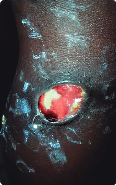
FIGURE 1. A Buruli ulcer lesion of the right lower limb showing undermined edges.
pig skin(8). In mice, mycolactones have been shown to be associated with vacuolar nerve tissue injury, which might explain the painlessness of BU lesions(9). There has been substantial progress made in characterising the activity of mycolactones, especially their necrotising and immunosuppressive effects(6) (7) (10) (11). The cytotoxic effects of mycolactones released by clusters of M. Ulcerans include the destruction of the skin and surrounding soft tissues. This results in the formation of devitalized, avascular tissue and necrotic slough at the wound bed, which is characteristic of BU(6) (7) (8). This devitalised tissue might provide an ideal medium for secondary bacterial infections, which consequently might interfere with wound healing(12). However, as mycolactones are macrolides that can have antibiotic activity against a broad spectrum of bacteria, it has been suggested that secretion of these compounds by M. ulcerans during active disease might prevent superinfection in BUD wounds(10) (12). This speculation was strengthened by a study demonstrating that high levels of mycolactones in BU patients correlated with clinical and bacteriological response to therapy(13).
Recent studies using synthetic mycolactones from
M. ulcerans showed that these compounds had no antimicrobial activity against any of the microorganisms tested(11). Another study recently demonstrated that M. ulcerans infection and mycolactone secretion do not prevent secondary bacterial infections in BU lesions as previously speculated, and indeed that these super infections might delay wound healing(12). Thus, to optimise BUD treatment in endemic settings, there is a need to characterise these infections and their antibiotic susceptibility patterns, especially in under-resourced populations. This will aid in the identifi cation of common secondary bacterial infections in BU, and suggest possible empirical regimens for treating these infections according to geographical regions/settings. Nigeria was previously thought to not be endemic for BUD due to very few cases being reported over four decades since it was fi rst detected in the country(14). However, in 2012, we piloted a case-search strategy in three rural districts of Southern Nigeria and found 36 BUD cases that were confirmed by polymerase chain reaction (PCR)(14). As a result, a scale-up has been underway since 2014. The aim of this study was to evaluate contaminating bacterial fl ora and their antibiotic susceptibility patterns from previously untreated BUD lesions from three states in Southern Nigeria.
METHODS
Study participants
This prospective analysis was conducted between January and June of 2015. The study population comprised pre-treatment patients with quantitative-PCR (q-PCR)-confi rmed BUD with active ulcers (Figure 1). Fifty-one study participants were recruited from three States in Southern Nigeria. The states included Cross River State (South-South zone), Ogun State (South-West zone), and Anambra State (South-East zone). Information about the gender and age of the patients was also provided. The ulcers were sampled for microbiological analysis before administration of any chemotherapy for BUD.
Sample collection
After superfi cial pre-cleansing of the ulcers with physiologic saline, specimens were collected by rotating a sterile, pre-moistened swab (Nuova Aptaca SRL, Canelli, Italy) across the ulcers. Two swabs were collected from the undermined edges of the BU lesions using the Levine method of specimen collection. The third swab was rotated over a small area with slight pressure to collect fl uid from the wound tissue(15). To preserve secondary bacteria, swabs were placed in Ames agar gel with charcoal and sent to the laboratory for analysis. The analysis was performed at the microbiology laboratory of the National Orthopaedic Hospital, Enugu, Nigeria.
Microbiological analysis
0 5 10 15 20 25 30
FIGURE 2.Percentage of bacterial species isolated from wound swabsamples collected from 51 patients with Buruli ulcer disease. The Other Gram-negative group includes Citrobacter koseri, Enterobacter agglomerans, Enterobacter sakazakii, Kluyvera ascorbata, Moraxella spp, Proteus mirabilis, and Providencia rettgeri.
Variable Number Percentage
Age (years)
≤10 13 25.6
11–20 7 13.7
21–30 6 11.8
31–40 10 19.6
41–50 7 13.7
51–60 4 7.8
≥61 4 7.8
Sex
female 27 52.9
male 24 47.1
Gram staining
Gram-negative 42 76.4
Gram-positive 13 23.6
Isolates per patient (number)
1 47 92.2
2 4 7.8
TABLE 1
Characteristics of 51 Buruli ulcer patients and associated bacterial isolates.
Bacterial species identification: bacterial isolates was identifi ed through morphological characteristics, gram staining, and biochemical reactions. Bacterial colonies were gram-stained(16) and identifi ed by biochemical tests. Gram-negative rods were characterized using substrates on the Microbact 24E Gram-negative identification system (Oxoid Limited, UK), according to manufacturer’s instructions. Gram-positive cocci were fi rst identifi ed by Gram staining and then analysed using the catalase test to differentiate between Staphylococcus spp and
Streptococcus spp. Staphylococcus spp were further differentiated into coagulase-positive and coagulase-negative strains.
Antibiotic susceptibility testing: susceptibility of the isolates to specifi c drugs was tested using the Kirby-Bauer disc diffusion method on Mueller Hinton agar(17). Antibiotics used for sensitivity testing included ampicillin, amoxicillin/clavulanic acid, ceftazidime, cefuroxime, gentamicin, ciprofl oxacin, and ofl oxacin. Gram-positive cocci were tested against ceftriaxone, erythromycin, and cloxacillin.
Ethics considerations
This study was approved by the Ethics and Research Advisory Board of the German Leprosy & TB Relief Association, Nigeria. All patients or legal guardians (for minors) gave written informed consent for all diagnostic processes and for publication of clinical photographs derived from the management of patients. All identifi ed BU cases were investigated and treated at no fi nancial cost to the patients.
Data analysis
The data collected were entered into a Microsoft Excel spreadsheet and analysed using Epi Info. Results are presented as absolute and relative frequencies.
RESULTS
Samples were collected from 51 patients; 27 (52.9%) were female and their ages ranged from 4 to 75 years. One or more bacterial species of clinical importance was isolated from a specimen from each patient (Table 1). A total of 17 different microbial species were isolated; 76.4% were Gram-negative and 23.6% were Gram-positive isolates. The most common bacterial species detected were Staphylococcus aureus (24%), followed by
Aeromonas hydrophila (13%) Pseudomonas aeruginosa (13%),
Klebsiella pneumoniae (11%), Enterobacter cloacae(6%),
Pseudomonas pseudomallei (6%), and Burkholderia cepacia
(6%) (Figure 2).
The presence of only one bacterial species isolated from each sample was the most frequent occurrence (92%). Polymicrobial infections were found in four (8%) of the lesions and consisted of two species per sample. The predominant species found in polymicrobial infections was S. aureus, occurring with either of P. aeruginosa, Pseudomonas pseudomallei, A. hydrophila, or Escherichia coli.
The antimicrobial susceptibility patterns of both Gram-positive and Gram-negative isolates from BU wounds are shown in Table 2. S. aureus was the only Gram-positive species isolated from the samples. S. aureus showed a high frequency
of resistance to different classes of antimicrobial agents including amoxicillin/clavulanic acid, cloxacillin, ciprofl oxacin, erythromycin, and cefuroxime (each with resistance rates exceeding 80%). The most active agents against S. aureus
Susceptible Resistant
Organism Antibiotic tested Number tested n % n %
Staphylococcus aureus Amoxi-Clav 13 1 7.7 12 92.3
Ceftazidime 12 7 58.3 5 41.7
Ciprofl oxacin 5 0 0.0 5 100.0
Cefuroxime 13 2 15.4 11 84.6
Ceftriaxone 12 8 66.7 4 33.3
Erythromycin 13 2 15.4 11 84.6
Gentamicin 13 10 76.9 3 23.1
Ofl oxacin 13 7 53.8 6 46.2
Cloxacillin 11 1 9.1 10 90.9
Aeromonas hydrophila Ampicillin 7 1 14.3 6 85..7
Amoxi-Clav 7 1 14.3 6 85.7
Ceftazidime 7 3 42.7 4 57.1
Ciprofl oxacin 7 6 85.7 1 14.3
Cefuroxime 7 2 28.6 5 71.4
Gentamicin 7 3 42.9 4 57.1
Ofl oxacin 7 5 71.4 2 28.6
Pseudomonas aeruginosa Ampicillin 7 0 0.0 7 100.0
Amoxi-Clav 6 0 0.0 6 100.0
Ceftazidime 7 6 85.7 1 14.3
Ciprofl oxacin 7 7 100.0 0 0
Cefuroxime 7 0 0.0 7 100.0
Gentamicin 7 2 28.6 5 71.4
Ofl oxacin 7 3 42.9 4 57.1
Klebsiella pneumoniae Ampicillin 5 0 0.0 5 100.0
Amoxi-Clav 6 2 33.3 4 66.7
Ceftazidime 6 4 66.7 2 33.3
Ciprofl oxacin 6 6 100.0 0 0
Cefuroxime 6 0 0.0 6 100.0
Gentamicin 6 5 83.3 1 16.7
Ofl oxacin 6 5 83.3 1 16.7
Other Gram-negatives Ampicillin 22 0 0.0 22 100.0
Amoxi-Clav 22 3 13.6 19 86.4
Ceftazidime 22 12 54.5 10 45.5
Ciprofl oxacin 22 21 95.5 1 4.5
Cefuroxime 22 7 31.8 15 68.2
Gentamicin 22 13 59.1 9 40.9
Ofl oxacin 20 20 90.9 2 9.1
TABLE 2
Antibiotic susceptibility pattern of bacterial isolates from Buruli ulcer wounds in Nigeria.
Amoxi-Clav: Amoxicillin-Clavulanic acid.
Furthermore, the antimicrobial sensitivity pattern of the three most common Gram-negative isolates from BU wounds is shown in Table 2. The most ineffective antibiotic against all Gram-negative isolates was ampicillin; P. aeruginosa and
K. Pneumonia were not susceptible to any antibiotics with a resistance rate of 100%. A. hydrophila had a high resistance rate (85.7%) to ampicillin and amoxicillin/clavulanic acid.
P. aeruginosa showed 100% resistance to ampicillin, amoxicillin/ clavulanic acid, and cefuroxime. K. pneumoniae showed 100% resistance to ampicillin and cefuroxime. The three most common Gram-negative isolates (P. aeruginosa, A. hydrophila, and
K. pneumoniae) showed high susceptibility to ciprofl oxacin
(rates ranged from 85.7-100%). In addition, K. pneumoniae
and P. aeruginosa had susceptibility rates of 66.7% and 85.7%, respectively, to ceftazidime. Additionally, among other Gram-negative bacteria isolates, resistance rates were highest to ampicillin (100%), amoxicillin/clavulanic acid (86.4%), and cefuroxime (68.2%). However, antimicrobial susceptibility rates were highest for ofl oxacin (90.9%) and ciprofl oxacin (95.5%).
DISCUSSION
antimicrobial activity against a wide spectrum of bacteria including Gram-positive, Gram-negative, and atypical bacteria like Rickettsia and Chlamydia(18) (19) (20). Therefore, mycolactones, like other macrolides, were suggested to have antimicrobial properties that could prevent secondary bacterial infections in BU lesions. This speculation was recently debunked by a pilot study(12). To confi rm this observation, we evaluated bacterial contamination of BU lesions as well as associated antimicrobial susceptibility patterns.
In this study, 17 bacterial species were isolated from BU lesions. The majority of the ulcers were colonised by a single bacterial specie, whereas fewer than 10% had polymicrobial infections. The most common and only Gram-positive isolate was
S. aureus. This is consistent with studies that surveyed different types of wounds and chronic ulcers including BU lesions and found S. aureus as the most common isolate(12) (18) (19) (20). Virulence in S. aureus is mediated by the release of several virulence factors like invasins, hyaluronidase, catalase, coagulase, hemolysins, leukotoxin, and leukocidin(19) (20). Furthermore, several Gram-negative bacteria were also isolated from BU wounds with the most common being A. hydrophila, P. aeruginosa, and
K. pneumoniae. Our study agrees with a recent study suggesting that M. ulcerans infection and mycolactone secretion does not prevent secondary bacterial infections in BU lesions, and that most contaminants are S. aureus and P. aeruginosa(12). The relative high rates of infection with A. hydrophila suggest environmental transmission of M. ulcerans as this organism has been found in a variety of aquatic environments, including lakes, rivers, streams, swamps, rain water, and swimming pools, and it has also been isolated from tap water and soil(21) (22).This organism has also been isolated from other water- or soil-associated traumatic wound infections(21) (22).
The bacterial isolates were evaluated for susceptibility to commonly used antibiotics in Nigeria. All tested S. aureus
strains were resistant to ciprofl oxacin, 92% were amoxicillin-clavulanic acid-resistant, and 91% were cloxacillin-resistant. The level of resistance of S. aureus isolates to cloxacillin is crucial; beyond being one of the most commonly prescribed antibiotics for treating skin and soft tissue infections in Nigeria, it can also indicate the proportion of methicillin-resistant
Staphylococcus aureus (MRSA) present. However, in Nigeria and Ghana, relatively low rates of MRSA have been reported compared to those in South Africa(12) (23) (24). S. aureus strains showed mild to moderate susceptibility to third-generation cephalosporins (ceftazidime/ceftriaxone) and gentamicin. This fi nding indicates that these agents are likely to be the most effective in managing BU lesions contaminated with
S. aureus. This is worrisome because despite being administered intravenously, the results indicate that S. aureus has become more resistant to third-generation cephalosporins(20).
Furthermore, the antibiotic resistance of Gram-negative isolates is also of great concern; the most common isolates, especially P. aeruginosa, showed relatively high resistance to the majority of the antibiotics tested, especially, ampicillin, amoxicillin/clavulanic acid, and cefuroxime. Multidrug-resistant isolates of P. aeruginosa, those fully resistant to all of these
agents, are of major concern(20). Additionally, the results indicate
that P. Aeruginosa is developing a high level of resistance to aminoglycosides and third-generation cephalosporins. Similar evidence was reported by Nicoletti et al.(25)in a study on severe
infections from different human sources and Yeboah-Manu et al.(12) in a study of secondary infections of BU wounds(12) (25).
In addition, our results showed that common Gram-negative bacterial isolates from the lesions (A. hydrophila, P. aeruginosa, and K. pneumoniae) demonstrated a high level of susceptibility
to ciprofl oxacin ,indicating that this antibiotic might be useful in
managing secondary infections. Moreover, other less common Gram-negative isolates from the BU lesions also showed high
susceptibility to quinolones [ciprofl oxacin (96%) and ofl oxacin
(91%)].
This study had some strengths and limitations. An important
strength is that it confi rms a recent observation that BU lesions
might be associated with secondary bacterial infections
that could interfere with wound healing, therefore requiring
additional treatment with antibiotics. Furthermore, the choice of drugs for treatment of secondary infections of BU lesions
with locally available antimicrobial agents requires a better understanding of the infecting fl ora and its drug susceptibility
patterns. Therefore, this study provides this information for Nigeria. One potential limitation of this study is that several samples were not collected longitudinally before and during BU
treatment to determine the causes, sources, and consequences
of secondary BU wound infections(12). Another limitation is
that we utilised wound swabs for specimen collection. Wound swab specimen collection for microbiological analysis has
recently been questioned. The current gold standard for microbiological analysis of wounds requires the use of biopsy
specimens(26) (27). However, as previously highlighted, limitations
include determining which health workers should collect biopsies, a lack of laboratories with expertise on microbiological culture testing of biopsies, higher costs involved with the performance of these tests, and the potential for further tissue injury and delays in wound healing when biopsies are taken. These factors precluded the use of this method in the present study(12). Moreover, wound swabs have been shown to be
associated with the highest recovery rates(28); in addition, it was
shown that microbiological analysis of biopsies does not offer additional prognostic information when compared with analysis
of the surface microfl ora(29).
In conclusion, secondary bacterial infections occur in BU lesions in Nigeria and are associated with a wide variation in the rates of susceptibility and resistance to commonly used
antibiotics in the country. The fi ndings of this study might be of
interest to clinicians, investigators, and public health authorities, and offer suggestions to guide empirical therapy. In addition, the findings of this study might also guide policymakers to implement specific intervention strategies to eliminate antibiotic-resistant bacterial infections and their transmission
in BU endemic settings. However, further studies are required
Acknowledgments
We acknowledge all health workers and programme and project staff who participated in the meticulous data collection and reporting for their contributions.
Confl ict of interest
The authors declare that there are no confl icts of interest.
Financial support
The project was funded by Kindermissionswerk “Die Sternsinger” Stephanstra ße 35, D-52064 Aachen/Germany.
REFERENCES
1. Huang GKL, Johnson PD. Epidemiology and management of Buruli ulcer. Expert Rev Anti Infect Ther 2014; 12:855-865.
2. Merritt RW, Walker ED, Small PL, Wallace JR, Johnson PD, Benbow ME, et al. Ecology and transmission of Buruli ulcer disease: a systematic review. PLoS Negl Trop Dis 2010;4:e911. doi: 10.1371/journal.pntd.0000911.
3. van der Werf TS, Stienstra Y, Johnson RC, Phillips R, Adjei O, Fleischer B, et al. Mycobacterium ulcerans disease. Bull World Health Organ 2005; 83:785-891.
4. Wang HS, Pancholi P. Mycobacterial skin and soft tissue infection. Curr Infect Dis Rep 2014; 16:438.
5. Walsh DS, Portaels F, Meyers WM. Buruli ulcer (Mycobacterium ulcerans infection). Trans R Soc Trop Med Hyg 2008; 102:969–978.
6. Kishi Y. Chemistry of mycolactones, the causative toxins of Buruli ulcer. Proc Natl Acad Sci USA 2011;108:6703-6708.
7. George KM, Chatterjee D, Gunawardana G, Welty D, Hayman J, Lee R, et al. Mycolactone: a polyketide toxin from Mycobacterium ulcerans required for virulence. Science 1999; 283:854-857.
8. George KM, Pascopella L, Welty DM, Small PL. A Mycobacterium ulcerans toxin, mycolactone, causes apoptosis in guinea pig ulcers and tissue culture cells. Infect Immun 2000;68:877-683.
9. En J, Goto M, Nakanaga K, Higashi M, Ishii N, Saito H, et al. Mycolactone is responsible for the painlessness of Mycobacterium ulcerans infection (buruli ulcer) in a murine study. Infect Immun 2008; 76:2002-2007.
10. Pahlevan AA, Wright DJ, Andrews C, George KM, Small PL, Foxwell BM. The inhibitory action of Mycobacterium ulcerans soluble factor on monocyte/T cell cytokine production and NF-kappa B function. J Immunol 1999; 163:3928-3935.
11. Scherr N, Gersbach P, Dangy JP,Bomio C, Li J, Altmann KH, et al. Structure-activity relationship studies on the macrolide exotoxin mycolactone of Mycobacterium ulcerans. PLoSNegl Trop Dis 2013;7:e2143. doi: 10.1371/journal.pntd.0002143.
12. Yeboah-Manu D, Kpeli GS, Ruf MT, Asan-Ampah K, Quenin-Fosu K, Owusu-Mireku E, et al. Secondary bacterial infections of Buruli ulcer lesions before and after chemotherapy with streptomycin and rifampicin. PLoSNegl Trop Dis 2013; 7:e2191. doi: 10.1371/journal. pntd.0002191.
13. Sarfo FS, Phillips RO, Zhang J, Abass MK, Abotsi J, Amoako YA, et al. Kinetics of mycolactone in human subcutaneous tissue during antibiotic therapy for Mycobacterium ulceransdisease. BMC Infect Dis 2014; 14:202.
14. Ukwaja KN, Meka AO, Chukwuka A, Asiedu KB, Huber KL, Eddyani M, et al. Buruli ulcer in Nigeria: results of a pilot case search in three rural districts. Infect Dis Poverty 2016; 5:39.
15. Levine NS, Lindberg RB, Mason AD, Pruitt Jr BA. The quantitative swab culture and smear: a quick, simple method for determining the number of viable aerobic bacteria in open wounds. J Trauma 1976; 16:89-94.
16. Brock TD. Milestones in microbiology 1546-1940. 2ndedition. ASM
press 1999; 215-218.
17. Bauer AW, Kirby WM, Sherris JC, Turck M. Antibiotic susceptibility testing by a standardized single disk method. Am J Clin Pathol 1966; 36:493-496.
18. Körber A, Schmid EN, Buer J, Klode J, Schadendorf D, Dissemond J. Bacterial colonization of chronic leg ulcers: current results compared with data 5 years ago in a specialized dermatology department. J Eur Acad Dermatol Venereol 2010; 24:1017-1025.
19. Halbert AR, Stacey M, Rohr JB, Jopp-McKay A. The effect of bacterial colonization on venous ulcer healing. Australas J Dermatol1992;33:75-80.
20. Bessa LJ, Fazii P, Di Giulio M, Cellini L. Bacterial isolates from infected wounds and their antibiotic susceptibility pattern: some remarks about wound infection. Int Wound J 2015; 12:47-52.
21. Vally H, Whittle A, Cameron S, Dowse GK, Watson T. Outbreak of Aeromonas hydrophila wound infections associated with mud football. Clin Infect Dis 2004; 38:1084-1089.
22. Semel JD, Trenholme G. Aeromonas hydrophila water-associated traumatic wound infections: a review. J Trauma 1990; 30:324-327.
23. Shittu AO, Okon K, Adesida S, Oyedara O, Witte W, Strommenger B, et al. Antibiotic resistance and molecular epidemiology of Staphylococcus aureus in Nigeria. BMC Microbiol 2011; 11:92.
24. Shittu AO, Lin J. Antimicrobial susceptibility patterns and characterization of clinical isolates of Staphylococcus aureus in KwaZulu-Natal province, South Africa. BMC Infect Dis 2006; 6:125.
25. Nicoletti G, Schito G, Fadda G, Boros S, Nicolosi D, Marchese A, et al. Bacterial isolates from severe infections and their antibiotic susceptibility patterns in Italy: a nationwide study in the hospital setting. J Chemother 2006; 18:589-602.
26. Bowler PG, Duerden BI, Armstrong DG. Wound microbiology and associated approaches to wound management. Clin Microbiol Rev 2001; 14:244-269.
27. Heggers JP. Defi ning infection in chronic wounds: methodology. An historical review of the quantitative assessment of microbial fl ora in wounds. J Wound Care 1998; 7:452-456.
28. Cooper RA, Ameen H, Price PE, McCulloch DA, Harding KG. A clinical investigation into the microbiological status of “locally infected” leg ulcers. Int Wound J 2009; 6:453-462.