Human Papillomavirus and Penile Cancers in Rio de Janeiro,
Brazil: HPV Typing and Clinical Features
Marcos A. Scheiner, Mercia M. Campos, Antonio A. Ornellas, Eduardo W. Chin, Maria H. Ornellas, Maria J. Andrada-Serpa
Hematology Service (MAS, MMC), Section of Urology (AAO, EWC) and Bone Marrow Transplant Center (MHO), National Cancer Institute (INCA) and Virology Service (MJAS), Evandro Chagas Institute of Clinical Research, Oswaldo Cruz Foundation, Rio de Janeiro, Brazil
ABSTRACT
Objective: To determine the prevalence of human papillomavirus (HPV) DNA in penile cancers in Rio de Janeiro, Bra-zil.
Materials and Methods: We studied, prospectively, 80 consecutive cases of patients with penile cancers who underwent surgical treatment at three different Hospitals in Rio de Janeiro between March 1995 and June 2000. Of these patients, 72 were diagnosed with invasive squamous cell carcinoma and 8 patients with verrucous carcinoma. The following parameters were observed: presence or absence of HPV DNA viral type, histological subtypes, clinical stage and overall survival. Results: HPV DNA was detected in 75% of patients with invasive carcinomas and in 50% of patients with verrucous carcinomas. High risk HPVs were detected in 15 of 54 (27.8%) patients with HPV positive invasive tumors and in 1 of 4 (25%) patients with HPV positive verrucous tumors. HPV 16 was the most frequent type observed. No correlation was
REVHUYHGEHWZHHQ+39VWDWXVDQGKLVWRORJLFDOVXEW\SHS DVZHOODV+39VWDWXVDQGVWDJHVWUDWL¿FDWLRQS +39VWDWXVZDVDOVRQRWVLJQL¿FDQWO\DVVRFLDWHGZLWKWKHSUHVHQFHRIUHJLRQDOPHWDVWDVHVS 7KHRYHUDOOVXUYLYDO
was related to the presence of lymph node metastases (p < 0.0001).
Conclusions: HPV infection may have contributed to malignant transformation in a large proportion of our penile cancer cases but only inguinal metastasis was a prognostic factor for survival in these patients with penile carcinoma.
Key words: human papillomavirus; penile cancer; Brazil Int Braz J Urol. 2008; 34: 467-76
INTRODUCTION
Penile cancer prevalence varies according to geographic region and ethnic origin (1). In Brazil, penile cancer represents 2% of all cancers in males and is more frequent in north and northeast regions with an incidence ranging from 1.3 to 2.7 per 100,000, according to the geographic region (2).
The mechanism by which Human Papilloma-virus (HPV) leads to malignant transformation is likely
HPV infections are associated with benign and malignant epithelial lesions and a high-risk HPV group is probably the major cause of anogenital can-cers. To date, more than 100 HPV genotypes have EHHQLGHQWL¿HG5HFHQWO\0XQR]HWDOSRROHG data from 11 case-control studies from nine countries DQGFODVVL¿HGWKHIROORZLQJ+39JHQRW\SHVLQWR a high-risk group: 16,18,31,33,35,39,45,51,52,56,58 ,59,68,73 and 82. Three other genotypes [26, 53 and @ZLOODOPRVWFHUWDLQO\DOVREHFODVVL¿HGDVSDUWRI the high-risk group (4).
The prevalence of HPV infections in penile cancer is similar to those observed in vulvar carci-QRPD$GGLWLRQDOO\VSHFL¿FKLVWRORJLFDOVXEW\SHV of penile cancer are consistently associated with HPV infection - basaloid and warty squamous cell carci-noma (3,5).
In Brazil, few studies have reported HPV infection in penile cancer. McCance et al. (6) showed a positivity of 49% using Southern blot for the detec-tion of HPV DNA, while Bezerra et al. (7) reported a SUHYDOHQFHRIIRU+39'1$LQSDUDI¿QHP-bedded material using the polymerase chain reaction (PCR).
The aim of the present study was to assess the prevalence of HPV infection in a large series of patients with invasive squamous cell carcinoma and verrucous carcinoma of the penis.
Verrucous carcinoma is a less aggressive variant of squamous cell penile carcinoma, which rarely metastasizes to regional nodes regardless of size, evidence of local invasion or duration of disease (8).
Invasive squamous cell carcinoma of the pe-nis usually metastasizes to the inguinal region though lymphatic channels and approximately 20% of the patients whose nodes appear to be clinically normal have inguinal metastasis at operation.
MATERIALS AND METHODS
Histopathological Specimens
Eighty penile cancer specimens were col-lected from patients from three different Hospitals in Rio de Janeiro: Brazilian National Cancer Institute,
Pedro Ernesto University Hospital and Mario Kröeff Cancer Hospital between March 1995 and June 2000. All patients were evaluated prospectively and gave their informed consent to participate in the study. Our Institutional Review Board also approved the study.
Specimen Processing and DNA Extraction
7ZR WR ¿YH FP IUDJPHQWV ZHUH FROOHFWHG from the tumor region during surgical procedure and frozen at -80oC or immediately processed. The specimens were washed in saline and cut into small pieces. Samples were then digested with 50 μL of proteinase K (10 mg/mL) in a volume of 3 mL of cell lyses solution (10 mM Tris-HCl pH 7.6, 10 mM EDTA pH 8.0 and 50 μL of SDS 10%) and incubated at 42oC for 14 to 16 hours. DNA was recovered after ethanol precipitation, dried at room temperature and dissolved in sterile water. HeLa cell line DNA was used as an HPV positive control (9). All samples were tested for '1$LQWHJULW\E\DPSOL¿FDWLRQRIDIUDJPHQWRIWKH ȕJORELQJHQHXVLQJ3&*+DVSULPHUV
PCR Reaction
$OOVDPSOHVZHUH¿UVWVXEMHFWHGWRDQDPSOL¿ -cation using a generic pair of primers (MY09/MY11) for HPV (10) that amplify a fragment of the conserved /UHJLRQ7KH'1$VDPSOHZDVDPSOL¿HGLQ/ reactions. DNA from a HeLa cell line infected with HPV-18, was used as a positive control. All positive VDPSOHVXVLQJWKHJHQHULFSULPHUVZHUHDPSOL¿HG ZLWKDVSHFL¿FSDLURISULPHUVIRU+39DQG+39 18. The expected PCR products were fragments of ESIRUJHQHULFSULPHUVESIRUȕJORELQ SULPHUVDQGESZLWK+39DQGVSHFL¿F primers, respectively. Amplicons were analyzed by electrophoresis in a 1.5% agarose gel stained with JP/RIHWKLGLXPEURPLGH6DPSOHVLGHQWL¿HGDV positive for HPV DNA were genotyped by restriction fragment length polymorphism (RFLP). Specimens were examined without prior knowledge of the histol-ogy of the lesions.
'1$VDPSOHVWKDWZHUHQHJDWLYHIRUWKH¿UVW
number of cycles was reduced to 30 and 2.5 μL of template was used (12). Strict laboratory conditions were followed in order to avoid contamination.
Restriction Fragment Length Polymorphism Analysis
The amplicons obtained by the MY9/11 prim-er sets wprim-ere submitted to RFLP using the enzymes BamHI, DdeI, HaeIII, RsaI and Sau3AI. The digested and non-digested PCR products were analyzed in 12% polyacrylamide gels. The gels were stained with ethidium bromide and photographed. Fragments were analyzed according to Bernard et al. (13).
Clinicopathological Data
Specimens from 72 patients histologically diagnosed with invasive squamous cell carcinoma and specimens from 8 patients diagnosed with verrucous carcinoma were analyzed. Patients with verrucous pe-nile carcinoma were studied to assess the prevalence of HPV infection. Patients with invasive squamous cell carcinoma of the penis were studied to assess the prevalence of HPV infection. Stage was based on FOLQLFDOJURXQG¿QDOVWDJHZDVGHWHUPLQHGDWWKHWLPH of presentation according to the 1978 TNM system DQGZHUHIXUWKHUFOLQLFDOO\FODVVL¿HGDFFRUGLQJWRWKH AJCC Cancer Staging Manual [AJCC, 2002] (14). Patients were clinically observed during a median of PRQWKV:HGH¿QHGVWDJHVWUDWL¿FDWLRQLQWRJURXS, (T1N0), II (T1N1,T2N0-1), III (T1N2,T2N2,T3N0-2) and IV (T1-3N3,T4N0-3).
The pathological material collected from patients with invasive squamous cell carcinoma was UHYLHZHGDQGDOOWXPRUVZHUHFODVVL¿HGDFFRUGLQJWR the Broder’s grading system.
Statistical Analysis
Patient follow-up was gathered from medical charts from the Brazilian National Cancer Institute and when necessary through contact with the patient’s family. The data obtained were recorded on standard UHVHDUFKIRUPVDQG¿OHGLQDGDWDEDVH$QDO\VHVZHUH performed using SPSS®. Association with clinical
stage, histopathological subtype and HPV status was done using the chi-square test. A “p” value < 0.05 ZDVFRQVLGHUHGVWDWLVWLFDOO\VLJQL¿FDQW$QDO\VLVRI actuarial survival rates was performed by the Kaplan-Meier method and log rank test. Differences were FRQVLGHUHGVLJQL¿FDQWZKHQWKH³S´YDOXHZDVOHVV than 0.05.
RESULTS
A total of 80 specimens of penile tumors from an equal number of patients were analyzed. Patients were clinically observed during a median of 15 months (1 to 80 months). The mean age of patients was 57.6 years (ranging from 36 to 86 years) and no statistical difference was observed between HPV positive and negative patients.
The glans was the most frequent site in-YROYHG DQG WXPRUV ZHUH FODVVL¿HG DV IROORZV (10%) cases of verrucous carcinoma and 72 (90%) invasive squamous cell carcinoma of penis. Of the 72 patients with invasive carcinoma, 16 (22.2%) patients presented well differentiated, 53 (73.6%) moderately differentiated and 3 (4.2%) poorly dif-ferentiated tumors.
The distribution category T in all 72 cases with invasive squamous cell carcinoma of the penis included 8 (11.1%) in clinical stage T1, 23 (31.9%) in clinical stage T2, 24 (33.3%) in clinical stage T3 and 15 (20.8%) in clinical stage T4. The remaining 2 patients (2.8%) in clinical stage TX were included because they had been referred to our institution after surgical treatment of the primary tumor. The N cat-egory distribution in the 72 cases included 32 (44.4%) in stage N0, 7 (9.7%) in stage N1, 19 (26.4%) in stage N2 and 13 (18%) in stage N3. The one remaining SDWLHQWZDVFODVVL¿HGDV71;07KHO\PSKQRGHV FRXOGQRWEHHYDOXDWHGLQRQHSDWLHQWFODVVL¿HG as stage NX.
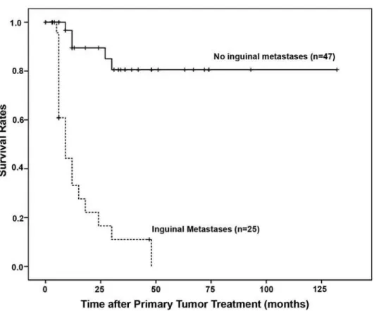
Of the 72 patients, 25 (34.7%) presented lymph node metastases. The 5-year disease-free survival rates for patients with negative and posi-tive lymph node involvement were 80% and 0% respectively. Differences between the 2 groups were VWDWLVWLFDOO\VLJQL¿FDQWS)LJXUH+39 VWDWXVZDVDOVRQRWVLJQL¿FDQWO\DVVRFLDWHGZLWKWKH presence of regional metastases (p = 0.89).
None of the 8 patients with verrucous carcino-ma developed local recurrences or distant metastases and 31/72 patients (43%) with invasive carcinomas died due to progression of the malignancy.
HPV DNA was detected in 44% (35 of 80) RISDWLHQWVXVLQJWKH0<¿UVWURXQG3&57KH overall detection of HPV DNA increased to 72.5% RIXVLQJWKHQHVWHG*33&5$IUDJPHQW RIWKHȕJORELQJHQHZDVDPSOL¿HGLQDOOVSHFLPHQV
as a control of DNA integrity. After RFLP typing RIWZHQW\WKUHH+39FDVHVKLJKULVN+39VZHUH detected in 69% (16 of 23) while low risk HPVs were found in 7 positive cases. The HPV 16 type was observed in 12 of 23 (52%) cases.
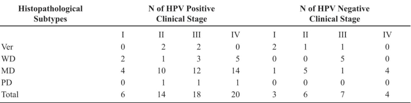
Table-1 shows HPV genotyping according to the histopathological subtype. The distribution of positive and negative HPV tumors, according to the histopathological grade of differentiation and clinical stage, is shown in Table-2. No statistical correlation was observed between HPV status and histopathologi-cal subtype (p = 0.51).
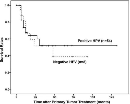
HPV positive patients had better 5-year disease-free survival rates than those with negative HPV results although the differences between the 2 groups were not significant (p = 0.779, Figure-2).
Table 1 – HPV genotyping of penile tumors according to the pathological subtype.
Histopathological
Subtype N HPV+ (%)
Genotypes
6 16* 18* 28 31* 33* 45* 71 NT
Ver 8 4/80 (5) 1 1* 2
WD 16 11/80 (13.7) 2 1* 1* 1 6
MD 53 40/80 (50) 1 8* 1* 1 1* 1* 27
PD 3 3/80 (3.8) 2* 1 0
Total 80 58/80 (72.5) 4 12* 1* 2 1* 1* 1* 1 35
Ver = verrucous carcinoma; WD = well differentiated squamous cell carcinoma; MD = moderately differentiated squamous cell carci-noma; PD = poorly differentiated squamous cell carcicarci-noma; NT not typed. HPV = human papillomavirus; Asterisks indicate high-risk HPV types.
Table 2 – HPV DNA positive and negative penile cancer according to the histopathological subtypes and clinical stage.
Histopathological Subtypes
N of HPV Positive Clinical Stage
N of HPV Negative Clinical Stage
I II III IV I II III IV
Ver 0 2 2 0 2 1 1 0
WD 2 1 3 5 0 0 5 0
MD 4 10 12 14 1 5 1 4
PD 0 1 1 1 0 0 0 0
Total 6 14 18 20 3 6 7 4
Ver = verrucous carcinoma; WD = well differentiated squamous cell carcinoma; MD = moderately differentiated squamous cell car-cinoma; PD = poorly differentiated squamous cell carcinoma. HPV = human papillomavirus; No correlation was observed between
+39VWDWXVDQGKLVWRORJLFDOVXEW\SHVS DVZHOODV+39VWDWXVDQGVWDJHVWUDWL¿FDWLRQS COMMENTS
Age-standardized HPV prevalence varied nearly 20 times between normal populations, from 1.4% (95% CI 0.5-2.2) in Spain to 25.6% (22.4-28.8) in Nigeria (15). The present study evaluated the prevalence of HPV DNA in the largest series of penile tumors, in Rio de Janeiro, Brazil. Squamous cell carcinoma represents 90% of penile cancers. In our series, 70.3% of cases were moderately dif-ferentiated SCC. Seventy-two percent (58 of 80) of specimens were HPV DNA positive. This frequency
The most frequent viral type detected in penile carci-noma was HPV 16, observed in 20.7% (12 of 58) of cases. This result was similar to that previous reported for anogenital cancers. Low risk HPVs were observed in 7 of 58 (12%) positive carcinomas and HPV 6 was the most frequent type, which was found in 4 cases. In Argentina, Picconi et al. (17) reported a very high prevalence of HPV in penile carcinomas (71%) and HPV 18 was the most frequent type found.
Although, our HPV positive patients had bet-ter 5-year disease-free survival rates than those with negative HPV results the differences between the 2 JURXSVZHUHQRWVLJQL¿FDQWS )LJXUH
Lont et al. (18) also detected high-risk HPV DNA in 29% of the tumors, with HPV 16 being the predominant type, accounting for 76% of high-risk HPV containing SCCs. Disease-specific 5-year survival in the high-risk HPV-negative group and high-risk HPV-positive group was 78% and 93%, respectively (log-rank test p = 0.03). In multivariate
analysis, the HPV status was an independent predictor IRUGLVHDVHVSHFL¿FPRUWDOLW\S 7KHVHUHVXOWV indicate that the presence of high-risk HPV (29%) confers a survival advantage in patients with penile carcinoma.
In our study, only one case of HPV 18 was REVHUYHG7KLUW\¿YH RI SDWLHQWV ZLWK non-16 and non-18 positive cases could not be typed. No multiple HPV infections were detected. Multiple infections were observed in 11.6% of Chinese women (19) with cervical cancer and in 8.5% of penile tumors (3). Although, multiple HPV types are less frequent, our results may be due to differences in the ability of the set of primers to amplify different amounts and combinations of HPV types within a sample as well as the capacity of RFLP analysis to identify multiple infections (20).
No correlation was observed among his-topathological subtypes of invasive squamous cell penile carcinomas and HPV infection (x2 = 0.43 and
Figure 2 –5HVSHFWLYH\HDUVXUYLYDOUDWHVDFFRUGLQJWRWKHSUHVHQFHQ RUDEVHQFHRI+XPDQ3DSLOORPDYLUXVQ .DSODQ
p=0.51), although the majority of HPV DNA positive cases (54) were observed among the moderately and poorly differentiated carcinomas.
Seven of 8 cases of verrucous carcinomas were HPV DNA positive and one presented high risk HPV 16. In a previous report, Rubin et al. (3) related 4 of 12 verrucous carcinomas as being HPV DNA positive including 2 with high-risk HPVs. On the other hand, previous studies showed that verrucous carcinoma is more often HPV negative or low-risk HPV positive (21,22).
The majority of penile cancer cases (40%) RFFXUUHGLQWKH¿IWKDQGVL[WKGHFDGHRIOLIH/\PSK node metastasis was associated with poor survival rate and no difference was observed in survival between HPV positive and HPV DNA negative cases.
Although the central role of HPV infection in the etiology of cervical cancer has been recognized, +39LQIHFWLRQDORQHLVLQVXI¿FLHQWIRUWKHPDOLJQDQW transformation in penile cancer. In addition, the pres-ence of HPV DNA could not be considered a prognos-tic factor. Several epidemiological reports indicate that other factors such as the lack of circumcision, hygiene practices, the presence of other sexually transmitted diseases, the number of sexual partners and cigarette smoking may predispose to penile carcinogenesis, and the exact role of HPV infection in the development of penile cancers remains to be elucidated.
On June 8, 2006, the U.S. Food and Drug Administration approved the use of a new vaccine to prevent infection from four types of HPV. Two of the HPV types targeted by the vaccine (16 and HPV-18) are responsible for about 70 percent of the cases of cervical cancer worldwide. The other two HPV types (HPV-6 and HPV-11) cause approximately 90 percent of the cases of genital warts. The vaccine is currently recommended for use in young females before they become sexually active, and its possible use in males is under scrutiny. One of several reasons that HPV vac-cines have focused on women rather than men is that cervical cancer accounts for 80 percent of HPV-related cancers. Male cancers are obviously in the minority, EXWSHUFHQWLVVWLOOVLJQL¿FDQWHVSHFLDOO\FRQVLGHULQJ the prevalence of HPV infection. While women have about 80 percent of the total burden of disease and death, it is certainly not inconsequential for men. Penile cancer affects 100,000 men a year worldwide, and the
numbers are increasing. As a global issue, penile cancer is a relevant problem. For heterosexual men, the main EHQH¿WVRIDQ+39YDFFLQHZLOOEHWKHSUHYHQWLRQRI genital warts and, potentially, cervicalcancer in women. While studies have not yet been carried out, the hope is that the vaccine may eventually help prevent cancers linked to HPV, including penile cancers. If the vaccine proves successful, the administration of HPV vaccines could eventually become a requirement for boys and girls in middle school or high school as a potential way to reduce HPV infection (23).
Improved sampling techniques of the male genitalia and cohort studies in progress should provide important information on the natural history of HPV infection and disease in men, including risk factors for HPV acquisition and transmission. The impact on HPV infection in males of the vaccination in women will also need to be assessed.
ACKNOWLEDGEMENTS
This work was supported by the Brazilian &RXQFLOIRU6FLHQWL¿FDQG7HFKQRORJLFDO'HYHORS -ment (CNPQ) and Ary Frauzino Foundation, National Institute of Cancer.
CONFLICT OF INTEREST
None declared
REFERENCES
'LOOQHU-YRQ.URJK*+RUHQEODV60HLMHU&-(WLRO -ogy of squamous cell carcinoma of the penis. Scand J Urol Nephrol Suppl. 2000; 205: 189-93.
2. Parkin DM, Whelan SL, Ferlay J, Raymond L, Young J: Cancer Incidence in Five Continents VII. Lyon, IARC
6FLHQWL¿F3XEOLFDWLRQVZZZLDUFIU 5XELQ0$.OHWHU%=KRX0$\DOD*&XELOOD$/
4XLQW:* HW DO 'HWHFWLRQ DQG W\SLQJ RI KXPDQ
papillomavirus DNA in penile carcinoma: evidence for multiple independent pathways of penile carcino-genesis. Am J Pathol. 2001; 159: 1211-8.
4. Muñoz N, Bosch FX, de Sanjosé S, Herrero R,
Castell-VDJXp;6KDK.9HWDO(SLGHPLRORJLFFODVVL¿FDWLRQ
%H]HUUD$//RSHV$/DQGPDQ*$OHQFDU*17RU -loni H, Villa LL: Clinicopathologic features and human papillomavirus dna prevalence of warty and squamous cell carcinoma of the penis. Am J Surg Pathol. 2001; 25: 673-8.
6. McCance DJ, Kalache A, Ashdown K, Andrade L, Menezes F, Smith P, et al.: Human papillomavirus types 16 and 18 in carcinomas of the penis from Brazil. Int J Cancer. 1986; 37: 55-9.
%H]HUUD$/ /RSHV$ 6DQWLDJR *+ 5LEHLUR .&
Latorre MR, Villa LL: Human papillomavirus as a prognostic factor in carcinoma of the penis: analysis of 82 patients treated with amputation and bilateral lymphadenectomy. Cancer. 2001; 91: 2315-21. 8. Seixas AL, Ornellas AA, Marota A, Wisnescky A,
Campos F, de Moraes JR: Verrucous carcinoma of the penis: retrospective analysis of 32 cases. J Urol. 1994; 152: 1476-8; discussion 1478-9.
%RVKDUW0*LVVPDQQ/,NHQEHUJ+.OHLQKHLQ]$
Scheurlen W, zur Hausen H: A new type of papillo-mavirus DNA, its presence in genital cancer biopsies and in cell lines derived from cervical cancer. EMBO J. 1984; 3: 1151-7.
10. Manos MM, Ting Y, Wright DK, Lewis AJ, Broker TR, Wolinsky SM: The use of polymerase chain
reac-WLRQDPSOL¿FDWLRQIRUWKHGHWHFWLRQRIJHQLWDOKXPDQ
papillomaviruses. Molecular Diagnostics of Human Cancer. Cancer Cells. 1989; 7: 209-14.
11. Jacobs MV, de Roda Husman AM, van den Brule AJ,
6QLMGHUV3-0HLMHU&-:DOERRPHUV-0*URXSVSH
-FL¿FGLIIHUHQWLDWLRQEHWZHHQKLJKDQGORZULVNKXPDQ
papillomavirus genotypes by general primer-mediated PCR and two cocktails of oligonucleotide probes. J Clin Microbiol. 1995; 33: 901-5.
5HPPHUEDFK7: %ULQFNPDQQ 8* +HPSULFK$ &KHNRO0.KQGHO./LHEHUW8*3&5GHWHFWLRQ
of human papillomavirus of the mucosa: comparison
EHWZHHQ0<DQG*3SULPHUVHWV-&OLQ
Virol. 2004; 30: 302-8.
13. Bernard HU, Chan SY, Manos MM, Ong CK, Villa
//'HOLXV+HWDO,GHQWL¿FDWLRQDQGDVVHVVPHQW
of known and novel human papillomaviruses by
SRO\PHUDVHFKDLQUHDFWLRQDPSOL¿FDWLRQUHVWULFWLRQ
fragment length polymorphisms, nucleotide sequence, and phylogenetic algorithms. J Infect Dis. 1994; 170: 1077-85. Erratum in: J Infect Dis 1996; 173: 516. 14. American Joint Committee on Cancer: AJCC Cancer
Staging Manual 6th. Penis. New York, Springer. 2002; pp. 303-308.
&OLIIRUG*0*DOOXV6+HUUHUR50XxR]16QLMGHUV
PJ, Vaccarella S, et al.: Worldwide distribution of
human papillomavirus types in cytologically normal women in the International Agency for Research on Cancer HPV prevalence surveys: a pooled analysis. Lancet. 2005; 366: 991-8.
16. Heideman DA, Waterboer T, Pawlita M, Delis-van Diemen P, Nindl I, Leijte JA, et al.: Human papil-lomavirus-16 is the predominant type etiologically involved in penile squamous cell carcinoma. J Clin Oncol. 2007; 25: 4550-6.
17. Picconi MA, Eiján AM, Distéfano AL, Pueyo S, Alonio
/9*RURVWLGL6HWDO+XPDQSDSLOORPDYLUXV+39
DNA in penile carcinomas in Argentina: analysis of primary tumors and lymph nodes. J Med Virol. 2000; 61: 65-9.
/RQW$3.URRQ%.+RUHQEODV6*DOOHH03%HUNKRI
J, Meijer CJ, et al.: Presence of high-risk human papil-lomavirus DNA in penile carcinoma predicts favorable outcome in survival. Int J Cancer. 2006; 119: 1078-81.
19. Chan PK, Li WH, Chan MY, Ma WL, Cheung JL, Cheng AF: High prevalence of human papillomavirus type 58 in Chinese women with cervical cancer and precancerous lesions. J Med Virol. 1999; 59: 232-8.
4X:-LDQJ*&UX]<&KDQJ&-+R*<.OHLQ56
et al.: PCR detection of human papillomavirus:
com-SDULVRQEHWZHHQ0<0<DQG*3*3SULPHU
systems. J Clin Microbiol. 1997; 35: 1304-10.
*UHJRLUH/&XELOOD$/5HXWHU9(+DDV*3/DQFDVWHU
WD: Preferential association of human papillomavirus with high-grade histologic variants of penile-invasive squamous cell carcinoma. J Natl Cancer Inst. 1995; 87: 1705-9.
22. Noel JC, Vandenbossche M, Peny MO, Sassine A, de
'REEHOHHU*6FKXOPDQ&&HWDO9HUUXFRXVFDUFL -noma of the penis: importance of human papillomavi-rus typing for diagnosis and therapeutic decision. Eur Urol. 1992; 22: 83-5.
*HLSHUW19DFFLQDWLQJPHQIRU+39QHZVWUDWHJ\IRU
preventing cervical cancer in women? J Natl Cancer Inst. 2005; 97: 630-1.
Accepted after revision: 0D\
Correspondence address: Dr. Antonio Augusto Ornellas Section of Urology
EDITORIAL COMMENT
This study is very welcome as it demonstrates the high rate of human papillomavirus (HPV) associ-ated with penile cancer. The detection rate of 75% approaches the rate of 90% seen in carcinoma in-situ. Other studies show lower HPV rates. It was unclear if this was due to differences in technique, such as immunohistochemistry or due to real variations in biology. Furthermore, the lower rate of HPV (50%) in verrucous type indicates a possible different etiol-ogy to this subset of cancer. Studying the subtypes of HPV associated with penile cancer is paramount currently, given the recent development of vaccines DJDLQVWVSHFL¿FVXEW\SHVRI+39,WLVKDUGWRSURMHFW the effect mass vaccination of females will have on males, but ought to reduce carriage of HPV among heterosexual males. Vaccinating males may have EHQH¿WVIRUIHPDOHVDOVRDVWRWDOLPPXQL]DWLRQRI the whole female population is unlikely.
Reduc-ing the overall population pool of these subtypes is OLNHO\WREHQH¿WERWKPDOHVDQGIHPDOHV7KHLPSDFW on the incidence and type of penile cancer is likely have a lead time of several decades, as only a quar-ter of men with penile cancer present under the age of 50. Projections must also take into account the increasing life-expectancies in developed countries with more men living into their 70s and 80s, where the disease is more prevalent (see http:// www.oecd. org/statsportal/). Thus in the absence of any vaccina-tion the prevalence is likely to increase, whereas even with successful vaccination changes in demographics PD\UHGXFHWKHEHQH¿WLQWKHLQLWLDO\HDUV)LQDOO\WKH subtypes of HPV being targeted may result in only some cancers being prevented. These factors must be brought together in modeling and scenario planning when formulating strategies for service provision in any healthcare system.
Dr. P. K. Hegarty
Institute of Urology and Pathology University College Hospital London London, UK E-mail: [email protected]
EDITORIAL COMMENT
The prevalence of penile cancer in Brazil is 2% and this is higher than in the USA and Eu-rope, where it accounts for 0.3 to 0.6% of cancers (1,2). Squamous cell carcinoma (SCC) is the most common histological type of penile cancer and represents 95% of cases. However the aetiology of penile cancer is unknown, risk factors include age and lack of circumcision (1,2). Other predispos-ing factors to the development of penile SCC are the chronic inflammatory disorder penile lichen sclerosus (LS) (also termed balanitis xerotica obliterans) (3-6) or human papillomavirus (HPV) infection (1,2,7,8). A common aetiology for penile and cervical cancer is suggested by the geographi-cal correlation between the incidence of penile
and cervical cancers worldwide (9). The persistent infection with sexually transmitted high risk HPV is the main cause of cervical cancer (10,11). The prevalence of HPV penile infections in healthy men is reported to be 39% in Brazil and 3-9% in Western Europe, where there is a lower incidence of penile cancer (12,13). However, HPV detection in penile cancer cases varies from 20-80%, depending on detection method and geographical location (1,2). This is unlike cervical cancer where HPV infection can be detected in almost all cases (10).
cases of patients who underwent surgery at Hospi-tals in Rio de Janeiro between 1995 and 2000. High risk HPV 16 was the predominate HPV type detect-ed and no correlation was observdetect-ed between HPV status (all types) and penile cancer subtype, stage, regional metastases or survival. HPV 16 has previ-ously been reported to predominate in penile lichen sclerosus and SCC and may be an aetiological agent LQ WKH GHYHORSPHQW RI D VLJQL¿FDQW SURSRUWLRQ RI penile cancers (7,14). However the importance of HPV status in penile cancer progression and patient survival is controversial, as high-risk HPV is asso-ciated with aggressive variants (8) but recent series examining the relationship of HPV infection with prognosis have revealed either no correlation sur-vival or a favourable sursur-vival (15,16). This study from Rio de Janeiro is consistent with only inguinal metastasis being a prognostic factor for penile can-cer survival. In summary, high risk HPV infection occurs in penile SCC and it is likely to be an ae-WLRORJLFDODJHQWLQWKHGHYHORSPHQWRIDVLJQL¿FDQW proportion of penile cancers. These results are im-portant as prophylactic HPV vaccines for preven-tion of cervical cancer in women could also prevent penile cancers in men. However, several studies, including this one, show that once penile cancer has developed poor prognosis is associated with the occurrence of lymph node metastasis and not HPV status.
REFERENCES
0LFDOL*1DVFD05,QQRFHQ]L'6FKZDUW]5$3H -nile cancer. J Am Acad Dermatol. 2006; 54: 369-91. 2. Kayes O, Ahmed HU, Arya M, Minhas S: Molecular
and genetic pathways in penile cancer. Lancet Oncol. 2007; 8: 420-9.
1DVFD 05 ,QQRFHQ]L ' 0LFDOL * 3HQLOH FDQFHU
among patients with genital lichen sclerosus. J Am Acad Dermatol. 1999; 41: 911-4.
4. Depasquale I, Park AJ, Bracka A: The treatment of bala-nitis xerotica obliterans. BJU Int. 2000; 86: 459-65. 5. Powell J, Robson A, Cranston D, Wojnarowska F,
Turner R: High incidence of lichen sclerosus in pa-tients with squamous cell carcinoma of the penis. Br J Dermatol. 2001; 145: 85-9.
6. Pietrzak P, Hadway P, Corbishley CM, Watkin NA: Is the association between balanitis xerotica obliterans
and penile carcinoma underestimated? BJU Int. 2006; 98: 74-6.
7. Prowse DM, Ktori EN, Chandrasekaran D, Prapa A, Baithun S: Human papillomavirus-associated increase in p16INK4A expression in penile lichen sclerosus and squamous cell carcinoma. Br J Dermatol. 2008; 158: 261-5.
*UHJRLUH/&XELOOD$/5HXWHU9(+DDV*3/DQFDVWHU
WD: Preferential association of human papillomavirus with high-grade histologic variants of penile-invasive squamous cell carcinoma. J Natl Cancer Inst. 1995; 87: 1705-9.
9. Parkin DM, Bray F: Chapter 2: The burden of HPV-related cancers.Vaccine. 2006; (Suppl 3): S11-25. 10. zur Hausen H: Papillomaviruses and cancer: from basic
studies to clinical application. Nat Rev Cancer. 2002; 2: 342-50.
11. de Villiers EM, Fauquet C, Broker TR, Bernard HU,
]XU +DXVHQ + &ODVVL¿FDWLRQ RI SDSLOORPDYLUXVHV
Virology. 2004;324: 17-27.
12. Franceschi S, Castellsagué X, Dal Maso L, Smith JS, Plummer M, Ngelangel C, et al.: Prevalence and de-terminants of human papillomavirus genital infection in men. Br J Cancer. 2002; 86: 705-11.
1DVFD05,QQRFHQ]L'0LFDOL*$VVRFLDWLRQRI
penile lichen sclerosus and oncogenic human papillo-mavirus infection. Int J Dermatol. 2006; 45: 681-3.
5XELQ0$.OHWHU%=KRX0$\DOD*&XELOOD$/ 4XLQW:* HW DO 'HWHFWLRQ DQG W\SLQJ RI KXPDQ
papillomavirus DNA in penile carcinoma: evidence for multiple independent pathways of penile carcino-genesis. Am J Pathol. 2001; 159: 1211-8.
/RQW$3.URRQ%.+RUHQEODV6*DOOHH03%HUNKRI
J, Meijer CJ, et al.: Presence of high-risk human papil-lomavirus DNA in penile carcinoma predicts favorable outcome in survival. Int J Cancer. 2006; 119: 1078-81.
%H]HUUD$/ /RSHV$ 6DQWLDJR *+ 5LEHLUR .&
Latorre MR, Villa LL: Human papillomavirus as a prognostic factor in carcinoma of the penis: analysis of 82 patients treated with amputation and bilateral lymphadenectomy. Cancer. 2001; 91: 2315-21.
Dr. David M. Prowse
Centre for Molecular Oncology, Institute of Cancer Bart’s and The London Queen Mary’s School of