Performance analysis of software for
identification of intestinal parasites
Análise de desempenho de software para identificação de parasitas intestinais
Andressa P. Gomes1; Luana Noguerol1; Marta R. Bez1; Rejane G. Tavares2
1. Universidade Feevale. 2. Universidade Federal de Pelotas.
First submission on 14/08/14; last submission on 30/05/15; accepted for publication on 21/06/15; published on 20/08/15
ABSTRACT
Introduction: Intestinal parasites are among the most frequent diagnoses worldwide. An accurate clinical diagnosis of human parasitic infections depends on laboratory conirmation for speciic differentiation of the infectious agent. Objectives: To create technological solutions to help parasitological diagnosis, through construction and use of speciic software. Material and method: From the images obtained from the sediment, the software compares the morphometry, area, perimeter and circularity, and uses the information on specific morphological and staining characteristics of parasites and allows the potential identification of parasites. Results: Our results demonstrate satisfactory performance, from a total of 204 images analyzed, 81.86% had the parasite correctly identiied by the computer system, and 18.13% could not be identiied, due to the large amount of fecal debris in the sample evaluated. Discussion: Currently the techniques used in Parasitology area are predominantly manual, probably being affected by variables, such as attention and experience of the professional. Therefore, the use of computerization in this sector can improve the performance of parasitological analysis. Conclusions: This work contributes to the computerization of healthcare area, and beneits both health professionals and their patients, in addition to provide a more eficient, accurate and secure diagnosis.
Key words: software; images; parasitological diagnosis; HPJ; Trichuris trichiura; Ascaris lumbricoides.
INTRODUCTION
The presence and the advancement of technology in health care are due to the recognition that processing and analysis of experimental data, whether clinical or epidemiological, require constant evolution(9). In clinical laboratories the process is the
same, each day they are demanding from their information systems, allowing for improvement to meet their needs.
The information system has numerous applications for clinical laboratory. A good example is a database with information on all exams, where the key elements of test are deined, such as name, identiication code, units of measurement, reference values and critical values, among others. Fast and preparation information provided to customers are based on them, as well as the examination price and locations where it is performed(14). Information technology
is also present in the customer care process from the registration, which assists in phonetic search, eliminating possible registration
duplicity errors. The utilities of this system are also present in the collection process, by using barcode for sample identiication, increasing eficiency, resulting in signiicant errors decrease, and reducing costs(15). Otherwise, electronic receiving of tests result,
through automatic interface with the equipment that performed the analysis, and compiling patient’s previous data to create a delta check, assist in the approval of data obtained in the analysis(2). The
latest developments in the laboratories management applications facilitated the access to the results of tests released by the technical area over the Internet, or automatically sent by email to the end user, either the patient or their physician(4).
According to the World Health Organization (WHO)(5), it
is estimated that 3.5 billion people worldwide are infected with one or more types of parasites. In Brazil, it is performed about 20 million parasitological examinations per year in public bodies, and large portion of infected population is in rural and urban poverty areas(6).
An accurate clinical diagnosis of parasitic infections in human depends on laboratory conirmation to differentiate the speciic infectious agent. Clinical examination is the irst step in the diagnosis, and the stool ova and parasites (O&P) test is
the standard test for this diagnosis(7, 8). Such test may be carried
out by several techniques standardized in clinical laboratories. Although different sensibilities and methodologies, all consist of two fundamental steps: microscopic and macroscopic analysis of samples(8). Macroscopic examination allows observing stool
consistency, smelling, presence of abnormal elements, such as mucus and blood, and adult worms or parts of them. The microscopic examination allows observing helminth eggs or larvae, cysts, protozoan trophozoites or oocysts.
Currently, exclusively manual techniques are used in Parasitology area, and are likely to be affected by variables, such as professional attention and experience. Furthermore, the sensitivity of conventional diagnostic ranges from 48% to 76%, since all the inal analysis process depends exclusively on human evaluation, automated protocols for Parasitology are not found, so far(9).
Knowing these limitations and seeking to bring greater reliability to O&P test, this study aimed to develop an information system to assist in the parasitological diagnosis, which would be able to meet speciicity, sensitivity and speed criteria. In this paper, we chose two types of roundworms to characterize the parasitological diagnosis aid system: Ascaris lumbricoides and Trichuris trichiura. The choice of the parasites was based on eggs
morphology, since they are signiicantly different, and also because they are commonly found in O&P test.
MATERIAL AND METHOD
The analytical data were initially obtained by selecting microscopic images in the Laboratório de Biomedicina da Universidade Feevale, which were captured from pools known positive for the selected parasites (Ascaris lumbricoides and Trichuris trichiura). The resources used were a color CCD camera with resolution of 410,000 pixels, connected to the microscope Nikon Eclipse E200, from which the images were subsequently, transferred to a computer, and thus stroing the images. From the images obtained, the software Diagnosis Support System (DSS), specially developed for the system, made the identiication of parasites eggs through morphometric analysis, area, perimeter and circularity, and use information on speciic morphology and coloration characteristics of the parasites in question.
The system was developed in partnership with the Computer Course of Universidade Feevale, so that it is free to use without patent or paid registration. This system was created using C# language, with Microsoft Visual Studio 2005 platform and MATrix LABoratory (MATLAB) tool. C# is a object-oriented programming language designed by Microsoft, it is part of.NET platform, and is based on C++ and Java languages. According to Microsoft, Visual Studio 2005 is included in the Software Development Kit. Mostly, this platform is used for development in VB.NET and C#. MATLAB is a tool originally designed to perform matrix calculations. Today, it has several features in relation to image processing. According to Mosmann (2007), it is an interactive easy handling system that allows formulating solutions to the most diverse problems, especially those involving matrices. Therefore, speciic solutions can develop quickly, compared with C or FORTRAN languages; also it has several libraries to work in different areas of scientiic computing. In academic environments, MATLAB is often used for research and development(10).
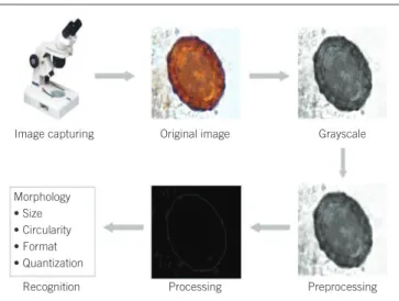
The analysis of images obtained from the slides prepared with sediment acquired by spontaneous sedimentation technique, initially the system obtained the sample image through a camera connected to a microscope. At irst, the prototype produced a new image from the original, but in grayscale. This was done because grayscale image is easier to manipulate. In preprocessing, and the image already in grayscale, a ilter was applied to reduce image noise level by blurring it, thus, causing blurry, so the software could distinguish the edges of objects contained in the image more easily. In the segmentation process, an edge detector was applied in the iltered image; with this procedure a new image with the contours of objects was obtained. Subsequently, the image was scanned for the region of interest, that is, objects similar to a parasite, generating a new image. Then, the software analyzed the characteristics of the images and its objects, checking color, size, shape and texture. The Ascaris lumbricoides and Trichuris trichiura eggs were identiied by eggs area, perimeter, and circularity characteristics; the area was deined as the number of black pixels in the segmented image. Then, to obtain the perimeter, an edge detector was applied in the image to identify the number of pixels in the edge. With the area and perimeter, the calculation was applied to determine the circularity (Figure).
RESULTS
After the initial development and testing of Parasitology DSS software, it was possible to define that the area of Ascaris lumbricoides eggs was in a range from 60,000-100,000 pixels; since the area of Trichuris trichiura was 20,000-41,000. It was also determined that Ascaris lumbricoides eggs had circularity between 0.79-1.1 pixels and Trichuris trichiura, between 0.9-1.2 pixels, these data
used for comparing the images (Table 1). These values were essential for the identifications, because the system initially conducted a comparative study verifying if the values found were in a possible range of area and circularity of the Trichuris trichiura eggs, otherwise, the same comparative study would be done for the Ascaris lumbricoides eggs.
For system tests, we obtained 54 Trichuris trichiura, 85 Ascaris lumbricoides and 65 artifacts, totaling 204 images. Table 2 shows the percentages of errors and accuracy for each egg of the evaluated parasites, as well as to artifacts, after selection and analysis of the images by the software.
By analyzing the images, it was realized that we obtained clear images and with good lighting, as well as bad images, i.e., dark, deformed and plenty of dirt, which affected the analysis by the software. Then, representative data for errors that occurred for both the artifacts identiication (Table 3) and Trichuris trichiura (Table 4) and Ascaris lumbricoides eggs (Table 5)
were identiied.
DISCUSSION
Usually, the identiication and quantiication of intestinal parasites, protozoa or helminths, are performed through microscopic inspection and manual sample, requiring technical expertise, as well as more time(9). However, advances in technology
and lower costs for implementation of computerized methods have opened opportunity to the emergence of new techniques, such as computerized image analysis. This can be used in different sectors of clinical laboratory, especially in the Parasitology sector routine, allowing the identiication of parasites by an automated mode and not subjective and overcoming some of the disadvantages of manual techniques(11).
FIGURE − Flow chart showing the steps of parasite eggs identiication using Parasitology software DSS
DSS: Diagnosis Support System. Morphology
• Size • Circularity • Format • Quantization
Image capturing Original image Grayscale
Preprocessing Processing
Recognition
TABLE 1 − Range of area, perimeter, and circularity values
of each parasite (expressed in pixels)
Ascaris lumbricoides (pixel) Trichuris trichiura (pixel)
Area 60,000-100,000 20,000-41,000
Perimeter 800-1,095 591-791
Circularity 0.79-1.1 0.9-1.2
TABLE 2 − Evaluation of ParasitologyDSS software performance
Type Total Error %Error %Accuracy
Trichuris trichiura 54 17 31.5 68.5
Ascaris lumbricoides 85 14 16.5 83.5
Artifacts 65 6 9 90.1
Total 204 37 18.1 81.9
DSS: Diagnosis Support System.
TABLE 3 − Representative data of erroneous identification of artifacts found ParasitologyDSS software. Area, perimeter
and circularity data are expressed in pixels Original Area Perimeter Circularity Result reported
by SAD
Actual result
36,198 687 1.04 Trichuris
trichiura Artifact
99,065 1,027 0.84 Ascaris
lumbricoides Artifact
90,635 1,007 0.89 Ascaris
lumbricoides Artifact
35,968 669 0.99 Trichuris
trichiura Artifact
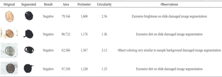
TABLE 5 −Misidentification of “Ascaris lumbricoides eggs” as “artifacts” by Parasitology DSS software. Area, perimeter and circularity data are expressed in pixels
Original Segmented Result Area Perimeter Circularity Observations
Negative 79,546 1,600 2.56 Excessive brightness on slide damaged image segmentation
Negative 80,722 1,176 1.36 Excessive dirt on slide damaged image segmentation
Negative 62,566 1,567 3.12 Object coloring very similar to sample background damaged image segmentation
Negative 97,250 1,238 1.25 Excessive dirt on slide damaged image segmentation
DSS: Diagnosis Support System.
TABLE 4 − Misidentification of “Trichuris trichiura eggs” as “artifacts” by Parasitology DSS software. Area, perimeter, and circularity data are expressed in pixels
Original Segmented Result Area Perimeter Circularity Observations
Negative 66,678 1,837 4.03 Excessive dirt on slide damaged image segmentation
Negative 69,195 891 0.91 Excessive dirt on slide damaged image segmentation
Negative 37,226 338 0.24 Identiied area bigger than the actual, due to dirtiness inclusion in egg image
Negative 33,508 793 1.49 Identiied area bigger than the actual, due to dirtiness inclusion in egg image
DSS: Diagnosis Support System.
Image analysis is not restricted to the process of analyzing, and yes, capturing the image, and to process it are also important. This technique allows improvement in the images obtained, and the automatic recognition and identiication of patterns using certain characteristics, and resulting in time and work reduction. This analysis has proven to be a potential alternative tool to overcome the disadvantages associated with visual identiication of microorganisms and parasites(9, 11, 12).
In this paper, the image analysis procedure allowed the identiication and differentiation of Trichuris trichiura and
Ascaris lumbricoides eggs, and artifacts. During the evaluation of Trichuris trichiura eggs image, from 54 truly positive images, 17 were not detected by the software, which corresponds to a percentage of 31.5% error and 68.5% accuracy. The lack of correct identiication could be justiied due to the interference of fecal debris present in large numbers in the slides. We emphasize the importance and the professional responsibility to
be careful in preparing the slides, minimizing interferences that could be a confounding factor when detected by the system, and still opting to use an O&P preparation method that produces fewer waste(13, 14). When the images of Ascaris lumbricoides eggs
due to the lack of commercially available equipment, plus the cost of implementation of automation, particularly in small to medium-sized laboratories. The search for standardization of a software dedicated to the identiication of eggs and parasite cysts, some studies have been conducted, both in Brazil and abroad. An important differentiation in our study compared to other studies found in the literature refers to the means of comparing images, since the developed Parasitology DSS software differs from the others for allowing the identiication of parasite species based on morphometric analysis, involving area, perimeter, circularity and also typical morphological structures of each species, and not only in a comparison of selected images in ile database. Two other parasites identiication systems were found in the literature, both are based on the comparison of images stored in image banks. Such systems acquire images automatically, scanning slide in different focuses, 400× increase, sending them to the computer analysis module, where the presence of parasites in samples is detected. In a similar study, Dogantekin et al. (2008), using a parasite identiication software, could identify 16 different species of parasites with an accuracy rate of about 95%(15). In the study by
Falcão et al. (2008), the developed software proved to be able to report the presence or absence of parasites with an index of 93%
accuracy, but without performing the their identiication(13). It
should be noted that in this latest work important improvements have been made, such as the use of more concentrated Lugol-based dye, followed by alkaline digestion of fecal micro debris, which led to greater clarity on parasitic structures and improved the contrast obtained.
Based on the results and implementing improvements to obtain samples with less debris, identiication of eggs of other parasites is presented as a perspective for using this software.
CONCLUSION
We conclude that the use of automation in the analytical phase of Parasitology is also possible, and, thus, therefore it is necessary to increase mechanical functions associated with the software, such as automatic prepare of sediment and slides, as well as scanning it through specialized microscopy. This system may bring important gains for laboratories and patients, as it may allows, for example, that the reports issued, today presented only in text form, they can be accompanied by conirmatory images, and allows a reducing the time spent for samples analysis.
RESUMO
Introdução: As parasitoses intestinais iguram entre os diagnósticos mais frequentes no mundo. Um diagnóstico clínico acurado das infecções parasitárias humanas depende da conirmação laboratorial para diferenciação especíica do agente infeccioso. Objetivos: Criar formas tecnológicas para auxiliar no diagnóstico parasitológico por meio da construção e da utilização de um software especíico. Material e método: A partir das imagens obtidas do sedimento, o software compara a morfometria, a área, o perímetro e a circularidade, além de utilizar informações sobre características especíicas de morfologia e coloração dos parasitos e permitir a identiicação dos possíveis parasitas. Resultados: Nossos resultados apontam desempenho satisfatório, sendo que do total de 204 imagens analisadas, 81,86% tiveram o parasita identiicado corretamente pelo sistema computacional e 18,13% não puderam ser identiicados, em função da grande quantidade de detritos fecais na amostra avaliada. Discussão: Atualmente, as técnicas realizadas no setor de Parasitologia são predominantemente manuais, sendo afetadas possivelmente por variáveis como atenção e experiência do proissional. Portanto, a utilização da informatização deste setor pode melhorar a performance das análises parasitológicas. Conclusão: O presente trabalho contribui para a informatização da área da saúde e beneicia tanto os proissionais da saúde como também seus clientes, além de proporcionar um diagnóstico mais eiciente, preciso e seguro.
Unitermos:software; imagens; diagnóstico parasitológico; HPJ; Trichuris trichiura; Ascaris lumbricoides.
REFERENCES
1. Jacob R. A importância da informática na administração hospitalar. Manaus: Centro Universitário Nilton Lins, Especialização em Administração Hospitalar e Serviços de Saúde; 2001.
2. Saccheta TEP. Informatização em laboratório clínico. Rev Soc Cardiol Est SP. 2003; 13(6): 1.
4. Fraser HSF, Kohane IS, Long WJ. Using the technology of the world wide web to manage clinical information. BMJ. 1997; 314: 1600.
5. Organização Mundial de Saúde. Working to overcome the global impact of neglected tropical diseases. First WHO report on neglected tropical diseases; 2010.
6. Alves ilho M. ‘Olho eletrônico’ obtém detecção automática de parasitos intestinais. Jornal da Unicamp. 2007; 361: 5.
7. De Carli GA. Parasitologia clínica: seleção de métodos e técnicas de laboratório para o diagnóstico das parasitoses humanas. São Paulo: Atheneu; 2001.
8. Neves DP. Parasitologia humana. 10 ed. São Paulo: Atheneu; 2000. 428 p. 9. Castanon CAB, Fraga JS, Fernandez S, Gruber A, Costa LF. Biological shape characterization for automatic image recognition and diagnosis of protozoan parasites of the genus Eimeria. Pattern Recognition. 2007; 40: 1899-1910.
10. Mossmann JB. Estudo de técnicas de processamento de imagens aplicadas ao apoio do diagnóstico de derrames serosos de etiologia benigna e maligna [Internet]. Feevale, Brasil. Available at: http://ead. feevale.br/tc/index.php?codcurso=1.
11. Ginoris YP, Amaral AL, Nicolau A, Coelho MAZ, Ferreira EC. Development of an image analysis procedure for identifying protozoa and metazoa typical of activated sludge system. Water Res. 2007; 41(12): 2581-99.
12. Suzuki CTN, Gomes JF, Falcão AX, Papa JP, Hoshino-Shimizu S. Automatic segmentation and classiication of human intestinal parasites from microscopy images. Biomedical Engineering, IEEE Transactions ON. 2012; 60(3): 803-12.
13. Falcão AX, Shimizu SH. Recentes avanços tecnológicos no exame parasitológico de amostras de fezes. Biofarma – Rev Tec Cient Farm Bioquim Anal Clin Toxicol. 2008; 3(6): 44-53.
14. Centers for Disease Control & Prevention (CDC). Diagnostic procedures - microscopic examination [Internet]. 2012. Available at: www.dpd.cdc.gov/dpdx/HTML/Frames/DiagnosticProcedures/body-dp-stoolexamin.htm>.
15. Dogantekin E, Yilmaz M, Dogantekin A, Avci E, Sengur A. A robust technique based on invariant moments – anis for recognition of human parasite eggs in microscopic images. Expert Syst Appl. 2008; 35(3): 728-38.
MAILING ADDRESS
Rejane G. Tavares