Follicular dendritic cell sarcoma:
report of two cases and literature review
Sarcoma de células dendríticas foliculares: relato de dois casos e revisão da literatura
Francisca E. M. M. Lima1; Iago F. Jorge2; Jacques Kauffman2; Gunter Gerson1; Maria do Patrocínio F. G. Beco1; Juliana M. Cavalcante3; Carlos Gustavo Hirth1;Raimundo N. L. Neto2
1. Hospital Haroldo Juaçaba, Fortaleza, Ceará, Brasil. 2. Universidade Federal do Ceará, Fortaleza, Ceará, Brasil. 3. Laboratório Argos, Fortaleza, Ceará, Brasil.
First submission on 15/03/15; last submission on 03/07/15; accepted for publication on 05/07/15; published on 20/08/15
ABSTRACT
Follicular dendritic cell sarcoma is a rare neoplasm, irst described in 1986 by Monda. Case 1: A female patient, 50-year-old performed abdominal computed tomography scan that detected a tumor lesion of 8.0 cm in the mesentery. She underwent resection of the lesion. Microscopic examination revealed epithelioid neoplasm, interspersed with lymphocytes, and positive immunohistochemical staining for CD21 and CD35. The patient underwent adjuvant chemotherapy. Case 2: A male patient, 21-year-old presented right-sided neck mass measuring 7.0 cm. The biopsy revealed proliferation of spindle cells, interspersed with inlammatory iniltrate and storiform arrangement, and positive immunohistochemical staining for CD21 and CD23. The patient underwent neoadjuvant radiotherapy and surgical resection.
Key words: follicular dendritic cell; dendritic cell neoplasm; follicular dendritic cell sarcoma; pathology.
INTRODUCTION
Follicular dendritic cells (FDC) are accessory cells of the lymphoid system. Their main function is to trap and present antigens to B cells and immune complexes. Tumor arising from this cell is very rare, and is named follicular dendritic cell sarcoma (FDCS), which was irst reported by Monda et al. in 1986(2). Since
then, more than 343 cases have been described in the literature, approximately 31% cases had only nodal disease, 58% isolated extranodal involvement, and 11% combined nodal and extranodal disease(3). The predominant site of tumor was cervical lymph
nodes. Recently, the diagnosis accuracy has been signiicantly improved, thanks to the aid of immunohistochemistry analysis and to more reliable FDC markers CD21 and CD35. Once FDCS is suspected, histologically, immunohistochemical stains for follicular dendritic cell differentiation should be performed to avoid misdiagnosis(4). Diagnosis of dendritic cell sarcoma (DCS)
is a challenge, even for experts of hematopathology, and we still do not have a well-designed treatment protocol for this unique tumor. The role of adjuvant therapy remains unclear(3). The aim
of this case report is to discuss the histopathological, clinical and therapeutic aspects of retroperitoneal FDCS.
CASE 1
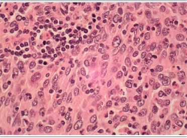
A 50 year-old asymptomatic woman was submitted to follow up abdominal computed tomography (CT) scanning, in June 2012, which revealed a 8.0 × 6.2 cm oval-shaped mesenteric mass, with well-deined limits and mild to moderate heterogeneous enhancement (Figure 1). Routine biochemical and hematological tests were within normal limits. There was a past medical history, in June 2008, of bowel obstruction due to colorectal adenocarcinoma, treated with left hemicolectomy, radiotherapy and chemotherapy. She underwent surgical resection of the mesenteric mass. Surgical exploration revealed a mass measuring 8.4 cm, adjacent to the transverse colon. No associated lymphadenopathy was observed. Cut sections revealed a tan, homogeneous solid mass with rough surface and some areas of necrosis. Multiple sections were taken from different areas of the tumor. Microscopic examination revealed that the tumor consisted of sheets of epithelioid cells, with oval nuclei, some with prominent nucleoli, eosinophilic cytoplasm and poorly deined cell outlines (Figure 2). Numerous small lymphocytes were interspersed between the tumor cells (Figure 3). The tumor exhibited necrosis, cellular
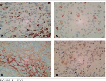
atypia, and high mitotic index (22 per 10 high-power ields) (Figure 4). Furthermore, the tumor cells exhibited positive immunohistochemical staining for vimentin, fascin, CD21 and CD35, focally positive for S100, CD68 and CD56, as well as negative staining for cytokeratin, CD45, CD117, epithelial membrane antigen, melan-A, and HMB-45 (Figures 4 and 5). Based on these histopathological and immunohistochemical indings, the patient was diagnosed with retroperitoneal FDCS. She received postoperative sequential chemotherapy with eight cycles of standard dose of cyclophosphamide, doxorubicin, vincristine and prednisone (CHOP), which initiated in May 2013. To evaluate the eficacy of adjuvant therapy, an abdominal CT scan was performed and resulted normal. She is in remission until today.
FIGURE 1 −Abdominal CT scan
A) post contrast axial CT image shows a well defined mass near the transverse colon measuring approximately 8.0 cm; B) post contrast sagittal CT scan with mild to moderate heterogeneous enhancement.
CT: computed tomography.
FIGURE 2 −FDCS
Sheets of epithelioid cells with eosinophilic cytoplasm.
FDCS: follicular dendritic cell sarcoma.
FIGURE 3 − FDCS
High magnification shows that the neoplastic cells are epithelioid with ovoid to elongated bland nuclei, distinct nucleolus and eosinophilic cytoplasm. There are numerous small lymphocytes interspersed between tumor cells.
FDCS: follicular dendritic cell sarcoma.
FIGURE 4 − FDCS
A) the tumor has numerous mitoses, some atypical; B) areas of necrosis; C) and D) immunohistochemical staining shows that this neoplasm is positive for CD21 (C) and for fascin (D).
FDCS: follicular dendritic cell sarcoma; CD21:
CASE 2
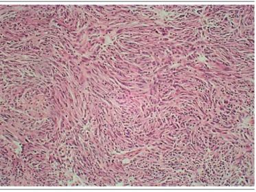
by his mother as unremarkable. Physical examination revealed a right-sided 5.8 × 4.4 cm, elastic hard, painless, ixed mass in the neck. The skin covering the neck swelling was normal with no evidence of ulceration. There was increase in the right amygdala and bulge in the soft palate. No other lesions were found in his head and neck examination. During the diagnose process, a CT scan was performed revealing multiple nodes level II and III on the right side extending to the soft palate (Figure 6). A biopsy was performed and revealed a tumor composed of oval to spindle-shaped cells arranged in sheets, fascicles, concentric whorls, and storiform patterns, with oval nuclei, some prominent nucleoli, eosinophilic cytoplasm, and poorly deined cell outlines (Figure 7). Numerous small lymphocytes are interspersed between the tumor cells (Figure 8). There was no necrosis and the mitotic index was 5 per 10 high-power ields. Immunohistochemically, the tumor cells were positive for CD21, CD23 (Figure 9) and CD45 but negative for cytokeratin, CD3, CD20, and CD68. Together, these results support the diagnosis of FDCS. In May 2012, the preoperative interdisciplinary tumor board conference decided to submit the patient to neoadjuvant radiotherapy. After signiicant reduction in mass volume to 3.0 cm and disappearance of changes in palate and amygdala, he was submitted to complete resection of the mass combined with a right selective neck dissection (level III). He received postoperative sequential chemotherapy with FIGURE 5 − FDCS
Immunohistochemical staining show that this neoplasm is focal positive for CD45 (A), D2-40 (B), CD68 (C), and S100 (D).
FDCS: follicular dendritic cell sarcoma; CD45: ; D2-40: ; CD68: ; S100: .
FIGURE 6 − Head and neck CT images
Post contrast CT images show a well-defined cervical mass stage II and III on the right side extending to the soft palate measuring approximately 6.0 cm, with moderate heterogeneous enhancement.
A) and B) axial CT image; C) sagittal CT image; D) coronal CT image.
CT: computed tomography.
FIGURE 7 − FDCS
Whorling bundles and fascicles of tumor cells are admixed with adjacent lymphoid infiltrates.
FDCS: follicular dendritic cell sarcoma.
DISCUSSION
FDC are accessory cells of the lymphoid system. Their main function is to trap and present antigens to B cells and immune complexes(1, 2). Tumor arising from this cell is very rare, and
is named FDCS, which was irst reported by Monda et al.(3). The
origin of FDC has been subject of heavy debates and speculations and remains unclear. In the past, it was believed that they had hematopoietic origin, however the ultrastructure, cytology, and immunophenotype of FDC do not support an hematopoietic origin, but favor a mesenchymal origin and raise the issue of whether they arise from further differentiation of local ibroblastic reticular cell(5)
or from migration of mesenchymal cell, probably from the bone marrow(5) or from vascular mural cells(6).
FIGURE 9 − FDCS
Immunohistochemical staining shows that this neoplasm is positive for CD21 (A) and CD23 (B).
FDCS: follicular dendritic cell sarcoma; CD21: ; CD23: . FIGURE 8 − FDCS
Spindle tumor cells are admixed with numerous lymphocytes.
FDCS: follicular dendritic cell sarcoma.
At present, 31% of patient had only nodal disease, 58% cases of isolated extranodal, and 11% had both nodal and extranodal involvement. In addition, the disease has been reported in a huge number of extranodal sites. Cervical lymph nodes were the most frequently affected nodal sites. Only 13 cases were described in the mesentery(7).
Most patients with FDCS present slowly enlarging, painless, asymptomatic mass(6). Approximately 10% of patients present
fever or weight loss(8). Abdominal pain was the most common
symptom for most patients with abdominal involvement, followed by systemic symptoms (i.e., fever, weight loss, fatigue, intestinal obstruction, rectal bleeding, and dyspepsia). We can highlight that 11 patients with abdominal disease were asymptomatic and almost always incidentally diagnosed(3).
The initial diagnosis of FDCS is based on clinical examination, imaging, and pathological assessment. Imaging investigation provides delineation of the extent of the mass and staging. Ultrasound and CT are usually the initial imaging modalities of choice used in the evaluation of patients. CT shows smaller relatively homogeneous masses; however, heterogeneity as a result of necrosis or hemorrhagic areas has been reported in more than 80% cases(9).
Etiopathogenesis of FDCS remains unclear. Castleman disease, which is a benign lymphoproliferative disorder, has been suggested as a precursor lesion for this tumor. As in the hyperplasia-dysplasia-neoplasia sequence proposed for development of some epithelial neoplasms, FDCS may occur in lymph nodes harboring dysplastic FDC in Castleman disease. Additionally, some studies have reported clonal expansion of FDC in Castleman disease(10). Epestein-Barr
virus is involved in pathogenesis of a small subset of FDCS cases, but most of them were reported as “inlammatory pseudotumor-like follicular dendritic cell sarcoma”(11). It is a variant of FDCS
with different clinical and pathologic features. Due to the rarity of EBV infection associated with classical FDCS(12), the pathogenesis
of this variant may be different. There are many reports suggesting an association between FDCS and autoimmunity.
Pathological diagnosis is challenging(12) and may require
a combination of morphological, immunophenotypical, cytochemical, and electron microscopic analyses(8, 13). On gross
pathology, FDCSs are well-circumscribed with a tan cut surface and areas of necrosis and cystic change in larger tumors. On microscopy, plump spindled to ovoid cells with eosinophilic cytoplasm and distinct cell borders are arranged in a fascicular, whorled or storiform pattern, typically iniltrated by scattered
small lymphocytes(10). Saygin et al.(5) describe morphological
features of 16 cases available for analysis. Among them, 75% cases demonstrated classical features with whorls, fascicular or storiform pattern. One case with this pattern presented nuclear pseudoinclusions which is far more common in FDCS. Fiveteen from the 16 patients had lymphoplasmacytic iniltration, epithelioid cells were observed in 18.7% cases (3 from 16). Giant cells were present in 25% cases (4 from 16).
IHC (immunohistochemistry) is required to confirm the diagnosis. FDCS usually show reactivity for CD21, CD23 and CD35(13). However, diagnosis may be difficult, mainly
because follicular dendritic cell markers are not included in a routine IHC panel and, it may require pathology expert review(8). They are identified by positive immunohistochemical
staining for CD21 (C3d receptor), CD23, CD35 (C3b receptor), R4/23, Ki-M4, Ki-M4p, and Ki-FDC1p(14). To summarize the
literature, CD21 and CD35 are the most widely markers used. Other useful markers are vimetin, CD23, CD68, S100 protein, fascin, Ki-M4p and Ki-FDC1p; however, these are unspecific. FDCS typically lacks expression of CD1a, desmin and CD45, which allows their differential diagnosis from interdigitating dendritic cell sarcoma, langerhans cell tumors, histiocytic and lymphoid neoplasias. Therefore, the expression of non-typical FDCS markers should be taken into account in the differential diagnosis from other neoplasms. The diagnosis of FDC tumor is established based on morphology and IHC findings. Ultrastructural studies may be helpful, but are not indispensable for accurate diagnosis. All neoplams in the differential diagnosis lack follicular dendritic cell differentiation and are easily excluded if FDCS is considered and IHC staining with FDC marker is applied(15).
Detailed review of all cases indicated that at least 18.6% of patients (64 from 343) were erroneously diagnosed at the presentation. Entities most commonly confused with FDCS included undifferentiated carcinoma, lymphoma, malignant ibrous histiocytoma, peripheral nerve sheath tumor, ectopic meningioma, inlammatory pseudotumor, granulomatous inlammation, gastrointestinal stromal tumor, and unclassiied sarcoma(16).
Local recurrence and/or distant metastasis occurred in 44.6% of patients after initial treatment. Sxty-three patients (28.1%) experienced local recurrence at a median time of 15 months, and 61 cases (27.2%) developed distant metastasis at a median time of 18.5 months. Twenty-four patients had local and
distant recurrence at the same time. Common sites of metastasis were lung (9.4%), lymph nodes (8.9%), liver (9.4%), and bone (3%)(17). Similar to other soft tissue sarcomas, large tumor size
(≥ 6 cm), presence of coagulative necrosis, high mitotic counts (≥ 5 per 10 high-power ields), and signiicant cytologic atypia were shown to be associated with poor prognosis(18). Younger age,
abdominal involvement, sparse inlammatory iniltrate are also poor prognostic factors(19).
In recent years, dendritic cell tumors have been increasingly recognized by pathologists with a dificult management for oncologists. Some treat FDS with an aggressive lymphoma regimen, which often includes chemotherapy, whereas others treat FDS as a soft tissue sarcoma, with wide resection and adjuvant radiotherapy(18). Surgery should be the mainstay of treatment for
early FDCS cases, since patients treated with surgery had better overall survival when compared to other treatment modalities. However, adjuvant radiotherapy did not have a signiicant inluence on overall survival. Prior studies have also demonstrated that adjuvant treatments had no signiicant effect on disease-free survival after a radical surgical resection(20). Therefore, meticulous
examination of excised specimens for surgical margins and extra-capsular iniltration is highly recommended. On the other hand, the number of patients with locally advanced and distant metastatic disease was low and the treatment received varied. In most of these cases, surgery was performed to reduce tumor burden. The role of surgery in late disease is not clear, only 2 from 23 patients who received combined adjuvant chemotherapy and radiotherapy succumbed to the disease (both had metastatic disease at onset). This highlights the importance of adjuvant therapies in advanced FDCS patients(20).
RESUMO
Sarcoma de células dendríticas foliculares é uma neoplasia rara, descrita pela primeira vez em 1986 por Monda. Caso 1: Paciente do sexo feminino, 50 anos, realizou tomografia computadorizada de abdômen que detectou lesão tumoral de 8,0 cm em mesentério. Foi submetida à ressecção da lesão. A microscopia revelou neoplasia epitelioide, com linfócitos de permeio e expressão imuno-histoquímica de CD21 e CD35. A paciente foi submetida à quimioterapia adjuvante. Caso 2: Paciente do sexo masculino, 21 anos, com massa cervical direita medindo 7,0 cm. A biópsia evidenciou proliferação de células fusiformes, com infiltrado inflamatório de permeio e arranjo estoriforme, com expressão imuno-histoquímica de CD21 e CD23. O paciente foi submetido a radioterapia neoadjuvante e ressecção cirúrgica.
Unitermos: células dendríticas foliculares; neoplasias de células dendríticas; sarcoma de células dendríticas foliculares; patologia.
REFERENCES
1. Vega F, Coombes KR, Thomazy VA, Patel K, Lang W, Jones D. Tissue-speciic function of lymph node ibroblastic reticulum cells. Pathobiology. 2006; 73(2): 71-81. PMID: 16943687.
2. Tew JG, Kosco MH, Burton GF, Szakal AK. Follicular dendritic cells as accessory cells. Immunol Rev. 1990; 117: 185-211. PMID: 2258191. 3. Monda L, Warnke R, Rosai J. A primary lymph node malignancy with features suggestive of dendritic reticulum cell differentiation. A report of 4 cases. Am J Pathol. 1986: 122: 562-72. PMID: 2420185.
4. Chan JK, Fletcher CD, Nayler SJ, Cooper K. Follicular dendritic cell sarcoma. Clinicopathologic analysis of 17 cases suggesting a malignant potential higher than currently recognized. Cancer. 1997; 79: 294-313. PMID: 9010103.
5. Saygin C, Uzunaslan D, Ozguroglu M, Senocak M, Tuzuner N. Dendritic cell sarcoma: a pooled analysis including 462 cases with presentation of our case series. Crit Rev Oncol Hematol. 2013; 88(2): 253-71. PMID: 23755890.
6. Krautler NJ, Kana V, Kranich J, et al. Follicular dendritic cells emerge from ubiquitous perivascular precursors. Cell. 2012; 150(1): 194-206. PMID: 22070220.
7. Karabulut B, Orhan KS, Guldiken Y, Dogan O. Follicular dendritic cell sarcoma of the nasopharynx. Int J Oral Maxillofac Surg. 2012; 41: 218-20. PMID: 21835593.
8. Perez-Ordoñez B, Erlandson RA, Rosai J. Follicular dendritic cell tumor: report of 13 additional cases of a distinctive entity. Am J Surg Pathol. 1996; 20(8): 944-55. PMID: 8712294.
9. Long-Hua Q, Qin X, Ya-Jia G, et al. Imaging indings of follicular dendritic cell sarcoma: report of four cases. Korean J Radiol. 2011; 12(1): 122-8. PMID: 21228948.
10. Jaffe ES, Harris NL, Stein H, Vardiman JW, editors. World Health Organization classiication of tumors. Pathology and genetics of tumors of hematopoietic and lymphoid tissues. Lyon: IARC Press; 2001.
11. Cheuk W, Chan JK, Shek TW, et al. Inlammatory pseudotumor-like folicular dendritic cell tumor: a distinctive low-grade malignant intra-abdominal neoplasm with consistente Epstein-Barr virus association. Am J Surg Pathol. 2001; 25(6): 721-31. PMID: 11395549.
12. Malik A, Veniyoor A, Fanthome B, Dutta V. Follicular dendritic cell sarcoma: a diagnostic challenge. J Cancer Res Ther. 2012; 8(2): 306-7. PMID: 22842383.
13. Hollowood K, Stamp G, Zouvani I et al. 1995 Extranodal follicular dendritic cell sarcoma of the gastrointestinal tract. Morphologic, immunohistochemical and ultrastructural analysis of two cases. Am J Clin Pathol. 103: 90-97.
14. Parwaresch MR, Radzun HJ, Hansmann ML, Peters KP. Monoclonal antibody Ki-M4 speciically recognizes human dendritic reticulum cells (follicular dendritic cells) and their possible precursor in blood. Blood. 1983; 62(3): 585-90. PMID: 6192857.
15. Shia J, Chen W, Tang LH, et al. Extranodal follicular dendritic cell sarcoma: clinical, pathologic, and histogenetic characteristics of an underrecognized disease entity. Virchows Arch. 2006; 449(2): 148-58. PMID: 16758173.
16. Chang KC, Jin YT, Chen FF, et al. Follicular dendritic cell sarcoma of the colon mimicking stromal tumour. Histopathology. 2001; 38(1): 25-9. PMID: 11135043.
17. Soriano AO, Thompson MA, Admirand JH, et al. Follicular dendritic cell sarcoma: a report of 14 cases and a review of the literature. Am J Hematol. 2007; 82(8): 725-8. PMDI: 17373675.
18. Amiri-Kordestani L, Priebat D, Chia SH. Follicular dendritic cell sarcoma of the neck: case report and review of current diagnostic and management strategies. Ear Nose Throat J. 2010; 89(7): E14-7. PMID: 20628972.
MAILING ADDRESS
Francisca Estefânia M. M. Lima
Hospital Haroldo Juaçaba; Rua 30 de Outubro, 501; Henrique Jorge; CEP: 60510-190; Fortaleza-CE, Brasil.
20. Shinagare AB, Ramaiya NH, Jagannathan JP, Hornick JL, Swanson RS. Primary follicular dendritic cell sarcoma of liver treated with