ORIGINAL
RES
EAR
CH
Correspondence to: Dirceu Costa – Programa de Pós-graduação em Ciência da Reabilitação, UNINOVE – Rua Vergueiro, 235 – Liberdade – CEP: 01504-001 – São Paulo (SP), Brazil – E-mail: [email protected]; [email protected]
ABSTRACT | The objective of this study was to evaluate the effect of transcutaneous electrical diaphragmatic stimulation (TEDS) on respiratory muscle strength and endurance, tho-racic-abdominal expansibility and spirometric variables of sub-jects with chronic as obstructive pulmonary disease (COPD). Eight COPD patients submitted to respiratory physiotherapy received treatment with TEDS twice a week for 06 weeks, to-taling 12 sessions. Before and after TEDS they were evaluated by the following parameters: maximal inspiratory pressure (MIP); maximal expiratory pressure (MEP); axillary, xiphoid and abdominal cyrtometry; and spirometry. After the Shapiro-Wilk test, the paired Student’s-t test and the Mann-Whitney test were applied for comparison of the two stages (before and after TEDS). For comparison of the before, after (post-1st ses-sion), 1st, 2nd, 3rd, 4th week stages, the ANOVA followed by Tukey test were applied (p<0.05). In accordance with the re-sults obtained it was observed that TEDS promoted significant increase in: MIP (47.3%); MEP (21.7%); axillary (55.5%); xiphoid (59.2%) and abdominal (74.2%) cyrtometry, but not in the spi-rometric variables. In longitudinal analysis (in the 4 following weeks) the increase found in MIP and in thoracic-abdominal expansibility was maintained. Thus, we conclude that TEDS promoted improvement in respiratory muscle strength and thoracic-abdominal expansibility in COPD patients without al-terations in spirometric variables, and some parameters were maintained in the following 4 weeks..
Keywords | Electric Stimulation; Pulmonary Disease, Chronic Obstructive.
The effect of transcutaneous electrical diaphragmatic
stimulation on respiratory parameters of Chronic
Obstructive Pulmonary Disease patients
Efeito da estimulação diafragmática elétrica transcutânea em parâmetros
respiratórios de pacientes com Doença Pulmonar Obstrutiva Crônica
Efecto de la estimulación diafragmática eléctrica transcutánea en parámetros
respiratorios de pacientes con Enfermedad Pulmonar Obstructiva Crónica
Karina Maria Cancelliero-Gaiad1, Daniela Ike1, Dirceu Costa1,2
Study conducted at the Universidade Federal de São Carlos (UFSCar) – São Carlos (SP) e Universidade Nove de Julho (UNINOVE) – São Paulo (SP), Brazil.
1Respiratory Physical Therapy Laboratory of the Graduate Program of Physical Therapy at UFSCar – São Carlos (SP), Brazil. 2Respiratory Functional Evaluation Laboratory of the Graduate Program of Rehabilitation Sciences at UNINOVE – São Paulo (SP), Brazil.
RESUMO | O objetivo do estudo foi avaliar o efeito da estimula-ção diafragmática elétrica transcutânea (EDET) sobre a força e endurance muscular respiratória, expansibilidade toracoabdo-minal e variáveis espirométricas de indivíduos com doença pul-monar obstrutiva crônica (DPOC). Oito pacientes com DPOC submetidos à fisioterapia respiratória receberam tratamento com EDET duas vezes por semana durante 06 semanas, totali-zando 12 sessões. Antes e depois do tratamento eles foram ava-liados pelos seguintes parâmetros: pressão inspiratória máxima (PImáx); pressão expiratória máxima (PEmáx); cirtometria axilar, xifoideana e abdominal; e espirometria. Após o teste Shapiro-Wilk, o teste t de Student pareado e o teste Mann-Whitney foram aplicados para a comparação dos dois estágios (antes e após a EDET). Para a comparação dos estágios antes, após (pós-1a sessão), 1ª, 2ª, 3ª e 4ª semana, a ANOVA seguida do teste de Tukey foram aplicados (p<0,05). De acordo com os resulta-dos obtiresulta-dos, foi observado que a EDET promoveu aumento significativo em: PImáx (47,3%); PEmáx (21,7%); cirtometria axilar (55,5%); xifoideana (59,2%) e abdominal (74,2%), mas não nas va-riáveis espirométricas. Na análise longitudinal (nas 4 semanas seguintes) o aumento encontrado na PImáx e na expansibilida-de toracoabdominal foi mantido. Assim conclui-se que a EDET promoveu melhora na força muscular respiratória e na expan-sibilidade toracoabdominal em pacientes com DPOC sem alte-ração nas variáveis espirométricas; e alguns parâmetros foram mantidos nas quatro semanas seguintes.
INTRODUCTION
Among diferent aspects of skeletal muscle dysfunction, muscle mass regulation has attracted the attention of many researchers1 due to their clinical relevance,
espe-cially in patients with Chronic Obstructive Pulmonary Disease (COPD) and other chronic afections.
In COPD, speciically, the inspiratory muscle dys-function is associated with unfavorable clinical conse-quences such as dyspnea, respiratory failure with hy-percapnia and even early mortality2,3, which justify the
exhaustion of techniques that may prevent it.
According to Testelmans et al.4, inspiratory muscle
weakening in COPD patients is clinically relevant, be-cause the maximal inspiratory pressure (MIP) is corre-lated to their survival. Some studies have reported that an adaption occurs in the diaphragm in this condition, being characterized by an increase in type I ibers and decrease in type II ibers4,8, becoming more resistant to fatigue due
to the increase in activity this clinical condition requires9,10.
However, this change also leads to a reduction in the muscle strength of the diaphragm, with decrease in myosin content and sensibility to calcium, which favors muscle weakness at submaximal activation11.
Respiratory Physical herapy may contribute with the improvement of inspiratory muscle strength with trans-cutaneous electric diaphragmatic stimulation (TEDS). Aiming at better understanding this dynamics that acts directly on the diaphragm, studies have been conducted with animals12,13 and humans, for instance, in the
postop-erative period of gastroplasty for obesity14.
However, we found no studies about TEDS in pa-tients with COPD, and as this tool could be useful for patients with inspiratory muscle weakness this is a rea-son to conduct studies that can produce evidence of the technique beneits.
herefore, this study was aimed at evaluating the efects of TEDS in respiratory muscle strength, tho-racoabdominal expansibility and spirometric variables in patients with COPD at short term (after treatment) and four weeks later (residual efects). he hypoth-esis raised is that TEDS improves respiratory muscle strength, mainly inspiratory, because TEDS stimulates the contraction of the diaphragm.
METHODOLOGY
Sample
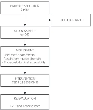
Eighteen patientes dignosed with COPD (accord-ing to criteria established by Rabe et al.15) were
se-lected, out of which eight were enrolled in the study sample. The number of subjects was 8 with 80% sample power.
Amongst all patients, those aged above 80 years (n=03), in home oxygen therapy (n=02), using cardiac pacemaker (n=02), presenting cancer (n=01) and gastro-intestinal disease (n=02) were excluded from the sam-ple. Some other exclusion criteria were: recent history of COPD exacerbation or respiratory tract infections,
RESUMEN | El objetivo del estudio fue evaluar el efecto de la estimulación diafragmática eléctrica transcutánea (EDET) so-bre la fuerza y endurance muscular respiratoria, expansibilidad toracoabdominal y variables espirométricas de individuos con enfermedad pulmonar obstructiva crónica (EPOC). Ocho paci-entes con EPOC sometidos a fisioterapia respiratoria recibieron tratamiento con EDET dos veces por semana durante 6 sema-nas, totalizando 12 sesiones. Antes y después del tratamiento fueron evaluados por los siguientes parámetros: presión inspi-ratoria máxima (PImáx); presión expiinspi-ratoria máxima (PEmáx); cirtometría axilar, xifoidea y abdominal; y espirometría. Después del test Shapiro-Wilk, el test t de Student pareado y el test Mann-Whitney fueron aplicados para la comparación de los dos niveles (antes y después de la EDET). Para la comparación de los niveles
antes, después (post-1a sesión), 1ª, 2ª, 3ª y 4ª semana, la ANOVA seguida del test de Tukey fueron aplicados (p<0,05). De acuerdo con los resultados obtenidos, fue observado que la EDET pro-movió aumento significativo en: PImáx (47,3%); PEmáx (21,7%); cir-tometría axilar (55,5%); xifoidea (59,2%) y abdominal (74,2%), pero no en las variables espirométricas. En el análisis longitudinal (en las 4 semanas siguientes) el aumento encontrado en la PImáx y en la expansibilidad toracoabdominal fue mantenido. Así se con-cluye que la EDET promovió mejora en la fuerza muscular respi-ratoria y en la expansibilidad toracoabdominal en pacientes con EPOC sin alteración en las variables espirométricas; y algunos parámetros fueron mantenidos en las cuatro semanas siguientes
uncontrolled arterial hypertension, allergic rhinitis and tuberculosis. Patients with COPD had their disease re-ported in a form, including medication related to the respiratory tract; and all of them were smokers or former-smokers, none presenting clinical or physi-ological characteristics of bronchial asthma.
The COPD group was clinically stable and par-ticipating in a program for pulmonary rehabilita-tion (PR) and attending respiratory physical ther-apy sessions twice a week. This program consisted of strength and endurance exercises (upper and lower limbs) associated with respiratory reeduca-tion, lengthening, aerobic exercises, orientations for daily life activities and techniques of energy con-servation. Respiratory muscle training was not per-formed in the period of TEDS or post-TEDS. All patients had been participating in the program for at least one year.
The study was approved by the Ethics commit-tee of the institution (Protocol 114/2009) and all patients signed the informed consent form before starting the research.
Intervention
To perform TEDS, we used the equipment Phrenics Dualpex 961 (Quark®; Brazil). The electrical cur-rent was pulsed, biphasic and symmetric, in the fol-lowing patterns: 30Hz frequency (cycles per second), 0.4 ms band phase, rise time of 0.7 seconds and fre-quency of 14 respirations per minute. The intensity of the current was the minimum required to provide the diaphragm with a muscle contraction that was comfortable for patients16.
Four carbon-silicone electrodes (3x5 cm) were put on patients’ skin — previously cleaned — with a conductor gel and micropore for fixation (3M®). Two electrodes were put on each side of the trunk, specifically on the third intercostals space near the middle part of the sternum and on the 7th
intercos-tals space, on the middle axillary line. Sessions lasted 30 minutes and patients were oriented to coordinate their breath (inspiration) with the contraction gen-erated by the electrical current.
Patients remained in semi-Fowler position (30º), with their lower limbs extended and upper limbs po-sitioned at their sides.
The intervention was performed twice a week for six weeks, totaling 12 sessions.
Assessment
Before and after intervention, the following assess-ments were made:
• Respiratory muscle strength: assessed by MIP and MEP by an analogical manovacuometer (Ger-Ar®), gauged in cmH2O, with variations of
±300 cmH2O, equipped with a bucal adaptor with a 2-mm diameter oriice, approximately, to avoid intraoral pressure that can be generated by un-welcome muscle contractions in the mouth cavity and, therefore, interference in results, in compli-ance with recommendations of the literature17,18.
Patients remained seated, with their trunk in a 90° position to the hips, using a nose clip during the maneuver. MIP was obtained from a maximal in-spiration sustained for at least two seconds, after a maximal expiration, at the level of residual volume (VR). MEP was obtained from a maximal expira-tory efort after a maximal inspiration at the level of total lung capacity (TLC), also sustained for at least two seconds18. hree technically satisfying
measures were taken at each pressure, avoiding air escaping through the mouth or the nose, and with similar values (without exceeding 10% diference between them). he highest value obtained for each pressure was considered for analysis19.
• Thoracoabdoinal expansibility: assessed by axil-lary, xiphoid and abdominal cyrtometry using a tape measure. Three anatomical reference points were considered — axillary fold, xiphoid ap-pendix, and umbilical line. The examiner asked the patients to perform maximal inspiration and expiration for each reference point three times. Data were registered in centimeters20.
• Spirometry: performed using a portable spi-rometer (Easy One®) for the following vari-ables: Slow Vital Capacity (SVC), Forced Vital Capacity (FVC), Maximum Voluntary Ventilation (MMV, muscle endurance), First Second Forced Ventilatory Volume (FVV1) and index FVV1/ FVD. Measures were all obtained while using a nose clip. For each maneuver, three acceptable and reproducible measures were taken, according to the guidelines by the American horacic Society21.
Volunteers remained seated in the 1st and 3rd
evaluations, and standing up in the 2nd evaluation.
Statistical analysis
After the application of Shapiro-Wilk normality test, the following procedure was adopted: comparison of two phases (pre and post-intervention) using the paired Student’s T (parametric) or Mann-Whitney (non-parametric) test, depending on the normality result; comparison between pre, post-intervention and 1, 2, 3 and 4 weeks after intervention using one-way ANOVA and Tukey tests. The significance level was set at 5% (p<0.05) and we used the software Graph Pad Prism 5® for data processing.
RESULTS
The general and respiratory characteristics of pa-tients with COPD are shown in Table 1.
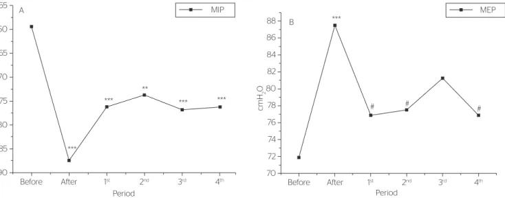
After TEDS, there was an increase in respira-tory muscle strength values (mean±SD; cmH2O), represented by 47.3% at MIP (before: -59.4±17.0; after: -87.5±22.4; p<0.001 compared to the pre-intervention phase) and by 21.7% in MEP (before: 71.9±18.1; after: 87.50±16.0; p<0.001 compared to the pre-intervention phase).
In the evaluation after intervention, MIP val-ues (cmH2O) (Figure 2A) still showed an increase, represented by: 28.4% (-76.3±15.1; p<0.001 com-pared to the pre-intervention phase) in the first week; 24.2% (-73.8±16.0; p<0.01 compared to the pre-intervention phase) in the second week; 29.5% (-76.9±16.7; p<0.001 compared to the pre-interven-tion phase) in the third week; and 28.4% (76.3±16.6; p<0.001 compared to the pre-intervention phase) in the fourth week. These values are percentages, com-pared to those of the pre-intervention phase.
MEP values (cmH2O) were not different in the following evaluations when compared to pre-inter-vention phase (71.9±18.1), but they did differ from those of the post-intervention phase (87.50±16.0), that is, values were lower in: first week (76.9±17.1; p<0.05 compared to the post-intervention phase); second week (77.5±19.6; p<0.05 compared to the post-intervention phase); and fourth week (76.9±17.1; p<0.05 compared to the post-interven-tion phase), as shown in Figure 2B.
In spirometry, there was no difference in the pre-dicted value percentages for the studies variables, including VC (before: 74.2±18.2; after: 72.9±22.3; p=0.65), FVC (before: 75.4±18.8; after: 75.1±20.9; p=0.95) and MMV (before: 50.4±22.1; after: 55.5±30.5; p=0.25).
Thoracoabdominal expansibility, assessed by cirtrometry (cm) presented difference, with increase by 55.5% at the axillary level (before: 3.6±1.2; after: 5.6±0.4; p=0.0004), 59.2% at the xiphoid level (be-fore: 2.7±1.3; after: 4.3±1.1; p=0.0022) and 87.1% at the abdominal level (before: 3.1±1.5; after:
Table 1. Anthropometric and spirometric variables of the Chronic Obstructive Pulmonary Disease Group (n=8)
Variable COPD
Age (years) 68.5±6.2 Gender (M/F) 6/2 Height (cm) 163.1±14.2 Weight (kg) 67.7±9.0 BMI (kg/m2) 25.6±3.3
HR (bpm) 84.8±9.9 SpO2 (%) 95.6±2.7
Spirometry
CVF (% previsto) 75.4±18.8 VEF1 (% previsto) 51.2±25.7 VEF1/CVF (% previsto) 64.2±16.8
Values expressed as mean±SD (standard deviation); COPD: Chronic Obstructive Pulmonary Disease; M: male; F: female; BMI: body mass index; HR: heart rate (bpm); SpO2: peripheral oxygen
saturation; FVC: forced vital capacity; FVV1: first-second forced ventilatory volume
PATIENTS SELECTION
(n=18)
EXCLUSION (n=10)
STUDY SAMPLE (n=08)
INTERVENTION TEDS (12 SESSIONS)
RE-EVALUATION
1, 2, 3 and 4 weeks later ASSESSMENT - Spirometric parameters - Respiratory muscle strength - Thoracoabdominal expansibility
5.8±1.5; p=0.0011). In the four following weeks, these increases were sustained at the three levels. No contralateral effects were observed in the stud-ied patients.
DISCUSSION
Our results show that TEDS increase inspiratory and expiratory muscle strength, as well as thora-coabdominal expansibility in patients with COPD without interfering in spirometric variables. This increase in inspiratory muscle strength and tho-racoabdominal expansibility was maintained in four weeks after the intervention. According to Sarlabous et al.22, the diaphragm plays a central role
in the mechanical inspiratory activity. Patients with COPD have their inspiratory muscle and thoracic mechanical functions severely affected. Muscle effi-ciency (the relation between electrical activities and muscle mechanics) is significantly reduced due to changes in the thoracic-diaphragmatic space caused by the disease and which cause the muscle contrac-tions to be ineffective, leading to energy loss.
his impaired contraction of the diaphragm in COPD may be explained by image studies such as magnetic resonance imaging, which shows a de-basement of the diaphragm and limitations of its movements compared to healthy individuals23,24.
According to Iwasawa et al.24, the exact mechanism
of the paradoxal movement of the diaphragm has not
been completely understood yet, but this can be re-lated to the low eicacy of its contraction in COPD. In addition to the debasement seen at imaging evaluation, many studies have reported an adaption of the diaphragm in COPD, with consequent in-crease of type I fibers, which makes the organ more resistant to fatigue. Due to the continuous overload to the muscle in this disease, some authors4,8,11 have
suggested that this adaption is considered beneficial to patients. In association with this changes in the type of muscle fibers, there is also a reduction in the cross-sectional area and in both fibers, type I and II, which characterizes atrophy of the muscle, even though it is submitted to continuous overload with the increase in this activity4.
Levine et al.25 studied two patients with severe
COPD and concluded that the fibers of the dia-phragm are less stronger than the fibers of healthy patients. Ottenheijm et al.26 studied the fibers
of this muscle as well, but in eight patients with COPD, and concluded that the strength of these muscles was reduced in COPD compared to healthy individuals. Stubbings et al.5, on their turn, reported
that type I fibers of the diaphragm are increased in COPD.
In agreement with the results reported by these authors, our study showed reduced inspiratory mus-cle strength, represented by lower values of MIP. Compared to the two equations — by Neder et al.19
and Costa et al.27 — for predicted values, patients
with COPD presented reduced MIP and MEP val-ues in our study.
A MIP
Before
Period
After 1st 2nd 3rd 4th
***
***
**
*** ***
-55
-60
-65
-70
-75
-80
-85
-90
cmH
2
O
B
MEP 88
Period 82
80
78
76
74
72
70
# ***
#
# 84
86
cmH
2
O
Before After 1st 2nd 3rd 4th
Based on the equations by Neder et al.19, the
studied sample had a MIP (cmH2O) (-59.4±17.0) below prediction (predicted MIP: -95.9±6.1) and, compared to the equations by Costa et al.27,
the values obtained were slightly lower than pre-diction (predicted MIP: 62.9±11.6). Expiratory muscle strength, represented my MEP (cmH2O) (71.9±18.1), was also lower compared to predictions by Neder et al.19 (100.5±14.3) and Costa et al.27
(89.9±9.4).
In view of the muscle changes, TEDS becomes important for patients with COPD as an alternative treatment, especially because it not only increases inspiratory muscle strength, but also sustains it for a period (four weeks on average). This result may be related to the alterations in the type of fibers caused by electrical stimulations. This has been reported in a previous study with rats, where the authors ob-served an increase in Type IID fibers of the dia-phragm, reduction of type I fibers and no changes in types IIA and IIB fibers12.
Although the effects of TEDS is specifically on the inspiratory muscle, this technique also helps to increase MEP values for a short period (right after intervention), and the stimulation of the expiratory muscles by the electrical field generated may have oc-curred, as reported by Cancelliero et al.13 in animals
and in other muscle groups with rats29. Associated
with the increase in respiratory muscle strength, an increase in thoracoabdominal expansibility was seen at cirtometry, a method considered to be reliable for the exploration of the dimensions and amplitudes of thoracoabdominal movements20 and that, despite
being hardly ever mentioned in literature, it has been widely used in clinical practice to assess tho-racic mobility at respiration movements29.
Our results agree with those by Costa et al.30,
who reported an improvement in thorcoabdominal mobility at cirtometry in women submitted to gas-troplasty and receiving TEDS, which played an im-portant role in the mechanical recovery of thoracic and abdominal movements in the bariatric postop-erative period.
According to the results of increase in thora-coabdominal mobility, one can infer that TEDS has effects similar to those obtained by physical exercis-es directed to thoracic mobility in casexercis-es of COPD, also assessed by cirtometry, according to the results reported by Paulin et al.31. Similarly, these authors
have not found changes in the spirometric results of
patients with COPD after they had been through a program of physical exercises.
The abdominal mobility was the feature most likely to be altered at the three levels (axillary, xi-phoid and abdominal) studied by Costa et al.30,
when a program of physical exercises was applied in obese patients, as well as in the study by Yamaguti et al.32, which showed an increase in the diaphragm
mobility, evaluated by pletismography after a pro-gram of respiratory exercises (three series of tem exercises in different positions) at short term in pa-tients with COPD. Their results are all in agree-ment with ours, once the increase observed was seen at these three levels.
Physiologically, inferior ribs are more oblique that the superior ones, and the more oblique, the greater the movement they are likely to do. This as-pect is even more relevant when we consider that in patients with COPD these ribs usually arrange themselves in an oblique position, hence more hori-zontal, due to hyperinsuflation caused by the loss of pulmonary elasticity. Therefore, the increase in abdominal mobility can also be characterized as in-dicative of improve in the mechanics of thoracoab-dominal movements and consequent improvement in the pulmonary ventilation.
Although the authors were using different tech-niques in this study, the results were in agreement, emphasizing the increase in thoracoabdominal ex-pansibility and respiratory strength, important fea-tures in the respiratory mechanics and dynamics.
Despite difficulties faced in maintaining patients’ compliance to treatment until the completion of the study, thus characterizing a possible limitation, the results provided important support for therapeutic alternatives in the process of pulmonary rehabilita-tion in patients with COPD. Our hypothesis was confirmed because TEDS improved respiratory muscle strength, notably inspiratory.
CONCLUSION
The maintenance of results for the period of four weeks may also be seen as a strong indication of success, but we suggest, however, that further stud-ies be conducted with longer follow-ups.
ACKNOWLEDGMENTS
The authors thank Quark®, the volunteers and CNPq (559018/2008-8) for the PNPD scholarship.
REFERENCES
1. Doucet M, Dubé A, Joanisse DR, Debigaré R, Michaud A, Paré MÈ, et al. Atrophy and hypertrophy signalling of the quadriceps and diaphragm in COPD. Thorax. 2010;65(11):963-70.
2. Bégin P, Grassino A. Inspiratory muscle dysfunction and chronic hypercapnia in chronic obstructive pulmonary disease. Am Rev Respir Dis. 1991; 143(5 Pt 1):905-12.
3. Gray-Donald K, Gibbons L, Shapiro SH, Macklem PT, Martin JG. Nutritional status and mortality in chronic obstructive pulmonary disease. Am J Respir Crit Care Med. 1996;153(3):961-66.
4. Testelmans D, Crul T, Maes K, Agten A, Crombach M, Decramer M, et al. Atrophy and hypertrophy signalling in the diaphragm of
patients with COPD. Eur Respir J. 2010; 35(3):549-56.
5. Stubbings AK, Moore AJ, Dusmet M, Goldstraw P, West TG, Polkey MI, et al. Physiological properties of human diaphragm muscle fibres and the effect of chronic obstructive pulmonary disease. J Physiol. 2008;586(10):2637-50.
6. Gosker HR, Wouters EF, van der Vusse GJ, Schols AM. Skeletal muscle dysfunction in chronic obstructive pulmonary disease and chronic heart failure: underlying mechanisms and therapy perspectives. Am J Clin Nutr. 2000;71(5):1033-47.
7. Levine S, Gregory C, Nguyen T, Shrager J, Kaiser L, Rubinstein N, et al. Bioenergetic adaptation of individual human
diaphragmatic myofibers to severe COPD. J Appl Physiol. 2002;92(3):1205-13.
8. Levine S, Kaiser L, Leferovich J, Tikunov B. Cellular adaptations in the diaphragm in chronic obstructive pulmonary disease. N Engl J Med. 1997;337(25):1799-806.
9. Nguyen T, Rubinstein NA, Vijayasarathy C, Rome LC, Kaiser LR, Shrager JB, et al. Effect of chronic obstructive pulmonary disease
on calcium pump ATPase expression in human diaphragm. J Appl Physiol. 2005;98(6):2004-10.
10. Bellemare F, Grassino A. Force reserve of the diaphragm in patients with chronic obstructive pulmonary disease. J Appl Physiol Respir Environ Exerc Physiol. 1983;55(1 Pt 1):8-15.
11. Ottenheijm CA, Heunks LM, Dekhuijzen RP. Diaphragm adaptations in patients with COPD. Respir Res. 2008;9:12.
12. Costa D, Cancelliero KM, Campos GE, Salvini TF, Silva CA. Changes in types of muscle fibers induced by transcutaneous electrical stimulation of the diaphragm of rats. Braz J Med Biol Res. 2008;41(9):809-11.
13. Cancelliero KM; Costa D, Silva CA. Transcutaneous electrical stimulation of the diaphragm improves the metabolic conditions of respiratory muscles in rats. J Phys Ther. 2006;10(1):59-65. 14. Forti E, Ike D, Barbalho-Moulim M, Rasera I Jr, Costa D. Effects of
chest physiotherapy on the respiratory function of postoperative gastroplasty patients. Clinics. 2009; 64(7):683-9.
15. Rabe KF, Hurd S, Anzueto A, Barnes PJ, Buist SA, Calverley P, et al.
Global strategy for the diagnosis, management, and prevention of chronic obstructive pulmonary disease: GOLD executive summary. Am J Resp Crit Care Med. 2007;176(6):532-55.
16. Geddes LA, Voorhees WD, Lagler R, Riscili C, Foster K, Bourland JD. Electrically produced artificial ventilation. Med Instrum. 1988;22(5):263-71.
17. Black LF, Hyatt, RE. Maximal respiratory pressures: normal values and relationship to age and sex. Am Rev Respir Dis. 1969;99(5):696-702.
18. Sobush DC, Dunning M. Assessing maximal static ventilatory muscle pressures using the “bugle” dynamometer. suggestion from the field. Phys Ther. 1984;64(11):1689-90.
19. Neder JA, Andreoni S, Lerario MC, Nery LE. Reference values for lung function tests II: maximal respiratory pressures and voluntary ventilation. Braz J Med Biol Res. 1999;32(6):719-27. 20. Caldeira VS, Starling CCD, Britto RR, Martins JA, Sampaio RF,
Parreira VF. Reliability and accuracy of cirtometry in healthy adults. J Bras Pneumol. 2007;33(5):519-26.
21. Miller MR, Hankinson J, Brusasco V, Burgos F, Casaburi R, Coates A, et al. ATS/ERS task force: standardisation of lung function
testing. Eur Respir J. 2005;26:319-38.
22. Sarlabous L, Torres A, Fiz JA, Gea J, Martinez-Llorens JM, Jane R. Evaluation of the respiratory muscular function by means of diaphragmatic mechanomyographic signals in COPD patients. Conf Proc IEEE Eng Med Biol Soc. 2009;2009:3925-28.
23. Etlik O, Sakarya ME, Uzun K, Harman M, Temizoz O, Durmus A. Demonstrating the effect of theophylline treatment on diaphragmatic movement in chronic obstructive pulmonary disease patients by MR-flu oroscopy. Eur J Radiol. 2004;51(2):150-4. 24. Iwasawa T, Kagei S, Gotoh T, Yoshiike Y, Matsushita K,
Kurihara H, et al. Magnetic resonance analysis of abnormal diaphragmatic motion in patients with emphysema. Eur Respir J. 2002;19(2):225-31.
25. Levine S, Nguyen T, Kaiser LR, Rubinstein NA, Maislin G, Gregory C, et al. Human diaphragm remodeling associated with chronic
obstructive pulmonary disease: clinical implications. Am J Respir Crit Care Med. 2003;168(6):706-13.
26. Ottenheijm CA, Heunks LM, Sieck GC, Zhan WZ, Jansen SM, Degens H, et al. Diaphragm dysfunction in chronic obstructive pulmonary disease. Am J Respir Crit Care Med. 2005;172(2):200-5. 27. Costa D, Gonçalves HA, Lima LP, Ike D, Cancelliero KM, Montebelo MI. New reference values for maximal respiratory pressures in the Brazilian population. J Bras Pneumol. 2010;36(3):306-12. 28. Durigan JLQ, Cancelliero KM, Bosi PL, Delfino GB, Montebelo
MIL, Guirro RRJ, et al. Metabolic and morphometric alterations
inherent to neuromuscular electric stimulation in the antagonist muscle submitted to ankle joint immobilization. Braz Arch Biol Technol. 2009;52(1):85-91.
30. Costa D, Forti EMP, Barbalho-Moulim MC, Rasera Jr. I. Study on pulmonary volumes and thoracoabdominal mobility in morbidly obese women undergoing bariatric surgery, treated with two diferent physical therapy methods. Braz J Phys Ther. 2009;13(4):294-300.
31. Paulin E, Brunetto AF, Carvalho CRF. Effects of a physical exercises program designed to increase thoracic mobility in
patients with chronic obstructive pulmonary disease. J Pneumol. 2003;29(5):287-94.