CLINICAL SCIENCE
I Postgraduate program in Physical Therapy - Federal University of São Carlos (UFSCar) - São Carlos/SP, Brazil.
II Bariatric Clínic of Piracicaba - Piracicaba/SP, Brazil. Email: [email protected]
Tel.: 55 16 3351.8448
Received for publication on February 11, 2009 Accepted for publication on April 26, 2009
EFFECTS OF CHEST PHYSIOTHERAPY ON THE
RESPIRATORY FUNCTION OF POSTOPERATIVE
GASTROPLASTY PATIENTS
Eli Forti,I Daniela Ike,I Marcela Barbalho-Moulim,I Irineu Rasera JrII Dirceu
CostaI
doi: 10.1590/S1807-59322009000700013
Forti E, Ike D, Barbalho-Moulim M, Rasera Jr I, Costa D. Effects of chest physiotherapy on the respiratory function of postoperative gastroplasty patients. Clinics. 2009;64(7):683-9.
INTRODUCTION: Bariatric surgery has become increasingly more recommended for the treatment of morbidly obese individuals for whom it is possible to identify co-morbidities other than alterations in pulmonary function. The objective of this study was to evaluate the effects of conventional chest physiotherapy (CCP) and of conventional physiotherapy associated with transcutaneous electrical diaphragmatic stimulation (CCP+TEDS) on pulmonary function and respiratory muscle strength in patients who have undergone Roux-en-Y gastric bypass.
METHODS: In total, 44 female patients with an average age of 37 ± 7.3 years and an average body mass index (BMI) of 47.4 ± 6.5 K/m² were selected as candidates for Roux-en-Y gastric bypass laparoscopy. They were evaluated for pulmonary volume and flow using spirometry and maximum respiratory pressure through manovacuometry during the preoperative period and on the fifteenth and thirtieth postoperative days.
RESULTS: No differences were detected between CCP and CCP+TEDS, and both factors contributed to the maintenance of pul-monary flow and volume as well as inhalation muscle strength. Exhalation muscle strength was not maintained in the CCP group at fifteen or thirty days postoperative, but it was maintained in patients treated with conventional chest physiotherapy + transcutaneous electric diaphragmatic stimulation.
DISCUSSION: These results suggest that both conventional chest physiotherapy and conventional chest physiotherapy + trans-cutaneous electric diaphragmatic stimulation prevent the reduction of pulmonary function during the Roux-en-Y gastric bypass postoperative period, and that transcutaneous electric diaphragmatic stimulation also contributes to expiratory muscle strength.
KEYWORDS: Electric stimulation; Bariatric surgery; Spirometry; Respiratory muscle strength; Physiotherapy.
INTRODUCTION
Obesity is an alteration in physical composition with genetic and environmental determinants that is defined by a relative or absolute excess of physical amounts of fat. In chronic obesity, more calories are consumed than are expended in physical energy, resulting in several significant health problems1 including high blood pressure, diabetes
mellitus, osteoarthritis, dyslipidemiaand sleep apnea, as well
as other morbid manifestations.2
According to Ladosky, Botelho and Albuquerque,3
Paisani et al.4 and Hamouni, Anthone and Crookes,5
This was also shown by Koenig,6 who analyzed the MVV of
obese and non-obese individuals.
Since obesity is a chronic condition of multifactorial etiology, its treatment involves many types of approach. Dietetic orientation, physical activity programs and the use of anti-obesity medicines are the main pillars of treatment. However, since conventional treatment for grade III obesity continues to produce unsatisfactory results in the clinical treatment of severely obese individuals, there is a need for more effective interventions such as bariatric surgery.7
However, bariatric surgery can cause additional alterations in pulmonary function due to the use of anesthesia and the actual surgical procedure, which results in a reduction in the residual functional capacity (RFC), the precocious closure of small airways, a greater degree of hypoxemia and a greater tendency for the development of atelectasy.8,9 Thus, obese
patients, who normally present alterations in respiratory muscle strength and function, become even worse as a result of abdominal surgery; the two factors combined have consequences in respiratory muscle mechanics. This occurs due to intra-operative handling, anesthesia, the incision and pain,10 and it is especially due to the reflected inhibition of
the diaphragm, which leads to diaphragmatic paresy and restrictive pulmonary behavior.11,12
In this context, respiratory physiotherapy has an important role in both the pre- and postoperative periods of bariatric surgery; it is therefore recommended for prophylaxis and in the treatment of complications arising during the postoperative period. Similarly, it is also recommended for the maintenance and fast restructuring of pulmonary function and respiratory muscle strength.13-22
However, not much is known about the effects of different respiratory physiotherapy techniques, which justifies comparative studies among them.
Some authors have studied the use of electrical stimulation to induce respiration. Sarnoff14 applied electrical
stimuli to a patient with respiratory failure caused by carbon asphyxiation. As this was successful, Sarnoff et al.15 and
Goldenthal16 subsequently used electrical stimulation for
victims of poliomyelitis. However, not enough information is available to support the application of this technique.
The application of electrical stimuli was also carried out in pulmonary function studies using healthy humans and animals, with the objective of determining responses with respect to the location of electrodes, types of equipment, types of current and stimulation parameters, as well as studying the influence of electrical stimulation on pulmonary function. Due to a lack of consensus between researchers and to technical difficulties in its application, many researchers have abandoned their studies on electrical stimulation.17
Nevertheless, diaphragmatic electrical stimulation can be safely and easily applied to normal individuals, where it has resulted in increased diaphragmatic muscle strength in all individuals analyzed.18
OBJECTIVE
The objective of this study was to evaluate the effects of conventional chest physiotherapy (CCP) and of conventional physiotherapy associated with transcutaneous electric diaphragmatic stimulation (CCP+TEDS) on pulmonary function and respiratory muscle strength in patients undergoing Roux-en-Y gastric bypass.
METHODS
In total, 44 patients operated on between February 2006 and April 2007 fulfilled the inclusion criteria; they were morbidly obese females who did not smoke, did not practice physical activities more than once a week, were free of acute or chronic pulmonary disease and had the capacity to perform the protocol evaluation tests properly.
All patients were informed about the objectives of the study and signed a formal, free and explanatory consent form. The experimental protocol was approved by the Ethics Committee for Research on Human Beings of the Institution under the protocol number 08/05. From then on, the patients were considered to be volunteers.
The 44 volunteers were randomly divided into 2 groups of 22. The randomization was carried out before making contact with the patients by drawing lots to determine which therapy (CCP or CCP+TEDS) would be carried out immediately after the operation. Those in the group that received conventional chest physiotherapy (CCP) were 37.6 ± 7.3 years old, 1.61 ± 0.06 meters tall and had an initial weight of 122.5 ± 18.26 Kg and a BMI of 47.43 ± 6.56 Kg/ m2. The volunteers in the group that received conventional
chest physiotherapy associated with TEDS (CCP+TEDS) were 37.2 ± 9.0 years old, 1.60 ± 0.07 meters tall, had an initial weight of 121.3 ± 15.9 Kg and a BMI of 47.4 ± 5.8 Kg/m2.
The volunteers who took part in the study were evaluated three times. To avoid any bias in the results, all measurements were taken by a researcher who was blinded as to which group a given volunteer belonged.
of respiratory muscle strength by measuring the maximum respiratory pressures. With the exception of the clinical history, the procedure was repeated for the other two evaluations.
An ultrasonic, computerized EasyOneTM spyrometer
(Model 2001, ndd Medizintechnik AG, Zurich, Switzerland) witha flow sensor and upgraded internal Win Spiro Software (version 1.04) that was connected to a PC was used to evaluate the pulmonary volumes and flows.
Three types of maneuvers were executed: Slow Vital Capacity (SVC), Forced Vital Capacity (FVC) and Maximum Voluntary Ventilation (MVV). Each maneuver was executed three times, according to the directions of the American Thoracic Society – ATS23 and the directives for the
pulmonary function test.24 The highest values obtained for
the Forced Vital Capacity (FVC), Forced Exhaling Volume in the first second (FEV1), FEV1/FVC ratio, Peak of Exhaling Flow (PEF), Vital Capacity (VC) and Maximum Voluntary Ventilation (MVV) were computed. For these procedures, all volunteers remained sitting down while using a nasal clip and were properly orientated to perform the maneuvers.
Maximum inspiratory pressure (MIP) and maximal
expiratory pressure (MEP) were measured using an
analogical manovacuometer (FAMABRAS, São Paulo, Brazil), with operating intervals from 0-300 cmH20. The manovacuometer was properly equipped with a hard plastic mouthpiece adaptation with a small 2 mm internal diameter hole that was used as a safety valve to prevent pressure from increasing in the mouth cavity and to ensure that the pressure was produced exclusively by the contraction of the facial musculature with the glottis closed. During theMEP measurements, volunteers were directed to inhale deeply at the TCLlevel and then carry out maximum expiration and maintain it. MIPwas measured after the volunteer made a maximum expiration that was close to the residual volume
(RV)and then inhaled their maximum volume through the
mouthpiece. Both were maintained for at least two seconds.25
A nasal clip was used to avoid air leakage during measurement of MIP and MEP; the volunteers remained sitting down and placed the mouthpiece firmly between the lips, avoiding perioralleakagewhile the examiner held the manuvacuometer.
After a training period that was long enough to learn the technique, each volunteer executed three measurements of maximum inhalation and expiration that were considered technically satisfactory [i.e., without perioral leakage and sustained for at least two seconds with values close to each other (≤10%)], and the greatest absolute value obtained was computed.25
In the execution of TEDS, Phrenix Dualpex (Quark, Piracicaba, São Paulo, Brazil) equipmentwas used with the
following parameters: pulse frequency of 30Hz, respiratory frequency of 14 rpm, ascent time (ramp) of 0.7 s, pulse width of 1.2 ms and an intensity sufficient to cause a tangible contraction of the diaphragm muscle.26
Two pairs of carbon electrodes were used: one pair located in the paraesternal region beside the xiphoid process and the other in the region corresponding to the motor points of the diaphragm muscle, between the sixth and seventh intercostal spaces in line with the right and left front armpits.26 The electrodes were fixed with a micropore
bandage onto skin previously cleaned with alcohol.
The placement of the electrodes was by hand-touch, with the volunteer in the dorsal recumbent position. After marking the position, the point of electrode placement was confirmed by visual observation of muscle contraction when the electric current was applied.
To execute the technique, the volunteers remained in the dorsal recumbent position with the bed head raised by 30º, the knees semi-inflected, the feet supported, the arms stretched alongside the body and the head on the pillow. The application time was 30 minutes in each session.
The CCP consisted of diaphragmatic respiratory exercises, deep inhalation exercises, inhalations fragmented two to three times and respiratory exercises associated with shoulder flexion movements and extension of the upper limbs. One series of 10 repetitions was carried out for each exercise. Walking sessions and preventive exercises for deep vein thrombosis were carried out.
The CCP and CCP+TEDS sessions were applied from the first to the third postoperative days in the morning and in the afternoon, for a total of five sessions. All volunteers stayed in the hospital for four days and received physiotherapy treatment until discharged.
Graph Pad StatMate, version 1.01i, was used for the sample calculation, taking the variable PImax into consideration since it is a reliable parameter for determining respiratory muscle strength. The level of confidence was 95%, and the power was also 95% for a total of 44 individuals.
GraphPad InStat for Windows, version 3.05, was used for the statistical analysis. Values were tested for normality using the Kolmogorov Smirnov test, but the normality hypothesis was rejected. Samples were therefore tested using non-parametric tests. The Friedman test was used to compare the intra-group repeated samples, and the Mann-Whitney test was used for inter-group comparisons. The significance level was 5%.
RESULTS
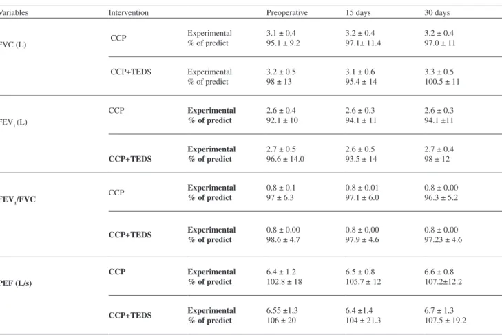
flow measurements (FVC, FEV1, FEV1/FVC and the PEF), volunteers treated with CCP and those treated with CCP+TEDS showed normal results in the three evaluations as shown in Table 1.
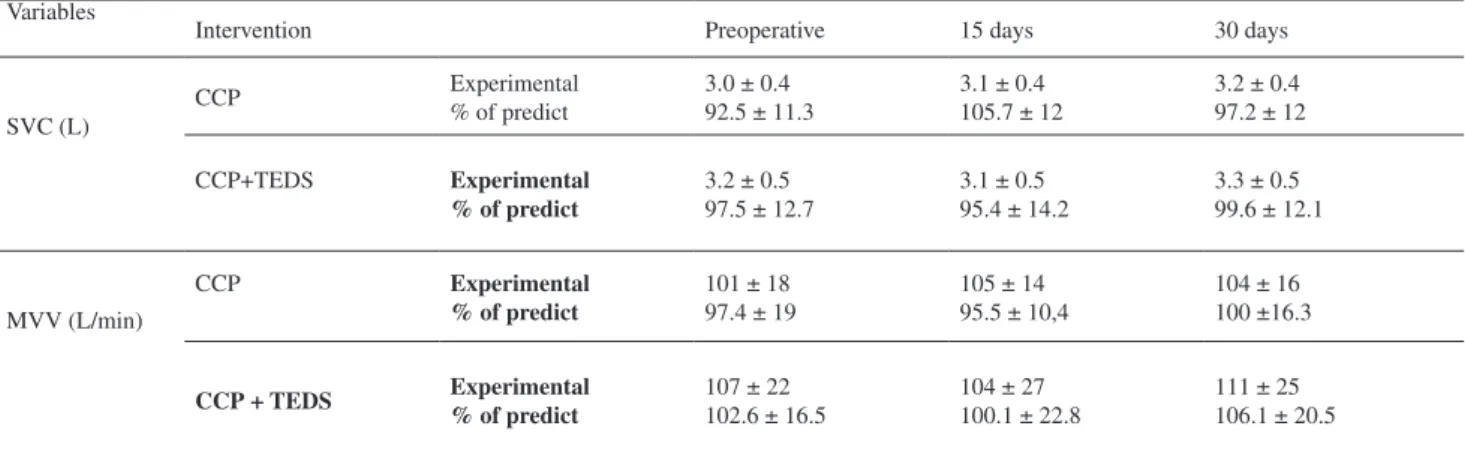
There were no significant differences in spirometric variables for the measurements of SVC and MVV between those that received CCP and those that received CCP+TEDS (Table 2).
As shown in Table 3, there were no significant differences in MIP amongst the three evaluations for either group. However, there was a significant decrease in MEP from the preoperative period to the 30th postoperative day in
the group that received only CCP. There was no change in MEP in the CCP+TEDS group.
DISCUSSION
There were no obstructions in the airways or thorax-pulmonary restrictions that could interfere with the results. Thus, the lack of evidence of spirometric differences
between the groups gives more reliability to the inclusion criteria as well as to the homogeneity of the sample used.
Because there were no significant differences between groups and because of the various evaluations of both groups studied, both treatments probably contributed to the prevention of decreases in pulmonary function parameters that are normally quoted in the literature.4,26 However, it is
necessary to emphasize that these authors did not use either CCP or CCP+TEDS in their studies.
Rubinstein et al.27 found limitations in respiratory flow
to between 50% and 75% of the vital capacity in obese patients. Although these authors either did not exclude or declined to mention that they did exclude patients with co-morbidities and/or with respiratory dysfunctions, volunteers with pulmonary dysfunction were excluded from the present study to avoid interference in the results.
Some authors such as Luce28 have shown decreases in
expiratory reserve volume, functional residual capacity (FRC) and tidal volume (TV) that have resulted in altered ventilation and perfusion or even shunts, causing subsequent
Table 1 - Means, standard deviations and statistical results for the pulmonary flow measurements: forced vital capacity (FVC), forced exhaling volume in the first second (FEV1), FEV1/FVC and the peak of exhaling flow (PEF) for both the experimental and predicted values, for the group treated with conventional chest physiotherapy (CCP) and the group treated with CCP + transcutaneous electric diaphragmatic stimulation (TEDS), in three evaluations.
Variables Intervention Preoperative 15 days 30 days
FVC (L) CCP
Experimental % of predict
3.1 ± 0,4 95.1 ± 9.2
3.2 ± 0.4 97.1± 11.4
3.2 ± 0.4 97.0 ± 11
CCP+TEDS Experimental
% of predict
3.2 ± 0.5 98 ± 13
3.1 ± 0.6 95.4 ± 14
3.3 ± 0.5 100.5 ± 11
FEV1 (L)
CCP Experimental
% of predict
2.6 ± 0.4 92.1 ± 10
2.6 ± 0.3 94.1 ± 11
2.6 ± 0.3 94.1 ±11
CCP+TEDS
Experimental % of predict
2.7 ± 0.5 96.6 ± 14.0
2.6 ± 0.5 93.5 ± 14
2.7 ± 0.4 98 ± 12
FEV1/FVC
CCP Experimental
% of predict
0.8 ± 0.1 97 ± 6.3
0.8 ± 0.01 97.1 ± 6.0
0.8 ± 0.00 96.3 ± 5.2
CCP+TEDS Experimental
% of predict
0.8 ± 0.00 98.6 ± 4.7
0.8 ± 0,00 97.9 ± 4.6
0.8 ± 0.00 97.23 ± 4.6
PEF (L/s)
CCP Experimental
% of predict
6.4 ± 1.2 102.8 ± 18
6.5 ± 0.8 105.7 ± 12
6.6 ± 0.8 107.2±12.2
CCP+TEDS Experimental
% of predict
6.55 ±1,3 106 ± 20
6.4 ±1.4 104 ± 21.3
hypoxemia in obese individuals; these alterations probably only affected obese patients with pulmonary dysfunction.
The present results are in agreement with the findings of Silva et al.22, who studied 26 patients given CCP and
found no spirometric alterations either in the preoperative period or on the 14th and 30th days after gastric bypass.
Rigatto29 and Barros30 concluded that during weight loss
subsequent to surgery, there was a decrease in respiratory muscular strength that was related to reduced lean mass and led to a reduced thickness of the diaphragm muscle after two weeks on a hypocaloric diet. It was also concluded that the obese individuals only completed the organization and adaptation process of the ventilatoryparameters six months after surgery. However, the present results demonstrated that treatment with CCP did not differ from treatment with CCP+TEDS in the maintenance of inhalation muscle strength. In addition, it was also shown that the expiratory muscle strength was not maintained after 15 and 30 postoperative days in the group treated with CCP, but was maintained in the group treated with CCP+TEDS (Table 3).
This confirms that CCP+TEDS is an important preventative measure during the postoperative period after bariatric surgery.
Despite the loss or maintenance of respiratory muscle strength, Rovina, Bouros and Tzanakis11 postulated that
reflexive inhibition of the phrenic nerve could cause diaphragmatic dysfunction. This suggests that the decrease in MEP during the postoperative period after open abdominal surgery occurred because the abdominal muscles were cut during surgery, which consequently made it difficult to generate expiratory pressures. Joris et al.31 considered
the reflexive inhibition of the phrenic nerve to be a more probable cause of the dysfunction of the diaphragm muscle than a contractile collapse due to muscular trauma occurring during surgery. Nevertheless, we would like to suggest that CCP, especially in conjunction with TEDS, should be used as a preventive technique in the postoperative period after bariatric surgery.
Toledo and Garcia32 detected MIP values on the 14th
postoperative day that were very close to those obtained in
Table 2 -Means, standard deviations and statistical results for the measurements of slow vital capacity (SVC) and maximum voluntary ventilation (MVV) for both the experimental and predicted values, for the group treated with conventional chest physiotherapy (CCP) and the group treated with CCP + transcutaneous electric diaphragmatic stimulation (TEDS), in three evaluations.
Variables
Intervention Preoperative 15 days 30 days
SVC (L)
CCP Experimental
% of predict
3.0 ± 0.4 92.5 ± 11.3
3.1 ± 0.4 105.7 ± 12
3.2 ± 0.4 97.2 ± 12
CCP+TEDS Experimental
% of predict
3.2 ± 0.5 97.5 ± 12.7
3.1 ± 0.5 95.4 ± 14.2
3.3 ± 0.5 99.6 ± 12.1
MVV (L/min)
CCP Experimental
% of predict
101 ± 18 97.4 ± 19
105 ± 14 95.5 ± 10,4
104 ± 16 100 ±16.3
CCP + TEDS Experimental
% of predict
107 ± 22 102.6 ± 16.5
104 ± 27 100.1 ± 22.8
111 ± 25 106.1 ± 20.5 There was no statistical difference between any of the variables
Table 3- Means and standard deviations in values obtained and statistical results for the measurements of MIP and MEP for the group treated with conventional chest physiotherapy (CCP) and the group treated with CCP + transcutaneous electric diaphragmatic stimulation (TEDS), in three evaluations
Pressures Preoperative 15 days 30 days
MIP (cmH2O) CCP -84.3 ± 20.3 -77.9 ± 17.2 -77.9 ± 16
CCP+TEDS -84 ± 21.7 -81.8 ± 21.5 -87.7 ± 22.8
MEP (cmH2O) CCP 100.2 ± 18.1 87.9 ± 16. 9 * 85.9 ± 16.5 #
CCP+TEDS 95.4 ± 21.9 83.6 ± 17.6 87.2 ± 19.8
*difference significant between the 1st and 2nd evaluations (p
≤0,5) # difference significant between the 1st and 3rd evaluations (p
the preoperative period, but did not mention the maintenance of MEP in volunteers who received physiotherapy. The present results reinforce the importance of respiratory physiotherapy in postoperative patients of bariatric surgery. This is especially true when compared with results obtained by Rovina, Bouros and Tzanakis,11 who did not use
respiratory physiotherapy and detected decreased MIP values during the postoperative period after abdominal surgery, and similarly when compared with the results of Paisani et al,4 who found decreases in MIP and MEP values from the
preoperative period to the 5th postoperative day after bariatric
surgery.
While Silva et al.33 concluded that respiratory muscle
function was invariably damaged after open abdominal surgeries, the present study showed that pulmonary function and respiratory muscle strength were maintained in patients treated with CCP and those treated with CCP+TEDS 15 and 30 days after bariatric surgery, with special emphasis on the effect of TEDS on MEP.
Despite weight loss in patients who have undergone gastroplasty and the consequent deterioration in respiratory muscle strength,29,30 the two postoperative evaluations made
in this study were carried out fifteen and thirty days after patients were released from the hospital. The delay was used to prevent any deleterious effects occurring immediately
after surgery, such as pain or diaphragmatic paresis,11,31,33
from interfering with the variables under evaluation. Geddes, Voorhes & Lagler34 noted that positioning of
the electrodes in the parasternal region at the height of the xiphoid process resulted in the liberation of the electric current to the phrenic nerve, which, for its part, penetrated the diaphragm and produced a contraction. However, the application of high intensity currents also caused contraction of the abdominal muscles. In an attempt to produce good contraction during the application of TEDS, the abdominal muscles were probably also stimulated, and this may have led to the maintenance of their strength in the postoperative period. Gray & Field also reported stimulation of the abdominal muscles by placement of the electrodes in the parasternal region (xiphoid).35
Finally, the results of this study suggest that both CCP and CCP+TEDS promote the maintenance of pulmonary function and therefore show an indispensable preventive character following bariatric surgeries. It was also concluded that inspiratory muscle strength was maintained within normal parameters by both of the proposed therapies, but the addition of TEDS also preserved expiratory muscle strength, which is fundamental to the recovery of obese patients submitted to RYGB.
REFERENCES
1. Brazilian Multi-society Consensus on obesity surgery – Salvador, 2006. 2. Latin American Consensus on Obesity . Rio de Janeiro, Oct 10, 1998. 3. Ladosky W, Botelho MAM, Albuquerque Jr. JP. Chest mechanics
in morbidly obese non-hypoventilated patients. Respirat Med. 2001;95:281-6.
4. Paisani DM, Chiavegatto LD, Faresin SM. Volumes, pulmonary capacities and respiratory muscle strength in postoperative gastroplasty. J Bras Pneum. 2005;31:125-32.
5. Hamoui N, Anthone G, Crookes PF.The Value of Pulmonary Function Testing Prior to Bariatric Surgery. Obesity Surgery. 2006;16:1570-3. 6. Koenig SM. Pulmonary complications of obesity. Am J Med Sci.
2001;321:249-79.
7. Segal A, Fandiño J. Indications & contra-indications for carrying out bariatric surgery. Rev Bras Psiquiatr. 2002;24;68-72.
8. Ogunnaike, BO, Jones SB, Jones DB, Provost D, Whitten C. Anesthetic considerations for bariatric surgery. Anesth Analg. 2002;95:1793-805. 9. Eichenberger A, Proietti S, Wicky S, Frascarolo P, Suter M, Spahn DR, et al. Morbid obesity and postoperative pulmonary atelectasis: an underestimated problem. Anesth Analg. 2002, n.95, p.1788-92. 10. Olsén, MF, Lönroth, H, Bake B,Effects of breathing exercises on
breathing patterns in obese and non-obese subjects. Clinical Physiology. 1999;19:251-7.
11. Rovina N, Bouros D, Tzanakis N, Velegraks M, Kandilakis S, Vlasserou F, et al. Effects of laparoscopic cholesystectomy on global respiratory muscle strength. Am J Respir Crit Care Med. 1996;153:458-61. 12. Chiavegato DL, Jardim JR, Faresin SM, Functional respiratory alterations
in laparoscopic cholesystectomy. J. Bras Pneumol. 2000;26:69-76. 13. Celli Br, Rodriguez Ks, Snider Gl. A controlled trial of intermittent
positive pressure breathing, incentive spirometry, and deep breathing exercises in preventing pulmonary complications after abdominal surgery. Am. Rev. Respir. Dis. 1984;130:12-15.
14. Sarnoff SJ, Maloney JV, Sarnoff IC. Eletrophrenic respiration in acute bulbar poliomyelitis. J Am Med Assoc.1950;143:1383-90.
15. Sarnoff SJ, Sarnoff CS, Whittenberger JL. Eletrophrenic respiration, Surg Gyn Obst. 1951;93:190-6.
16. Goldenthal, S. Bilateral and unilateral activation of the diaphragm in the intact human. External electrical stimulation by capacitive coupling as recorded by cineradiography. Conn Med. 1961;25:236-38. 17. Geddes LA, Voorhees WD, Babbs CF. Electroventilation. Amer Journ
Emer Med, 1985;3:337-9.
19. Thomas JA, Mcintosh MJ, Dean E. Are incentive spirometry, intermittent positive pressure breathing, and deep breathing exercises effective in the prevention of postoperative pulmonary complications after upper abdominal surgery? A systematic overview and meta-analysis. Phys Ther.1994;74:1-9.
20. Olsén MF, Hahn I, Nordgren S, Lönroth H, Lundholm K. Randomized controlled trial of prophylactic chest physiotherapy in major abdominal surgery. Br J Surg. 1997;84:1535-8.
21. Mackay MR, Ellis E. Physiotherapy outcomes and staffing resources in open abdominal surgery patients Physiother Theory Pract. 2002;18:75-93.
22. Silva AMO, Boin IFS, Pareja JC, Magna LA. Analysis of respiratory function in obese patients submitted to fobi-capella surgery.Rev. Col. Bras.Cir. 2007; 34:314-20.
23. ATS\ERS. Task Force: Standardisation of lung function testing. Standardisation of Spirometry. Eur Respir J. 2005;26:319-38. 24. Pereira CAC. Directives for pulmonary function tests. J Pneumol
2002;28 Supl 3:1-82.
25. Black LF, Hyatt RE. Maximal respiratory pressures: normal values and relationship to age and sex.Am Rev Respir Dis. 1969;103:641-50. 26. Geddes LA, Voorhees WD, Bourland CE, Riscilli CE.Optimum stimulus
frequency for contracting the inspiratory muscle with chest-surface electrodes to produce artificial respiration. Rev Ann Biomed Eng. 1990;18:103-8.
27. Ramos GC, Pereira ENSG, Oliveira EC. Evaluation of pulmonary function after laparoscopic and conventional cholesystectomies. Rev. Col. Bras. Cir. 2007;34:326-30.
28. Rubisntein I, Zamel N, DuBarry L, Hoffstein V. Airflow limitation in morbidly obese nonsmoking men. Ann Intern Med. 1990;112:828-32. 29. Luce JM. Respiratory Complication of Obesity. Chest.1980;78:626-31. 30. Rigatto AM. Ventilatory performance in obesity. Saúde em Rev.
2005;17:57-62.
31. Barros SE, Moura LSA, Carvalho MJC. Correlation between maximum respiratory pressures and the nutritional status of patients with lung disease. RevBras Nutr Clin. 2003;18:123-9.
32. Joris JL, Hinque VL, Laurent PE, Desaive CJ, Lamy ML. Pulmonary function and pain after gastroplasty performed via laparotomy or laparoscipy in morbidly obese patients. BR J Anaesth. 1998;80:283-8. 33. Toledo RC, Garcia RC. Complicações pulmonares e alterações na mecânica respiratória e na ventilometria em pós-operatório de gastroplastia / Pulmonary complications and alterations in the respiratory mechanics and ventilometry in post-operative gastroplasty.Reabilitar. 2005;29:32-40.
34. Geddes LA, Voorhees WD, Lagler R, Riscili C, Foster K, Bourland JD. Electrically produced artificial ventilation. Med Instrum. 1988;22:263-71.