PESQUIS
A
ORIGINAL
Correspondence to: Rodrigo Pegado de Abreu Freitas – Faculdade de Ciências da Saúde do Trairí – Universidade Federal do Rio Grande do Norte – Rua Trairí, s/n – Centro – CEP: 59200-000 – Santa Cruz (RN), Brazil – E-mail: [email protected]
ABSTRACT | This study aimed to investigate if there are
differences between the associated and isolated therapies from the laser and micro current on healing of burn wound healing in rats. A total of 40 male rats were randomly allocat-ed into four groups: control group (CG), micro current group (MG), laser group (LG) and laser/micro current group (LMG), treated with associated laser and micro current. Thermal damage was done on the back of the animal and a total of ten days therapy was performed. After treatment samples were taken from the lesions to perform semi quantitative his-topathological study using Hematoxylin Eosin and Masson Trichrome. The Kruskal-Wallis and Dunn’s Test were used for statistical analyses. We observed a significant difference between groups for production of fibroblasts (p=0.0003), collagen (p=0.0153), neoangiogenesis (p=0.0031) and skin annexes (p=0.0004). In semi-quantitative histological analy-sis, the LMG showed lower values in presence of collagen, fibroblasts and number of skin appendages, only for neo-angiogenesis, the associated therapy showed similar values to single modality therapy groups. Laser and microcurrent have beneficial effects on tissue healing. However, it is sug-gested that the association of these two therapies reduces the effectiveness of the treatment when compared to single mode treatment.
Keywords | combined modality therapy; lasers; electric stimulation; wound healing.
Low-level laser therapy and micro current in burn
wound healing in rats. Associated or isolated therapy?
Laserterapia e microcorrente na cicatrização de queimadura em ratos. Terapias
associadas ou isoladas?
Laserterapia y microcorriente en la cicatrización de quemaduras en ratas. ¿Terapias
asociadas o aisladas?
Rodrigo Pegado de Abreu Freitas1, Ana Paula Medeiros de Barcelos2, Brenda Medeiros da Nóbrega2,
Aline Barbosa Macedo3, Anderson Rodrigues de Oliveira4, Ana Maria de Oliveira Ramos5, Wouber
Hérickson de Brito Vieira6
Study conducted at the Physical therapy program at Universidade Potiguar (UNP) – Natal (RN), Brazil.
1Physical therapist; MSc, professor of Physical therapy at Universidade Federal do Rio Grande do Norte (UFRN) – Santa Cruz (RN), Brazil. 2Physical therapist graduated at UNP – Natal (RN), Brazil.
3MSc in Physical therapy, Universidade Metodista de Piracicapa (UNIMEP) – Piracicaba (SP), Brazil. 4Physical therapist; Professor with Specialization at Universidade Estácio de Sá – Natal (RN), Brazil. 5Doctor; PhD, professor at the post-graduate program in Health Sciences at UFRN – Natal (RN), Brazil. 6Physical therapist; PhD, professor of Physical therapy at UFRN – Natal (RN), Brazil.
RESUMO | Este estudo teve o objetivo de investigar se
INTRODUCTION
In physiotherapy, several resources are being used to accelerate and improve the quality of the regenerative process, such as lasers, microcurrents, ultrasounds, and ultraviolet radiation1-3. hese resources accelerate the
healing process, acting upon the sequence of physi-ological and biochemical events of this process through inlammation, collagen synthesis, formation of granu-lation tissue, and re-epithelization1,2.
hrough the analysis of cellular structures that are activated and/or inhibited by these resources, studies2,4,5
performed on laboratory animals have contributed to the elucidation of which physiotherapy resources might promote a regenerative process of quality. he therapeu-tic efects have been attributed to the interaction be-tween external energetic stimuli and the biological tis-sue (biostimulation), promoting an increase in cellular activities during the healing process.5,6
Among the modalities used, the low-intensity laser therapy is highlighted6-8; it shortens the time for
remod-eling, and improves the quality of the tissue in neofor-mation1,6. he basic biological mechanism promoted by
this eletrophysical resource seems to be the absorption of red and infrared light by chromophores contained in the protein components of the respiratory chain located in the mitochondria, which, in turn, initiate a torrent of biochemical events upon absorbing energy, and this results in an increase of enzymatic activity, production
of triphosphate adenosine, protein synthesis, cellular proliferation, deposition, and collagen organization9-10.
Other authors11-12 suggest that the electric
stimula-tion by microcurrents also accelerates ATP synthesis, has antioxidant efect, stimulates transmembrane trans-port, and reestablishes tissue bioelectricity, promoting reduction of the inlammatory process, pain relief, and healing acceleration. In this way, the modality would aim at normalizing the low of the currents, which may be interrupted when tissue lesion occurs due to burning13-14.
hus, laser and microcurrent therapies have positive efects on the acceleration of the healing process when used separately. In physiotherapists’ clinical practice, the associated use of these therapies as a way of potenti-ating the efects mentioned above has been observed. However, few studies describe this associated efect, and, in this way, the real results have yet to be discussed. Studies7,15 describe that when multi-therapy is used in
healing processes, the beneicial efects might neutral-ize each other. Among the several experimental models, our study used one of thermic lesion in rats because of the possibility of easily applying the therapies, and of assessing all the necessary histological parameters for the comparison of the treatments.
herefore, the aim of the present study was to in-vestigate if there are diferences between associated and isolated laser and microcurrent therapies in tissue heal-ing in a model of burn wounds in rats.
recursos parece diminuir os efeitos do tratamento quando se comparam os grupos de modalidade única.
Descritores | terapia combinada; lasers; estimulação elétrica; cicatrização.
RESUMEN | Este estudio tiene el objetivo de investigar si hay
diferencias entre las terapias asociadas y aisladas del láser y mi-crocorrientes en la reparación de lesión por quemadura en ra-tas. Un total de 40 animales fueron divididos aleatoriamente en cuatro grupos: grupo control (GC), grupo microcorriente (GM), grupo láser (GL) y grupo láser/microcorriente (GLM), tratados con láser asociado a microcorrientes. Después de inducidas las lesiones térmicas en el dorso del animal, fueron realizados en total diez días de tratamiento. Las muestras de tejido fueron recolectadas para el estudio histopatológico semicuantitativo usando Hematoxilina Eosina y Tricómico de Masson. Fueron utilizados los tests de Kruskal-Wallis y post-hoc de Dunn’s. Hubo diferencia significativa entre los grupos para la producción de
fibroblastos (p=0,0003), colágeno (p=0,0153), neoangiogénesis
(p=0,0031) y anexos cutáneos (p=0,0004). En el análisis histoló-gico semicuantitativo, el GLM presentó valores menores en los
parámetros histológicos de presencia de colágeno, número de
fibroblastos y anexos cutáneos (p<0,05) en relación a las tera-pias aisladas, excepto para la neoangiogénesis, cuyos valores
de la terapia asociada fueron semejantes a los grupos de terapia
con modalidad única. A pesar de que el láser y la microcorriente
de forma aislada tienen efectos benéficos para la cicatrización del tejido, la asociación de las modalidades parece haber
dismi-nuido la acción de la reparación. El láser y las microcorrientes
son efectivos en acelerar el proceso de reparación del tejido. Sin embargo, se sugiere que la asociación de estos recursos parece
disminuir los efectos del tratamiento cuando son comparados
con los grupos de modalidad única.
Palabras clave | terapia combinada; Terapia laser de baja
MATERIAL AND METHODS
Forty Wistar rats (Rattus norvegicus) were used, weighing between 250 and 300 grams, chosen random-ly, provided by the UNP bioterium. Random allocation into four groups was performed (n=10): microcurrent group (MG), laser group (LG), control group (CG), and laser/microcurrent group (LMG). All rats were submit-ted to the same environment and biological day/night cycle with 10 to 12 hours of controlled exposure to light, temperature and illumination, humidity maintained by air conditioning, and minimum noise. he animals re-mained in individual polypropylene cages lined with laboratory-grade pine shavings as bedding, and received Labina® chow and water. he therapies were performed in the afternoon (2 to 5 pm). For the four groups, the procedures were performed in the following sequence: (a) administering of dissociative anesthetic Zoletil® with dosage of 50 mg/kg intramuscularly in the quad-riceps muscle; (b) trichotomy on the animal’s back; (c) induction of a second-degree burn wound2 with a
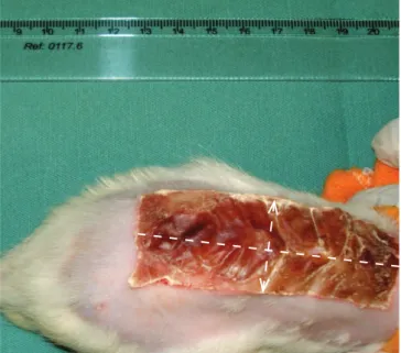
6x3 cm aluminum plate pressured against the animal’s back 4 cm away from the skull base during 10 sec-onds; the plate was previously warmed in becker with water at 100°C for a period of 10 minutes, as used by Meyerholtz et al.2 and Meireles et al.16 (Figure 1). he
proposed treatments were initiated immediately after the lesions, and performed daily during 10 days. he CG was submitted to the same experimental protocol, with exception of the exposition to the treatments.
For the application of laser, the device Photon Laser III of the brand DMC® of São Carlos, SP, was utilized; it carries the following features: visible laser (AlGaInP) in the 660 nm range, continuous mode, 30 mW power, 10 J/cm2 dosage, and energy of 0.3 J per application
spot during 9 seconds per spot inside the burn wound. In the region adjacent to the wound (borders) the same power was used but with dosage of 12 J/cm2 and energy
of 0.33 J per spot during 11 seconds. he application was performed through spot technique in direct contact with the wound, where the probe was positioned with light pressure at an angle of 90 degrees. he interval of 1.5 cm between the spots was respected, totalizing 6 spots on the burn wound and 14 spots in its adjacent region, totalizing 20 spots per animal. For the microcur-rent, the device Physiotonus Microcurrent Stimulator of the brand Bioset® was used through the application of a square monophasic pulsated current of reversible polarity each 2.5 seconds, with an intensity of 160 μA and frequency of 60 Hz during 15 minutes through two
adhesive silicon electrodes (Valutrode®) with 3.2 cm2
of diameter positioned at the extremities of the wound. For the MLG group, laser therapy was performed irst, and then the microcurrent treatment. he two in-struments were previously calibrated and warranted by their suppliers.
his study was approved by the Research Ethics Committee of the Potiguar University (UNP), ile number 062/2008.
Histology
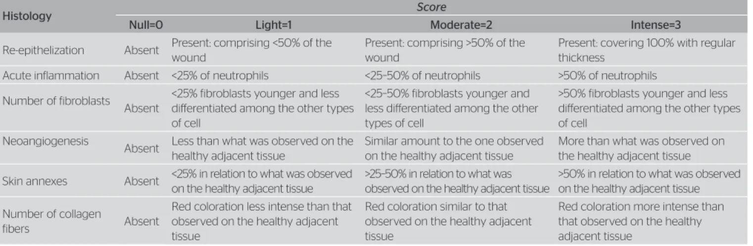
After 24 hours from the last therapy application, the animals were sacriiced in a closed chamber with lib-eration of CO2. In the instant following the sacriice, the biopsy of the skin tissue was performed 7 cm away from the skull base for the purposes of a histologi-cal study, including the wound in its healing process, the wound border, and part of the skin adjacent to the wound border. he samples were ixated in formalin, inserted in parain blocks, taken to the microtome, and cut in sections of 5 µm of thickness. he sagittal sections were kept in a drying chamber, and the cuts were posteriorly submitted to coloration by hematox-ylin-eosin and Masson’s trichrome. he tissue analysis was performed by a blind evaluator with the use of a Nikon® (Nikon, Tokyo, Japan) optical microscope. All the criteria applied in the semi-quantitative histologi-cal analysis16-17 were veriied through scores in a scale
from 0 to 3 (Table 1).
Figure 1. Burn model used. Adapted from Meireles et al.16 Wound in the
Statistical analysis
For the data analysis the software IBM SPSS® 19 and the GraphPad Prism® 5 were used. For the compari-son of the intergroup non-parametrical averages, the Kruskal Wallis and the Dunn’s post-test were used. We considered p<0.05 signiicant.
RESULTS
On the tenth day after the burn wound was inlicted, the appearance of the skin lesion in the LG, MG, and MLG pointed to loss of epidermis and hypodermis, representing a second-degree burn2, with moderate
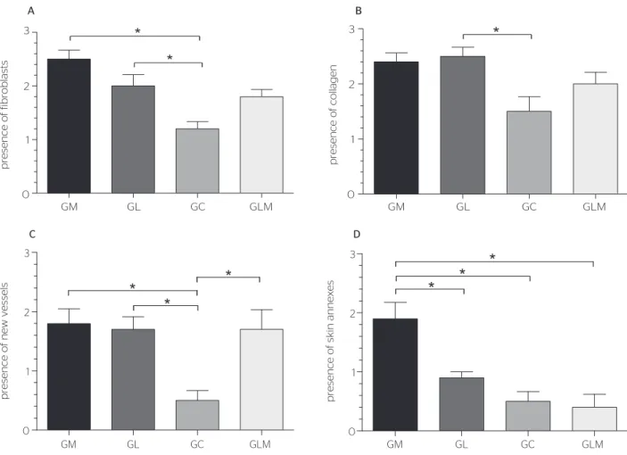
acute inlammatory reaction but with a clean and un-infected wound. here was no signiicant diference among the groups in relation to epithelial regeneration (p=0.0568), and inlammatory process (p=0.9640). We observed a signiicant diference among the groups in the number of ibroblasts (p=0.0003), collagen (p=0.0153), neoangiogenesis (p=0.0031), and skin an-nexes (p=0.0004).
For an increase in the presence of ibroblasts, the application of only one of the modalities was more ef-icient than associated therapy (Figure 2A). he MG did not present any diference to the MLG in relation to the presence of collagen ibers (Figure 2B). here was a signiicant increase in neoangiogenesis in all groups treated in comparison to CG (Figure 2C); however, the associated therapy (MLG) did not present a signiicant diference when compared to the therapies applied sep-arately (LG and MG). he MG registered signiicant improvement in comparison to others groups in relation
to the presence of skin annexes (Figure 2D). he associ-ated therapy had similar values to the groups of single-modality therapy only in relation to neoangiogenesis.
DISCUSSION
hrough the use of a burn model on Wistar rats, it was observed that, when applied in association, the visible laser AlGaInp (660 nm and 30 mW power) and the microcurrent (160 μA and 60 Hz frequency) promot-ed signiicant improvement only in the formation of new blood vessels in comparison to the single-modal-ity therapy. In all the other parameters evaluated, the individual use of one of the isolated therapies was bet-ter (ibroblasts, collagen, and skin annexes) than the joint therapy.
Evidence suggests that the use of red or infrared wave lengths in a series of dosage parameters (median of 4.2 J/cm2), including the ones used in the present
study, results in signiicant beneits on the healing of wounds in animal models and pathological processes in humans18,19. he use of laser in diferent wave lengths
is capable of accelerating epidermal formation, increas-ing the thickness of epidermal layers, and promotincreas-ing neovascularization and reorganization of collagen
i-bers1,2,20-25. he result of the treatment varies according
to the parameters; the visible laser is utilized more often for being more supericial and for interacting specii-cally with supericial chromophores, adapting itself to the treatment of epithelial lesions18,19,23.
Microcurrent therapy is also an eicient resource in the healing process13-15. In their study of the
heal-ing process in guinea pigs with the use of an electric
Table 1. Criteria for histological analysis
Adapted from Meireles et al.16 and Iordanou et al.17.
Histology Score
Null=0 Light=1 Moderate=2 Intense=3
Re-epithelization Absent Present: comprising <50% of the wound
Present: comprising >50% of the wound
Present: covering 100% with regular thickness
Acute inflammation Absent <25% of neutrophils <25–50% of neutrophils >50% of neutrophils
Number of fibroblasts Absent
<25% fibroblasts younger and less diferentiated among the other types of cell
<25–50% fibroblasts younger and less diferentiated among the other types of cell
>50% fibroblasts younger and less diferentiated among the other types of cell
Neoangiogenesis
Absent Less than what was observed on the healthy adjacent tissue
Similar amount to the one observed on the healthy adjacent tissue
More than what was observed on the healthy adjacent tissue
Skin annexes Absent <25% in relation to what was observed on the healthy adjacent tissue
>25–50% in relation to what was observed on the healthy adjacent tissue
>50% in relation to what was observed on the healthy adjacent tissue
Number of collagen
fibers Absent
Red coloration less intense than that observed on the healthy adjacent tissue
Red coloration similar to that observed on the healthy adjacent tissue
current of 50 μA, Agne et al.24 described an increase
in ibroblasts and inlammatory cells migration, and a greater alignment of collagen ibers, which contributed to healing. Using a microcurrent of 50 μA in the treat-ment of burn wounds in rats, Santos et al.5 observed a
number of ibroblasts and collagen superior to that of the control group. With microcurrents of 300 μA for 30 minutes/day, Demir et al.15, observed improvement
in cell proliferation and maturation, which stimulated ibroblast growth. hese discoveries, positive in rela-tion to the number of ibroblasts and the increase in the amount of collagen ibers, were also veriied in our research. In vitro studies suggest that a microcurrent of 100 μA and laser promote the migration26 and
prolif-eration23 of human dermal ibroblasts.
Laser and microcurrent represent an excellent ther-apy target in the promotion of neoangiogenesis during the healing process. he endothelial cells of the micro vessels seem to be sensitive to laser stimulation through the expression of gene proteins that regulate the cell cycle and the proliferation of these cells27. Bai et al.28
describe that electric ields of 150 to 400 mV/mm also perform migration, reorientation, and extension of the endothelial cells of micro-circulation vessels.
he endothelial cells of micro-circulation present diferent behaviors when compared to macrovascular tissues, which suggests that each cellular type has a dis-tinct disposition of receptors, and tolerance to diferent electric ields, contributing or not contributing to the activation of the vascular endothelial growth factor28.
Even with the positive action of laser and micro-current upon the healing process in the various model types, such as skin1, diabetic ulcers6, and temperature
burns5,16,24, their associated use still deserves more
discussion.
Gum et al.7 report the idea that the combined
ther-apy might prompt an overdose of stimuli upon the cells, which leads to the annulment of therapeutic efects. In their study with laser and microcurrent, an improve-ment in the strength, elasticity, tension, and maximum efort in rabbit tendons was observed. he improve-ments brought by multi-therapy were consistent but less *Significant when p<0.05. MG: Microcurrent group; LG: laser group; CG: control group; MCG: laser/microcurrent group.
Figure 2. Intergroup comparison of the histological variants through Dunn’s post-test
* *
*
*
*
*
*
*
*
GM GL GC GLM
0 1 2 3
GM GL GC GLM
presence of collagen
presence of skin annexes
presence of fibroblasts
presence of new vessels
0 1 2 3
B
GM GL GC GLM
0 1 2 3
GM GL GC GLM
0 1 2 3
D A
noticeable when compared to protocols of single mo-dality. It is possible that the electric stimulation might have hampered the occurrence of cellular and molecular reactions involved in the healing process, such as the gene expression of cellular growth factors, errors in the process of cell diferentiation, and alteration in the be-havior of receptors and ionic channels. Considering that these act upon the cellular metabolism, we point to the hypothesis of cellular fatigue, and alterations in cellular signalization or in the metabolic ways of the cells27,28.
Laser and microcurrent seem to act directly upon the expression of cellular growth factors in several types of cell (ibroblast, vascular endothelium, epithelial cells) related to the healing process. However, each type seems to possess a certain threshold (necessary dosage for positive efects) and tolerance (maximum dose to produce positive efects) to energetic stimuli.
Although laser and microcurrent are beneicial to tissue healing when used separately, their combi-nation seems to decrease therapeutic action. he re-sults recommend attention during the treatment of dermal burn lesions, and the suggestion of a therapy with these modalities used independently might be the best course of action. he biophysical and cellular action mechanisms that involve the combined use of therapeutic resources deserve broader investigation in order to obtain a more complete explanation of the phenomena analyzed.
CONCLUSION
his study concludes that, when applied separately, laser and microcurrent accelerate the healing process of burn wounds. However, when associated, they promote an improvement in neoangiogenesis only, and do not present signiicant improvement of the epithelial regen-eration, the inlammatory process, collagen, ibroblasts, and skin annexes. We suggest that the association of both resources decreases the efects of treatment when compared to the single-modality groups.
ACKNOWLEGEMENTS
We thank UNP for the use of its venue, and DMC® Importation and Exportation of Equipment Ltd., from São Carlos (SP), for the donation of the laser device.
REFERENCES
1. Maiya GA, Kumar P, Rao L. Efect of low intensity helium-neon (He-Ne) laser irradiation on diabetic wound healing dynamics. Photomed Laser Surg. 2005;23(2):187-90.
2. Meyerholz DK, Piester TL, Sokolich JC, Zamba GK, Light TD. Morphological parameters for assessment of burn severity in an acute burn injury rat model. Int J Exp Pathol. 2009;90(1):26-33. 3. Okuni I. Phototherapy in rehabilitation medicine. Masui. 2012;
61(7):700-5.
4. Hussein AJ, Alfars AA, Falih MA, Hassan AN. Efects of a low level laser on the acceleration of wound healing in rabbits. N Am J Med Sci. 2011;3(4):193-7.
5. Santos VNS, Ferreira LM, Horibe EK, Duarte IS. Electric microcurrent in the restoration of the skin undergone a trichloroacetic acid peeling in rats. Acta Cir Bras. 2004;19(5):466-70.
6. Reddy GK, Stehno-Bittel L, Enwemeka CS. Laser photostimulation accelerates wound healing in diabetic rats. Wound Repair Regen. 2001;9(3):248-55.
7. Gum SL, Reddy GK, Stehno-Bittel L, Enwemeka CS. Combined ultrasound, electrical stimulation, and laser promote collagen synthesis with moderate changes in tendon biomechanics. Am J Phys Med Rehabil. 1997;76(4):288-96.
8. Stadler I, Lanzafame RJ, Evans R, Narayan V, Dailey B, Buehner N, et al. 830-nm irradiation increases the wound tensile strength in a diabetic murine model. Lasers Surg Med. 2001;28(3):220-6.
9. Karu TI, Kolyakov SF. Exact action spectra for cellular responses relevant to phototherapy. Photomed Laser Surg. 2005;23(4):355-61. 10. Karu TI, Pyatibrat LV, Kalendo GS. Photobiological modulation of cell
attachment via cytochrome c oxidase. Photochem. Photobiol Sci. 2004:3(2);211-6.
11. Konikof JJ. Electrical promotion of soft tissue repairs. Ann Biomed Eng. 1976;4(1):1-5.
12. Lee BY, Al-Waili N, Stubbs D, Wendell K, Butler G, Al-Waili T, et al. Ultra-low microcurrent in the management of diabetes mellitus, hypertension and chronic wounds: report of twelve cases and discussion of mechanism of action. Int J Med Sci. 2009;7(1):29-35. 13. Lee BY, Wendell K, Al-Waili N, Butler G. Ultra-low microcurrent therapy:
a novel approach for treatment of chronic resistant wounds. Adv Ther. 2007;24(6):1202-9.
14. Cheng N, Van Hof H, Bockx E, Hoogmartens MJ, Mulier JC, De Dijcker FJ, et al. The efects of electric currents on ATP generation, protein synthesis, and membrane transport in rat skin. Clin Orthop Relat Res. 1982;171:264-72.
15. Demir H, Balay H, Kirnap M. A comparative study of the efects of electrical stimulation and laser treatment on experimental wound healing in rats. J Rehabil Res Dev. 2004;41(2):147-54.
16. Meireles GC, Santos JN, Chagas PO, Moura AP, Pinheiro AL. Efectiveness of laser photobiomodulation at 660 or 780 nanometers on the repair of third-degree burns in diabetic rats. Photomed Laser Surg. 2008;26(1):47-54.
17. Iordanou P, Lykoudis EG, Athanasiou A, Koniaris E, Papaevangelou M, Fatsea T, et al. Efect of visible and infrared polarized light on the healing process of full-thickness skin sounds: an experimental study. Photomed Laser Surg. 2009;27(2):261-7.
19. Peplow PV, Chung TY, Baxter GD. Photodynamic modulation of wound healing: a review of human and animal studies. Photomed Laser Surg. 2012;30(3):118-48.
20. Güngörmüş M, Akyol U. The efect of gallium-aluminum-arsenide 808-nm low-level laser therapy on healing of skin incisions made using a diode laser. Photomed Laser Surg. 2009;27(6):895-9. 21. Reddy GK, Gum S, Stehno-Bittel L, Enwemeka CS. Biochemistry
and biomechanics of healing tendon: part II. Efects of combined laser therapy and electrical stimulion. Med Sci Sports Exerc. 1998;30(6):794-800.
22. Zhao M, Bai H, Wang E, Forrester JV, McCaig CD. Electrical stimulation directly induces pre-angiogenic responses in vascular endothelial cells by signaling through VEGF receptors. J Cell Sci. 2004;117(Pt 3):397-405. 23. Enwemeka CS, Parker JC, Dowdy DS, Harkness EE, Sanford LE,
Woodruf LD. The eficacy of low-power lasers in tissue repair and pain control: a meta-analysis study. Photomed Laser Surg. 2004;22(4):323-9.
24. Agne JE, Lorenzini S, Bechman L, Hamerski Romero C, Casagrande R. Uso de microcorrientes en ratones Wistar con úlceras diabéticas: resultados histológicos. Fisioterapia. 2004;26(3):164-9.
25. Kloth LC. Electrical stimulation for wound healing: a review of evidence from in vitro studies, animal experiments, and clinical trials. Int J Low Extrem Wounds. 2005;4(1):23-44.
26. Sugimoto M, Maeshige N, Honda H, Yoshikawa Y, Uemura M, Yamamoto M, et al. Optimum microcurrent stimulation intensity for galvanotaxis in human fibroblasts. J Wound Care. 2012;21(1):5-6, 8,10.
27. Feng J, Zhang Y, Xing D. Low-power laser irradiation (LPLI) promotes VEGF expression and vascular endothelial cell proliferation through the activation of ERK/Sp1 pathway. Cell Signal. 2012;24(6):1116-25.