fistulas ranges from 0 to 28%,(1,2) and the mortality rate ranges from 16 to 72%.(3) Non-neoplastic tracheoesophageal fistulas can be congenital,(4,5) iatrogenic(6) or related to thoracic trauma. Once such a fistula has been diagnosed, surgical closure is formally indicated.
Introduction
Fistulas in the tracheobronchial tree have a multifactorial etiology and present a variable incidence in the literature. The related morbidity and mortality are high. Bronchopleural fistulas usually result from surgical procedures involving pulmonary resection; the incidence of these
Endoscopic treatment of tracheobronchial tree fistulas using
atrial septal defect occluders: preliminary results*
Tratamento endoscópico de fístulas da árvore traqueobrônquica com dispositivos para a correção de defeitos do septo interatrial: resultados preliminares
Paulo Rogério Scordamaglio, Miguel Lia Tedde, Hélio Minamoto, Carlos Augusto Cardoso Pedra, Fábio Biscegli Jatene
Abstract
Fistulas in the tracheobronchial tree (bronchopleural and tracheoesophageal fistulas) have a multifactorial etiology and present a variable incidence in the literature. In general, the related morbidity and mortality are high. Once such a fistula has been diagnosed, surgical closure is formally indicated. However, the clinical status of affected patients is usually unfavorable, which precludes the use of additional, extensive surgical interventions. In addition, attempts at endoscopic closure of these fistulas have seldom been successful, especially when the fistula is large in diameter. We report the cases of three patients submitted to endoscopic closure of fistulas, two of which were larger than 10 mm in diameter, by means of the insertion of atrial septal defect occluders. The procedure was minimally invasive, and the initial results were positive. The results indicate that this is a promising technique for the resolution of tracheobronchial tree fistulas.
Keywords: Bronchial fistula; Tracheoesophageal fistula; Bronchoscopy; Respiratory therapy.
Resumo
As fístulas da árvore traqueobrônquica, sejam elas broncopleurais ou traqueoesofágicas, apresentam etiologia multifatorial, com incidência variável na literatura. Em geral, apresentam alta morbidade e mortalidade, com indicação formal de correção cirúrgica. Entretanto, a condição clínica dos pacientes muitas vezes não permite uma reintervenção cirúrgica de grande porte. Além disso, as tentativas de fechamento endoscópico raramente têm sucesso, principalmente em fístulas de grande diâmetro. Relatamos os casos de três pacientes submetidos ao fechamento endoscópico de fístulas, sendo duas maiores que 10 mm, com a aplicação de dispositivos oclusores utilizados na cardiologia intervencionista, de forma minimamente invasiva e com resultados iniciais positivos. Esses dados sinalizam que essa pode ser uma técnica promissora na resolução de fístulas da árvore traqueobrônquica.
Descritores: Fístula brônquica; Fístula traqueoesofágica; Broncoscopia; Terapia respiratória.
* Study carried out at the University of São Paulo School of Medicine Hospital das Clínicas, São Paulo, Brazil.
Correspondence to: Paulo Rogério Scordamaglio. Rua Artur Prado, 449, apto. 11, Bela Vista, CEP 01322-000, São Paulo, SP, Brasil. Tel 55 11 3283-0657. E-mail: [email protected]
Financial support: None.
and open drainage of the residual pleural cavity was maintained. Bronchoscopy revealed that the fistula measured 12 mm, and a 15-mm atrial septal defect occluder (Occlutech Figulla; Occlutech GmbH, Jena, Germany) was therefore chosen. We began catheterization of the fistula using a polytetrafluoroethylene-coated guide wire and then advanced the occluder-sheath set to the bronchial stump, the distal disc being released in the pleural cavity and the proximal disc being released in the bronchial tree. There was an immediate reduction in air leak, and the patient was discharged from the hospital after 12 h. The evaluation performed after a follow-up period of 180 days revealed that granulation tissue had closed the fistula almost completely (Figure 1).
Case 2
A 69-year-old male patient presented with hypertension and atypical carcinoid tumor. The patient was a smoker (50 pack-years). The patient underwent right pneumonectomy. The patient developed a complete bronchial stump fistula of approximately 18 mm with severe air leak. An additional surgical intervention was ruled out due to surgical risk. A 20-mm atrial septal defect occluder (GORE-Helex; Gore, Flagstaff, AZ, USA) was therefore placed under general anesthesia, closing the fistula completely. There was clinical stabilization of the profile, and the patient was However, the clinical status of affected patients
is usually unfavorable, which precludes the use of additional, extensive surgical interventions.
In an attempt to avoid surgical closure, various endoscopic techniques have been described. However, endoscopic closure of these fistulas has seldom been successful, especially when the fistula is large in diameter.(7-12)
In the three cases described next, we report our initial experience in performing endoscopic closure of fistulas, one of which was a chronic tracheoesophageal fistula and two of which were complete bronchial stump fistulas, by means of the insertion of atrial septal defect occluders. This study was approved by the Research Ethics Committee of the University of São Paulo School of Medicine Heart Institute.
Case reports
Case 1
A 53-year-old male patient presented with hepatitis C, abdominal aortic aneurysm, carotid aneurysm, systemic arterial hypertension and grade II heart failure. The patient was a smoker (70 pack-years) and had a history of treated pulmonary tuberculosis and right upper lobe aspergilloma with hemoptysis.
The patient underwent right upper lobec-tomy and developed a bronchial stump fistula. Numerous attempts at closure were unsuccessful,
Case 3
A 73-year-old male patient presented with malnutrition, a history of severe acute pancrea-titis and a diagnosis of distal tracheoesophageal fistula after prolonged intubation. The patient also presented persistent cough, worsened by the ingestion of solids and liquids and by the change of decubitus. The patient had refused the initial indication for surgical closure, an option that was then discarded due to the high surgical risk.
Bronchoscopy revealed a tracheoesopha-geal fistula of approximately 5 mm in the distal trachea. We chose to place a 20-mm GORE-discharged from the hospital 5 days after the
procedure.
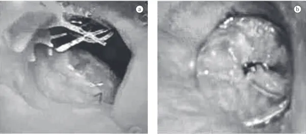
The evaluation performed after a follow-up period of 30 days revealed that 40% of the occluder had been covered by epithelium. On day 45, the device migrated to the pleural cavity. The patient, however, remained oligo-symptomatic. Bronchoscopy showed granulation tissue that reduced the diameter of the fistula to 7 mm. Another bronchoscopy, performed on day 75, revealed granulation tissue and repair with fibrosis, which reduced the diameter of the fistula to 4 mm and suggested that the healing process was still in progress despite the absence of the occluder (Figure 2).
Figure 2 - In a), complete stump fistula after right pneumonectomy. In b), appearance of the site immediately after the device had been placed.
We therefore used two devices that are employed in cardiac interventions and that are also widely used clinically.
The Occlutech Figulla device is composed of two nitinol discs that are sequentially released from the applicator. Thus, the distal disc can be placed at the extremity of the stump in the pleural cavity, whereas the proximal disc is released in the bronchial lumen; the connecting waist between the two discs becomes firmly attached to the fistula and avoids the displace-ment of the device.(13)
The GORE-Helex device consists of a single nitinol wire attached to a continuous tape of polytetrafluoroethylene that forms two hybrid discs in the shape of a spiral that, when released, attach themselves to each side of the fistula.(14) Because the GORE-Helex device is more malle-able than is the Oclutech Figulla device, we believed the former would be more appropriate to close the tracheoesophageal fistula for two reasons: first, because the two organs involved presented motility; and second, because we were concerned that a rigid device might widen the fistula.(14)
If our initial results are corroborated by future studies, this might be a promising tech-nique for the endoscopic treatment of fistulas in the tracheobronchial tree.
References
1. Taghavi S, Marta GM, Lang G, Seebacher G, Winkler G, Schmid K, et al. Bronchial stump coverage with a pedicled pericardial flap: an effective method for prevention of postpneumonectomy bronchopleural fistula. Ann Thorac Surg. 2005;79(1):284-8.
2. Kramer MR, Peled N, Shitrit D, Atar E, Saute M, Shlomi D, et al. Use of Amplatzer device for endobronchial closure of bronchopleural fistulas. Chest. 2008;133(6):1481-4. 3. Gursoy S, Yapucu MU, Ucvet A, Yazgan S, Basok O, Ermete
S. Fibrin glue administration to support bronchial stump line. Asian Cardiovasc Thorac Ann. 2008;16(6):450-3. 4. Watanabe T, Okuyama H, Kubota A, Kawahara
H, Hasegawa T, Ueno T, et al. A case of tracheal agenesis surviving without mechanical ventilation after external esophageal stenting. J Pediatr Surg. 2008;43(10):1906-8.
5. Gutiérrez C, López J, Barrios JE, Valdés E, Ayuso L, Cousello M, et al. Endoscopic treatment of recurrent tracheoesophageal fistula [Article in Spanish]. Cir Pediatr. 2008;21(3):130-4.
6. Oksuz H, Senoglu N, Zencirci B, Ezberci M, Yuzbasioglu MF. Pneumothorax, pneumomediastinum, tracheo-esophageal fistula presenting with endotracheal intubation in post-cesarean period: A case report. Cases J. 2008;1(1):134.
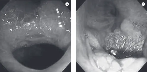
Helex atrial septal defect occluder. After having been released, however, the device did not lock properly into place, which resulted in flapping of its tracheal face during exhalation and led to accumulation of secretion. We decided to substi-tute the 20-mm occluder for a 15-mm occluder. The 20-mm occluder locked into place prop-erly, and the profile improved significantly. The patient was discharged from the hospital after 5 days, without cough (even during the inges-tion of liquids). The evaluainges-tion performed after a follow-up period of 120 days revealed that the esophagus was normal and that the disc placed in the esophagus had been completely incorpo-rated. Bronchoscopy showed epithelialization of the tracheal disc, which protruded partially toward the tracheal lumen. We decided to remove the occluder. Local repair and complete closure occurred after 7 days (Figure 3).
Discussion
One of the forms of endoscopic closure of fistulas in the tracheobronchial tree is the appo-sition of different materials with the purpose of forming a closing plug. However, it is difficult to securely affix these materials at the site of the fistula, and their displacement can, in addition to reopening the fistula, release a foreign body into the airway.
Endoscopic techniques have been used to inject different substances (alcohol, tetracycline, hypertonic glucose) in the submucosa, with the purpose of inducing inflammation and closing the fistula. Part of the problem with these tech-niques is that the aggression is not modulated and can result in ischemia and necrosis and therefore worsen the problem.
A recent study described the closure of a 5-mm bronchopleural fistula by means of a device used in the treatment of heart septal defects.(13)
Our objective is to use this type of device, the biocompatibility of which has been confirmed by its extensive use in cardiac interventions.
11. Lois M, Noppen M. Bronchopleural fistulas: an overview of the problem with special focus on endoscopic management. Chest. 2005;128(6):3955-65.
12. West D, Togo A, Kirk AJ. Are bronchoscopic approaches to post-pneumonectomy bronchopleural fistula an effective alternative to repeat thoracotomy? Interact Cardiovasc Thorac Surg. 2007;6(4):547-50.
13. Halabi A, Hijazi ZM. A new device to close secundum atrial septal defects: first clinical use to close multiple defects in a child. Catheter Cardiovasc Interv. 2008;71(6):853-6.
14. Pedra CA, Pedra SF, Esteves CA, Chamiê F, Ramos S, Pontes SC Jr, et al. Initial experience in Brazil with the Helex septal occluder for percutaneous occlusion of atrial septal defects. Arq Bras Cardiol. 2003;81(5):435-52. 7. Paul S, Talbot SG, Carty M, Orgill DP, Zellos L.
Bronchopleural fistula repair during Clagett closure utilizing a collagen matrix plug. Ann Thorac Surg. 2007;83(4):1519-21.
8. Mora G, de Pablo A, García-Gallo CL, Laporta R, Ussetti P, Gámez P, et al. Is endoscopic treatment of bronchopleural fistula useful? [Article in Spanish]. Arch Bronconeumol. 2006;42(8):394-8.
9. Keckler SJ, Spilde TL, St Peter SD, Tsao K, Ostlie DJ. Treatment of bronchopleural fistula with small intestinal mucosa and fibrin glue sealant. Ann Thorac Surg. 2007;84(4):1383-6.
10. Singh SS, Pyragius MD, Shah PJ, Stubberfield J, Jurisevic CA, Chaloob S. Management of a large bronchopleural fistula using a tracheobronchial stent. Heart Lung Circ. 2007;16(1):57-9.
About the authors
Paulo Rogério Scordamaglio
Attending Physician. Department of Bronchoscopy, University of São Paulo School of Medicine Hospital das Clínicas Heart Institute, São Paulo, Brazil.
Miguel Lia Tedde
Attending Physician. Department of Thoracic Surgery, University of São Paulo School of Medicine Hospital das Clínicas Heart Institute, São Paulo, Brazil.
Hélio Minamoto
Attending Physician. Department of Thoracic Surgery, University of São Paulo School of Medicine Hospital das Clínicas Heart Institute, São Paulo, Brazil.
Carlos Augusto Cardoso Pedra
Chief of the Medical Section of Surgical Interventions for Congenital Heart Diseases. Dante Pazzanese Institute of Cardiology, São Paulo, Brazil.
Fábio Biscegli Jatene