Arq Neuropsiquiatr 2004;62(3-B):832-834
1M.D., PhD. Head of Neurosurgical Clinic, Hospital da Força Aérea do Galeão, Rio de Janeiro RJ, Brazil (HFAG), Neurosurgeon of Neurosurgical Department, Hospital Universitário Antonio Pedro, Universidade Federal Fluminense, Niterói RJ, Brazil (UFF); 2M.D. Neurosurgeon, HFAG; 3Residents in Neurosurgery, HFAG
Received 19 December 2003, received in final form 20 April 2004. Accepted 2 May 2004.
Dr. José Alberto Landeiro - Avenida Monsenhor Ascâneo 591/202 - 22621-060 Rio de Janeiro RJ - Brasil. E-mail: [email protected]
REMOTE HEMORRHAGE FROM THE
SITE OF CRANIOTOMY
José Alberto Landeiro
1, Marlo Steiner Flores
2, Mário Alberto Lapenta
2,
Alexandre C. Galdino
3, Bruno C.R. Lázaro
3ABSTRACT - Postoperative intracranial hemorrhage is a serious and sometimes a fatal neurosurgical com-plication. Hemorrhage occurring at regions remote from the site of intracranial operations comprises an uncommon affection, most ignored by the assistant physicians. It bares a still incomprehensive pathophys-iology, despite several theories trying to explain it. Looks like a common sense that the presence of the remote site hemorrhage cannot be related to concomitant presence of hypertension, coagulopathy or undis-covered lesions. We report three cases of postoperative hemorrhages occurring in a remote site of supra-tentorial craniotomies, two patients presented cavernous sinus meningeoma and one patient was submit-ted to intracranial vascular surgery.
KEY WORDS: remote site hemorrhage, craniotomy, postoperative hemorrhage.
Hemorragia à distância da área da craniotomia
RESUMO - Hemorragia intracraniana de ocorrência em pós-operatório é grave complicação das cirurgias intracranianas. O aparecimento de foco hemorrágico em regiões distantes ao sítio operatório original é considerado incomum, e muitas vezes ignorado pelos médicos assistentes. A fisiopatologia envolvida no processo não é de todo compreendida, apesar das diversas teorias já propostas. São apresentados três casos de hemorragia á distancia da área cirúrgica, no pós-operatório de dois pacientes portadores de meningeo-ma do seio cavernoso e de um submetido à clipagem de aneurismeningeo-ma intracraniano.
PALAVRAS-CHAVE: craniotomia, hemorragia no pós-operatório.
One of the major complications of craniotomy and the fearing of most surgeons is hemorrhage. Postoperatively hemorrhages usually occur at the site of the operation but surprisingly it can be pre-sented in sites remote from the original surgical access as epidural, subdural, or intracerebral hem-orrhage. Although there are few reports in the lit-erature sometimes characterized as an incidental finding, remote site hemorrhage generally bares significant morbidity and mortality.
Nowadays, with the facility of images methods such as computer tomography scan (CT) and mag-netic resonance images (MRI), remote site hemor-rage plays a new role as a nosological entity in the chapter of neurosurgical complications. Despite of few previous reports, remote site hemorrhage can be appreciated as an independent pathology. We report 3 cases of remote hemorrhage from the site of craniotomy.
CASES
Arq Neuropsiquiatr 2004;62(3-B) 833
Case 2 -A 58-year-old woman with a long history of headache was admitted for neurosurgical evaluation . There was no alteration on the neurological exam. Brain CT and MRI revealed a large meningioma located on the left frontal convexity causing important mass effect and another meningioma in cavernous sinus on the opposite side. Surgical treatment was proposed. The tumor locat-ed on the convexity was totally resectlocat-ed leading to im-provement on patient’s general sintomatology. Three months later, the patient presented progressive right facial pain in territory of ophthalmic branch of trigem-inal nerve associated with III nerve palsy. The patient was submitted to a right fronto-orbito-zygomatic (FOZ) cran-iotomy. Brain CT performed about 12 hours after surgery revealed a small lobar parietal hematoma located ros-tral to the previous craniotomy on the opposite side, with no associated signs of intracranial repercussion. These remote site hemorrage was presented as an incidental
finding. The patient have had an uneventful clinical course, presenting a good outcome (Figs 1,2,3)
Case 3 - A 42-year-old woman with a meningioma in the left cavernous sinus was submitted to FOZ ap-proach. Postoperatively, the patient presented headache, oculomotor palsy and lethargy. Brain CT performed on the next day showed an adequate ressection of the tu-mor with the presence of frontal pneumocephalus bel-low the site of craniotomy and a small hematoma placed on the tentorial surface of the cerebellum. No mass ef-fects were visualized on the posterior fossa, but the frontal pneumocephalus was associated to displace-ment of the frontal lobe. She was submmited to a pneu-mochephalus drainage, with the cerebellar lesion cared in a conservative fashion. The patient had a successful post operative course (Figs 4,5).
Fig 1. MR showing a volumous meningio-ma on the left side and meningio-mass effect. On the right side, a cavernous sinus tumour in a patient with trigeminal neuralgia.
Fig 2. Immediatelly postoperative CT view showing a left side temporoparietal he-matoma.
Fig 3. A CT scan reveals a subaracnoid he-morrhage in the same patient.
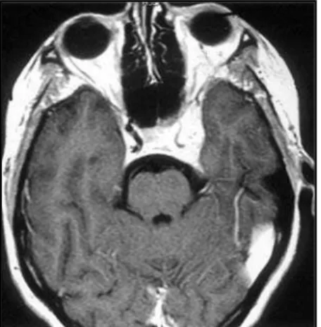
Fig 4. MRI T1, weigthed image with ga-dolinium enhancement showing a left ho-locavernous sinus meningioma. Note the involvement of the internal carotid artery.
834 Arq Neuropsiquiatr 2004;62(3-B)
and postoperatively. All patients were operated in supine position.
Cerebellar remote hemorrhage is an under-rec-ognized complication after supratentorial neuro-surgical procedures, specially those involving the opening of CSF cisterns or the ventricular system1,2,7.
Although the occurrence of RCH is associated with some risk of death or major morbidity, in most cases a benign course is exhibited. Cerebellar “sag” as a result of CSF volume loss, causing tran-sient occlusion of superior bridging cerebellar veins and consequent hemorrhagic venous infarction, is the most likely cause of RCH 1,2.
Not one single presurgical or surgical factor can reliably predict the occurrence of cerebellar he-morrhage after supratentorial craniotomy or re-mote hemorrhages at all, and the etiology of this entity still remains unclear. The most important keys to minimize the hazardous sequelae are to be aware of this potential complication, attempt to a precocious diagnosis and to provide prompt treatment in all cases.
REFERENCES
1. Marquardt G, Setzer M, Schick U, Seifert V. Cerebellar hemorrhage after supratentorial craniotomy. Surg Neurol 2002;57:241-251.
2. Friedman M, Piepgras PG, Duke DA, et al. Remote cerebellar hemor-rhage after supratentorial surgery. Neurosurgery 2201;49:1327-1340. 3. Konig A, Laas R, Hermann HD. Cerebellar hemorrhage as a complication
after supratentorial craniotomy. Acta Neurochir (Wien) 1987;88:104-108. 4. Cloft HJ , Matsumoto JA, Lanzino G, Cail WS. Posterior fossa hemorrha-ge after supratentorial surhemorrha-gery. Am J Neuroradiol 1997;18:1573-1580. 5. Honegger J, Zetner J, Spreer J, Carmona H, Shulze-Bohage A. Cerebellar
hemorrhage arising postoperatively as a complication of supratentor-ial surgery: a retrospective study. J Neurosurg 2002;96:248-254. 6. Lefranc F, De Witte O, David P, Brotchi J. Cerebellar hemorrhage
com-plicating a supratentorial craniotomy: a case report and review of the literature. Neurochirurgie 2000;46:395-397.
7. Tomii M, Nakajima M, Ikeuchi S, Ogawa T, Abe T. Infratentorial hem-orrhage following supratentorial surgery. No Shinkei Geka 1999;27: 921-925.
8. Brisman MH, Bederson JG, Sen CN, Germano IM, Moore F, Post KD. Intracerebral hemorrhage occurring remote from the craniotomy site. Neurosurgery 1996;39:1114-1121.
9. Kuroda R, Nakatami J, Akai F, et al. Remote subarachnoid hemorrhage in the posterior fossa following supratentorial surgery: clinical obser-vation of six cases. Acta Neurochir (Wien) 1994;12:158-165. 10. Yoshida S, Yonekawa Y, Yamashita K, Ihara I, Morooka Y. Cerebellar
hemorrhage after supratentorial craniotomy: report of three cases. Neurol Med Chir (Tokyo) 1999;30:738-743.
11 Koller M, Ortler M, Langmayr J, Twerdy K. Posterior-fossa hemor-rhage after supratentorial surgery: report of three cases and review of the literature. Acta Neurochir (Wien) 1999;141:587-592.
12. Kalfas IH, Little JR. Postoperative hemorrhage: a survey of 4992 intracra-nial procedures. Neurosurgery 1998;23:343-357.
DISCUSSION
Postoperative hemorrhage is a well known but serious complication of intracranial procedures and usually occurs at the site of the operation. Re-mote cerebellar hemorrhage (RCH) occurring after supratentorial craniotomy is the most commonly
described pattern of remote hemorrhage1-6 and
may occasionally cause significant neurological mor-bidity or even death. Mortality may reach 25% of
the cases1. Most of the cases described occurred
within hours after the surgery, sometimes imme-diately afterwards, suggesting that most of these hemorrhages develop during or soon after sur-gery2,7. The findings suggest that this clinical pic-ture is unrelated to age, previous arterial hyperten-sion, inherent or induced coagulopathies, type of primary underlying lesion, intraoperative
position-ing of the pacients or type of anesthesia8.
The loss of substantial CSF volume during sur-gery must play a central role in the pathophysiolo-gical development of remote cerebellar hemor-rhage1,2,4,7-9. Suction of the CSF may cause nial hypotension. Further reduction of intracra-nial pressure leads to an increased transluminal venous pressure2,7,10,11.
A growing consensus suggests that RCH is like-ly to be a manifestation of cerebellar venous infarc-tion and hemorrhage. Typical locainfarc-tion, in the supe-rior cerebellar and vermian cortex, corresponds to the territory drained by superior cerebellar veins3. The timing for the appearance of the symptoms and apparent “blossoming” on CT in the first 24 hours after surgery are not specific but are consistent
with evolution of venous hemorrhage2. With regard
to neoplastic lesions, meningiomas have been described to be most frequently complicated by hemorrhagic postoperative manifestations, prob-ably because of abnormal hyperfibrinolysis, but
not directly related to remote hemorrhage1,12.