RODRIGUES FG ETAL.
818 REV ASSOC MED BRAS 2016; 62(9):818-821
IMAGE IN MEDICINE
Endoscopic ultrasound in the diagnosis of foreign bodies of the
colon and rectum
FABIO GONTIJO RODRIGUES1*, JOÃO BATISTA CAMPOS2, GIOVANNA DASILVA3, STEVEN D. WEXNER4
1MD, PhD, Department of Colorectal Surgery, Cleveland Clinic Florida, Weston, FL, USA. Endoscopia Clínica e Cirúrgica, Belo Horizonte, MG, and Researcher from the CNPq, Brazil 2MD, Endoscopia Clínica e Cirúrgica, Belo Horizonte, MG, Brazil
3MD, Department of Colorectal Surgery, Cleveland Clinic Florida, Weston, FL, USA 4MD, PhD (Hon), Department of Colorectal Surgery, Cleveland Clinic Florida, Weston, FL, USA
S
UMMARYStudy conducted at the Department of Co-lorectal Surgery, Cleveland Clinic Florida, Weston, FL, USA, and at Endoscopia Clínica e Cirúrgica, Belo Horizonte, MG, Brazil
Article received: 11/24/2015
Accepted for publication: 12/1/2015
*Correspondence:
Address: 1641, Sorrento Drive Weston, Florida – USA Postal code: 33326 [email protected]
http://dx.doi.org/10.1590/1806-9282.62.09.818
Although the ingestion of foreign bodies is a common clinical problem, severe complications such as perforation are rare and occur in less than 1% of cases. Different types of foreign bodies and the various affected regions within the gastrointestinal tract make foreign body ingestion a complex entity, with a wide range of presentation requiring different diagnostic modalities. We report two cases of patients who underwent endoscopic ultrasound for evaluation of sub-epithelial lesions consisting of foreign body granulomas in the colon and rectum. Colorectal foreign body granuloma is a rare complication after accidental inges-tion. Endoscopic ultrasound can be a useful diagnostic tool and can avoid the need for more invasive procedures.
Keywords: foreign bodies, colon, rectum, granuloma, endoscopic ultrasound.
I
NTRODUCTIONAlthough the ingestion of foreign bodies is a common clinical problem, severe complications like perforation are rare and occur in less than 1% of cases.1,2 Different types of foreign bodies and the various affected regions within the gastrointestinal tract make foreign body inges-tion a complex entity, with a wide range of presentainges-tion requiring different diagnostic modalities.
In the last three decades, endoscopic ultrasound has gained importance both as a diagnostic and as a thera-peutic tool for gastrointestinal tract disorders. Ten years ago, 60% of gastroenterologists were already using endo-scopic ultrasound in the United States.3-10 However, in other parts of the world such as Brazil, this method is still not widespread, especially within the public health system and in rural regions.
We report two cases of patients who each underwent endoscopic ultrasound for evaluation of colorectal sub-epithelial lesions consisting of foreign body granulomas.
C
ASE1
A 62-year-old healthy white male was referred for endo-scopic ultrasound after a screening colonoscopy that
showed two elevated lesions adjacent to each other in the ascending colon. The lesions measured approximately 10 mm and 7 mm in their greater diameter, had normal ap-pearing covering mucosa, which was firm to compression with closed biopsy forceps (no pillow-sign); biopsies were not performed (Figure 1).
FIGURE 1 Colonoscopic view of subepithelial lesions in the
ENDOSCOPICULTRASOUNDINTHEDIAGNOSISOFFOREIGNBODIESOFTHECOLONANDRECTUM
REV ASSOC MED BRAS 2016; 62(9):818-821 819
Laboratory examinations were normal. Endoscopic ultrasound showed hypoechoic oval lesions arising from the submucosal layer, with a linear hyperechoic image within the lesions. These were suggestive of granulomas secondary to a foreign body reaction, probably due to a fishbone (Figure 2). The patient was informed of the findings and expectant management was indicated. CT scan was performed to exclude other abnormalities and future follow-up by colonoscopy was indicated.
C
ASE2
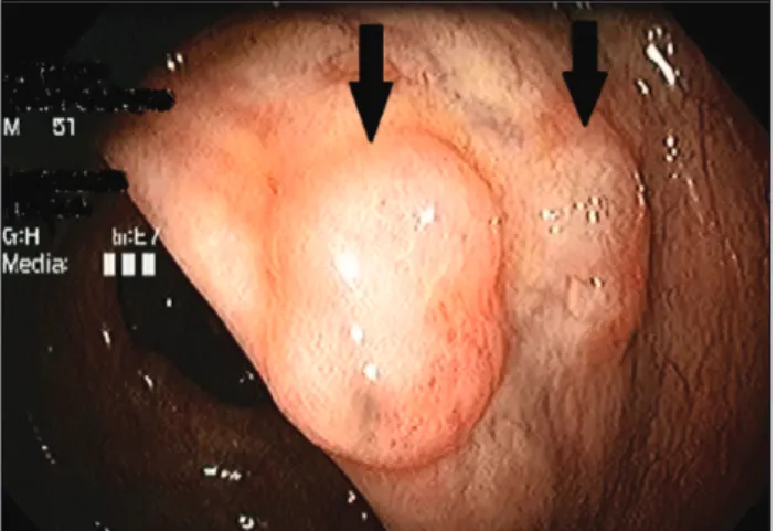
A 51-year-old healthy white male was referred for endo-scopic ultrasound after the finding of a subepithelial rectal lesion, measuring approximately 10 mm in its greater diameter, with discrete mucosal enanthema and central umbilication on screening colonoscopy (Figure
3). Biopsy has shown mild unspecific inflammatory chang-es in the mucosa. A concomitant lchang-esion was seen in the ascending colon with a pale yellow color and soft consis-tency to the touch (pillow sign present), considered to be a lipoma.
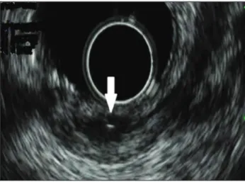
Endoscopic ultrasound showed a lesion with mixed echogenicity (predominantly hypoechoic) and a hyper-echoic linear structure in its interior (Figure 4). The lesion was considered a granuloma due to foreign body reaction (the patient reported ingestion of a fishbone a few months prior). The patient underwent expectant management and will be followed up with subsequent proctologic screening.
D
ISCUSSIONPerforation secondary to ingested foreign bodies has been reported in all segments of the gastrointestinal tract. Acute perforation with extraluminal contamination and sepsis can require emergent surgical treatment. Sharp pointed objects such as toothpicks, needles, dental plates, and fish and chicken bones are usually associated with perforation or penetration in the gastrointestinal organ walls. Foreign body complications can also arise after sutures and in some unusual situations such as formation of a barium granuloma after radiologic examination.11-13 Most recently, with the advent of endoscopic therapeutic modalities, foreign body reactions are becoming more frequent after positioning of stents (gastrointestinal and biliary) and after injection of materials such as cyanoac-rylates for the treatment of varices.14 Furthermore, the impaction of foreign bodies in the rectum can cause ano-rectal abscesses and fistulas.
FIGURE 2 Endoscopic ultrasound image – hypoechoic nodule with
a linear hiperechoic image in its interior (blue arrow).
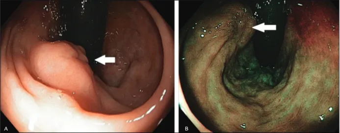
FIGURE 3 A. Colonoscopic view of the rectal subepithelial lesion with central umbilication and mild hyperemia (white arrow). B. Same
lesion – NBI view (white arrow).
RODRIGUES FG ETAL.
820 REV ASSOC MED BRAS 2016; 62(9):818-821
Diagnosis of foreign body complications can be dif-ficult due to the varied presentation, different types of foreign bodies, unsuspected or a long interim after inges-tion, and a wide range of differential diagnosis in the gastrointestinal tract. Foreign body granulomas have been confused with inflammatory bowel disease and tumors throughout the gastrointestinal tract.11,12,15 Plain radio-graph is the most commonly used modality to detect ingested foreign bodies; radiographs can reveal radio-opaque objects and also assess the presence of pneumo-peritoneum in cases of perforation.16 In cases of radiolu-cent objects, computed tomography appears to be the modality of choice and can show the presence of abscess, free air, and any involvement of the surrounding struc-tures.17 Endoscopic ultrasound images can vary according to the type of foreign body. Wood and plastic materials tend to produce acoustic shadow, metals are typically hyperechoic and cause reverberation, and glass is hyper-echoic but does not cause reverberation. The inflamma-tory reaction can lead to a hypoechoic ring around the foreign body. Parasites produce hyperechoic images with a hypoechoic band in its interior and transverse images show a targeted lesion.
Gastrointestinal lesions such as gastrointestinal stro-mal tumors (GIST) and neuroendocrine tumors usually present as hypoechoic lesions. Some gastrointestinal tu-mors can show a hyaline deposition under the mucosa and increased refringence; this is an important differential diagnosis. A definitive diagnosis of a small foreign body
granuloma is typically made by the pathologist after its removal, but sometimes an invasive procedure can be avoided.
The follow-up regimen of patients with foreign body granulomas is unclear. Since most published studies are case reports, there are no algorithms to monitor these patients. A long chronic course has been reported such as in a patient who required colonic resection secondary to foreign body ingestion 10 years prior.18 The patient had an inflammatory tumor after perforation of the trans-verse colon. He underwent examinations for abdominal pain and a palpable mass was found on physical exam-ination. Conversely, resolution without any long-term consequences has also been reported. Kikuchi et al. re-ported the case of a patient with an esophageal foreign body causing a granuloma. The authors repeated esoph-agogastroduedonoscopy, esophagography and CT after one year and reported a progressively smaller lesion and no identifiable foreign body.19
C
ONCLUSIONColorectal foreign body granuloma is a rare complication after accidental foreign body ingestion. Endoscopic ul-trasound can be a useful diagnostic tool and can avoid the need for more invasive procedures.
R
ESUMOUltrassonografia endoscópica no diagnóstico de corpos estranhos no cólon e no reto
Embora a ingestão de corpos estranhos seja uma condição clínica frequente, complicações graves como perfuração são raras e ocorrem em menos de 1% dos casos. Tipos diferentes de corpos estranhos e as diversas regiões afeta-das do trato gastrointestinal fazem da ingestão de corpos estranhos uma entidade complexa, com uma variada gama de apresentações, demandando várias modalidades diag-nósticas. Nós reportamos dois casos de pacientes que foram submetidos à ultrassonografia endoscópica para avaliação de lesões subepiteliais, consistindo em granu-lomas de corpo estranho no cólon e no reto. Granugranu-lomas de corpo estranho colorretais são uma complicação rara após ingestão acidental. Ultrassonografia endoscópica pode ser uma ferramenta diagnóstica útil e pode evitar procedimentos mais invasivos.
Palavras-chave: corpos estranhos, colo, reto, granuloma, ultrassonografia endoscópica.
FIGURE 4 Endoscopic ultrasound images of the rectal lesion
ENDOSCOPICULTRASOUNDINTHEDIAGNOSISOFFOREIGNBODIESOFTHECOLONANDRECTUM
REV ASSOC MED BRAS 2016; 62(9):818-821 821
R
EFERENCES1. Huang CH, Chiang CC, Yan YH, Tsai TJ, Chen CY. Role of endoscopic sonography in the diagnosis of fish bone perforation of the gastric wall resulting in submucosal pseudotumor. J Clin Ultrasound. 2011; 39(7):415-7. 2. Maleki M, Evans WE. Foreign body perforation of the intestinal tract. Report
of 12 cases and revision of the literature. Arch Surg. 1970; 101(4):475-7. 3. Dussik KT, Dussik F, Wit L. Auf dem wege zur hiperphonographie des
gehirnes. Wien Med Wschnschr. 1947; 97(38-39):425-9.
4. Ludwig GD, Struthers FW. Considerations underlying the use of ultrasound to detect gallstones and foreign bodies in tissue. U. S. Naval Med Research Inst Project 1949. NM004:001, report nº 4.
5. Wild JJ. The use of ultrasonic pulses for the measurement of biologic tissues and detection of tissues density changes. Surgery. 1950; 27(2):183-8. 6. Hildebrandt U, Feifel G, Ecker KH. Rectal endosonography. Baillière’s Clin
Gastroenterol. 1989; 3(3):531-41.
7. Lutz H, Rosch W. Transgastroscopic ultrasonography. Endoscopy. 1976; 8(4)203-5.
8. Strohm WD, Phillip I, Hagenmüller F, Classen M. Ultrasonic tomography by means of an ultrasonic fiberendoscope. Endoscopy. 1980; 12(5):241-4. 9. DiMagno EP, Buxton JL, Regan PT, Hattery RR, Wilson DA, Suarez JR, et
al. Ultrasonic endoscope. Lancet. 1980; 1(8169):629-31.
10. Ahmad NA, Kochman ML, Ginsberg GG. Practice patterns and attitudes toward the role of endoscopic ultrasound in staging of gastrointestinal malignancies: a survey of physicians and surgeons. Am J Gastroenterol. 2005; 100(12):2662-8.
11. Hizawa K, Aoyagi K, Suekane H, Mibu R, Yao T, Fujishima M. Suture granuloma in rectal anastomosis mistaken for locally recurrent cancer. J Clin Gastroenterol. 1996; 23(1):78-9.
12. Kimura H, Shima Y, Kinoshita S, Takahashi I, Okai T. Endosonographic misdiagnosis of tumor recurrence after surgery for malignant GIST. Endoscopy. 2002; 34(3):238.
13. Jung IS, Kim JO, Lee JS, Lee MS, Shim CS. Barium granuloma of rectum diagnosed by EUS. Gastrointest Endosc. 2003; 57(6):718-9.
14. Sato T, Yamazaki K, Toyota J, Karino Y, Ohmura T, Suga T. Inflammatory tumor in pancreatic tail induced by endoscopic ablation with cyanoacrylate glue for gastric varices. J Gastroenterol. 2004; 39(5):475-8.
15. Tee CT, Chen HC, Yusoff IF. Delayed dysphagia from a foreign body granuloma mimicking a subepithelial neoplasm. Clin Gastroenterol Hepatol. 2013; 11(7):e51.
16. Pinto A, Reginelli A, Pinto F, Sica G, Scaglione M, Berger FH, et al. Radiological and practical aspects of body packing. Br J Radiol. 2014; 87(1036):20130500.
17. Pinto A, Lanza C, Pinto F, Grassi R, Romano L, Brunese L, et al. Role of plain radiography in the assessment of ingested foreign bodies in the pediatric patients. Semin Ultrasound CT MR. 2015; 36(1):21-7.
18. Nambu K, Sato K, Tamegai Y, Imai S, Uchiyama S, Shibuya T. [A case of intraabdominal inflammatory tumor caused by perforation of the transverse colon dueto a fish bone which was preoperatively diagnosed]. Nihon Rinsho Geka Gakkai Zasshi (J Jpn Surg Assoc). 1998; 59(2):423-7. Available from: https://www.jstage.jst.go.jp/article/ringe1998/59/2/59_2_423/_pdf. 19. Kikuchi K, Tsurumaru D, Hiraka K, Komori M, Fujita N, Honda H. Unusual