INTRODUCTION
The expanded use of CT to assess abdomi-nal disorders has identified a large number of un-suspected small renal masses, less than 1 cm in size (1,2). However, to date, there are no data available of their outcome and pathologic diagnosis.
Unless diagnosed as cysts (based on cor-tical location, contours, base density of - 5 to +10 HU and lack of enhancement) these masses could not be further categorized by CT.
Surgical data from the recent urologic lit-erature suggest that only 19.6 - 43% of up to 4 cm lesions are benign (3-5). However, the differentia-Purpose: We evaluated the outcome and etiologies of small renal masses (less than
1 cm in size) discovered incidentally on 2 consecutive CTs that investigated non-urologic abdominal complaints.
Materials and Methods: A retrospective search for incidentally discovered small renal masses, less then 1 cm in size, was carried out in the files of 6 major US medical centers. 4822 such lesions had been reported over a 12 year period. A search of these patients’ records revealed 1082 subsequent new CTs for non uro-logic complaints, allowing the assessment of the fate of the masses. Lesions enlarg-ing, of ambivalent contour or enhancement were examined by a third multiphasic MDCT. The findings were interpreted by 2 blinded radiologists.
Results: Six hundred and four masses could no longer be identified, 231 were significantly smaller, 113 unchanged in size and 134 larger. Of the disappearing lesions 448 were located in the medulla, 94 both in medulla and cortex and 62 in cortex. Multiphasic MDCTs obtained in 308 masses enlarging, unchanged in size or of ambivalent appearance, revealed 7 neoplasms, 45 inflammatory lesions, 8 abscesses and 62 renal medullary necrosis. Concurrent antibiotic therapy of GI conditions may have caused some of the 496 lesions to disappear.
Conclusion: It is questionable whether the small number of malignant neoplasms (0.4%), inflammatory lesions (5%) and renal medullary necrosis (6%) justify rou-tine follow-up CTs and exposure to radiation. The delay in intervention in neo-plastic lesions probably didn’t influence tumor-free survival potential and clinical symptoms would soon have revealed inflammatory conditions. With exception of ambivalent lesions, clinical surveillance appears adequate.
The fate of small renal masses, less then 1 cm size:
outcome study
_______________________________________________
Erich K. Lang, Amer Hanano, Ernest Rudman, Raju Thomas, Leann Myers, Quan Nguyen, Richard
J. Macchia
Departments of Radiology and Urology SUNY, Downstate Medical School, Brooklyn NY (EKL,AH,ER,QN,RJM), Departments of Urology and Biostatistics, Tulane Health Science Center and School Public Health (EKL,RT,LM), New Orleans and Department of Radiology, Johns Hopkins Medical Institutions (EKL), Baltimore, USA
ABSTRACT
ARTICLE
INFO
_______________________________________________________________ _____________________
Key words:
kidney; neoplasms; outcome assessment
Int Braz J Urol. 2012; 38: 40-48
________________
Submitted for publication: December 15, 2010
________________
41
tion of benign and malignant lesions is difficult. Neither size nor growth rate are reliable predic-tors of malignancy (1, 6-10). In fact, even in le-sions with a “0” growth rate, an 83% incidence of malignancy has been reported (7). Recent reports have shown similar tumor-free survival rates of patients treated by surgery regardless whether the size of the lesion was 1.5 cm or 4 cm. This favored the option of surveillance of small lesions par-ticularly in elderly patients with co-morbidities (1,3,11-14). Moreover, histologically aggressive behavior of lesions can be identified by biopsy, which is advocated by some when pursuing sur-veillance (15,16).We have undertaken this retrospective study of our files to identify underlying pathol-ogy and particularly neoplastic etiolpathol-ogy as well as treatable conditions of these masses.
MATERIALS AND METHODS
Study Design
Waivers were obtained from the respective Institutional Review Boards for a retrospective review of the charts to identify lesions of inter-est. The reports of all CTs obtained to evaluate non-urologic abdominal complaints and condi-tions were reviewed for the incidental diagnosis of small renal mass (less then 1 cm).
Four thousand eight hundred and twenty-two such renal lesions were observed in the files of Tulane Health Science Center, (1995-2005) LSU Medical Center, Charity Hospital, VA Hospital, all in N.O, (1995-2000) SUNY Downstate Medi-cal School, Brooklyn, N.Y, (1998-2007) and Johns Hopkins Medical Institutions, Bayview, Baltimore, (2005-2007). All patients with a history of prior or existing neoplastic disease were excluded. To establish the fate of these mass lesions we then searched the files of the 4822 patients for any subsequent CTs obtained for non-urologic ab-dominal conditions, and identified 1082 such CTs carried out in the ensuing 4-36 months period (13.2 months mean).
Since these studies were investigating abdominal symptoms and complaints (mostly gastrointestinal (41 %), biliary (27%), pancreatic
(26%) and retroperitoneal & vascular (6%)), oral contrast had been administered in at least one of the two CTs in 93% of the patients; intravenous contrast in 91%. Eighty-four percent of the stud-ies were 2 phase CTs, 9% 3 phase and 7% single phase nonenhanced CTs. Since the two CTs that were compared may have been obtained for dif-ferent reasons, the techniques often differed. The accuracy of diagnosis may vary for different en-hancement phases. Variability in equipment and interpreters may be another shortcoming of our study design. A third follow-up, 3 or 4 phase MDCT was performed to assess 308 renal mass lesions which were larger, unchanged in size or of ambivalent appearance (ill-defined margins, rim enhancement) on the second CT. These CTs were limited to the kidneys, generating 2.5, 3.7 mm thick slices, some with 1.5 mm axial and coronal reconstructions. Technical factors: 100 ml non-ionic contrast medium at a flow rate of 4-5 ml/ sec, 100-140 KV, 180-320 MAS, 6 cm table move-ment. Phases: Pre-enhancement, late arterial cor-tico-medullary phase at 12-16 second scan delay, parenchymal phase with 40-60 second scan delay, and sometimes excretory phase with a 4-12 min-ute delay. An identical protocol was performed in all our institutions. The studies were interpreted by 2 experienced uro-radiologists, blinded to pri-or interpretations and clinical findings.
Statistical analysis was carried out using X 2 test to assess outcomes for different locations.
The lesions varied from 2-10 mm in size (6.2 mm mean), 805/1082 (75%) were solitary, 277/1082 (25%) multiple (2-5); their sum-total volume never exceeded 2.9 ml, 28 were located in both kidneys. Seven hundred fifty-seven (70%) patients were male, 308 (28%) female, and 17 (2%) children, aged 16 to 82 years (39 years mean).
RESULTS
le-sions had been located in the medulla, 94 (8.7%) in both medulla and cortex and 62 (5.7%) in cor-tex (Figure-1). Almost 73% of solitary medullary masses disappeared compared to 22% of solitary cortical masses (p = 0.0001); a significant finding. Among the lesions unchanged in size, 52 (4.8%) were located in the medulla, 30 (2.7%) in both cor-tex and medulla and 31 (2.9%) in corcor-tex; of those
larger, 69 (6.4%) were located in medulla, 21 (1.9%) in both cortex and medulla and 44 (4.1%) in cor-tex (Table-1). There were no significant differences in outcomes for different locations of small renal masses (X 2 test, p = 0.0001).
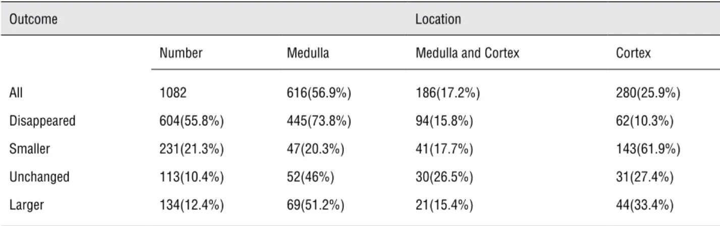
Location in the medulla, pre-enhancement density of 14-26 HU, lack of or heterogeneous en-hancement in the late arterial, cortico-medullary Table 1 - Location and Outcome of 1082 Small Renal Masses on Follow-up CT (after 4-26 months).
Outcome Location
Number Medulla Medulla and Cortex Cortex
All 1082 616(56.9%) 186(17.2%) 280(25.9%) Disappeared 604(55.8%) 445(73.8%) 94(15.8%) 62(10.3%) Smaller 231(21.3%) 47(20.3%) 41(17.7%) 143(61.9%) Unchanged 113(10.4%) 52(46%) 30(26.5%) 31(27.4%) Larger 134(12.4%) 69(51.2%) 21(15.4%) 44(33.4%)
43
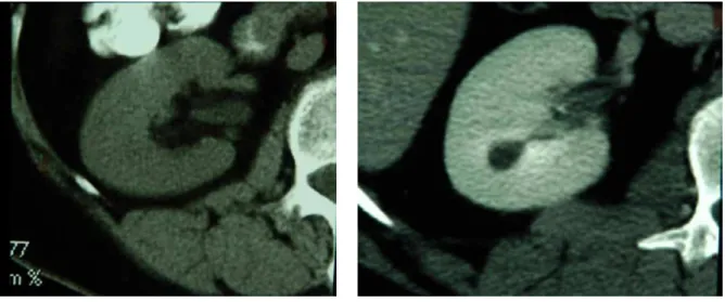
and parenchymal phase, and demonstration of an ill-defined enhancing perimeter rim, led to the presumptive diagnosis of renal medullary necrosis (RMN) in 22 of the enlarging, 4 of unchanged size and 36 of shrinking mass lesions (Figures 1 and 2).Enhancement of the rim of lesions in the cortico-medullary and parenchymal phase,
some-times layering debris in its center, prompted the diagnosis of abscess in 8 mass lesions, 4 in the medulla, 2 in cortex and 2 involving both cortex and medulla. Five decreased while under antibiot-ic therapy and 3 increased in size until treated by percutaneous drainage. Histologic and bacterio-logic proof was available in all 8 lesions (Table-2).
Table 2 - Significant and Potentially Treatable Conditions Observed Among 1082 Small Renal Masses.
Pathologic Diagnosis
Location Size Inflammatory Abscess RCC AML Metastasis Fibroma RMN
Cortex Smaller 4 1 - - - -
-Larger 12 1 2 2 - 1
-Medulla Smaller 4 1 - - - - 36
Unchanged 14 1 - - - -
-Larger - 2 - - - - 22
Cortex and Medulla
Smaller 8 1 - - - -
-Larger 3 1 - - 2 -
-Unchanged - - - 4
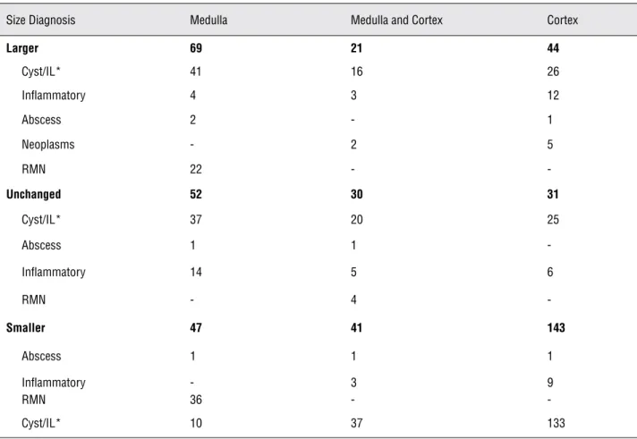
Table 3 - Outcome of 247 Enlarging or Unchanged In Size Small Renal Masses Reexamined by Multiphase MDCT.
Size Diagnosis Medulla Medulla and Cortex Cortex
Larger 69 21 44
Cyst/IL* 41 16 26
Inflammatory 4 3 12
Abscess 2 - 1
Neoplasms - 2 5
RMN 22 -
-Unchanged 52 30 31
Cyst/IL* 37 20 25
Abscess 1 1
-Inflammatory 14 5 6
RMN - 4
-Smaller 47 41 143
Abscess 1 1 1
Inflammatory - 3 9
RMN 36 -
-Cyst/IL* 10 37 133
*IL = Indeterminate Lesion, features suggestive of cyst.
While none of these patients presented with find-ings of urinary tract pathology, urine WBC/hpf was 8-12 in 3 at the time of the second CT work-up. This finding was ignored because of the estab-lished diagnosis of inflammatory sigmoid-colon disease. However, within 10 days, urine culture (N = 8) and blood culture (N = 3) became positive.
Poor and heterogeneous enhancement of 45 renal masses suggested an underlying inflam-matory etiology. A pale or non-enhancing center on the cortico-medullary phase suggested edema or early necrosis, while enhancement in the pe-riphery and immediately adjacent tissues in the parenchymal phase was consistent with inflam-matory hyperemia. WBC was elevated in 28 pa-tients, urine analysis showed 5-10 WBC/hpf in 3 patients. The laboratory findings seemed consis-tent with the diagnosed inflammatory disease of bowel, pancreas or gallbladder, and a urinary tract
infection was not considered. Within 2 weeks, urine cultures turned positive in 12 patients; urine analysis (WBC/hpf) was positive in 44 patients.
Eighteen of these lesions were found in the medulla, 16 in cortical location and 11 involved both cortex and medulla (Table-3). Fifteen of 45 (33.3%) had decreased in size, while 30 (66.6%) increased or remained unchanged in size (Tables 2 and 3). Antibiotic therapy used to treat inflamma-tory abdominal conditions between the first and second CT could have contributed to shrinkage of 15 of these lesions (Table-3). Ultimately, all pa-tients were treated with urinary tract antibiotics and the masses disappeared, though a small py-elonephritic scar persevered in 14 patients.
45
164HU respectively, two renal cell carcinomas were identified on 4 phase MDCTs (Table-2). A rapid growth rate (1.44 cm/year) of 2 mass lesions involving cortex and medulla, enhancing from 30 and 32 to 120HU in parenchymal phase, prompted exploration and segmental resection. Metastatic carcinomas (lung = 1, colon = 1) were found (Table-3). Presence of fat on MDCT in lesions enlarging at a rate of 0.2 & 1.1 cm/year respectively led to diagnosis of angiomyolipoma in 2 patients. Hemorrhage into the lesions may have obscured the fat on the first 2 CTs (Table-3). One enlarging cortical mass (32 HU) with morphology favoring a benign neoplasm was biopsied and a fibroma diagnosed (Table-3). The diagnoses were confirmedin 6 patients by histopathology of the resected specimen, and in one by core biopsy specimen.
Benign cysts were diagnosed in 87 enlarg-ing or stationary cortical and cortical and med-ullary masses, and 72 shrinking cortical masses based on established CT criteria. Two enlarging cysts, with 34 and 42 HU were thought to repre-sent hemorrhagic cysts. The diagnosis was con-firmed by unroofing in one, aspiration in 8 and follow-up imaging in the remainder.
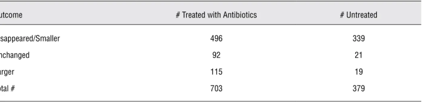
Seven hundred and three of 1082 (65%) patients had been placed on broad-spectrum an-tibiotics in the period between the first and the second CT, to treat intestinal and biliary infections (Table-4). Collateral impact on GU tract organisms could not be excluded. Four hundred and ninety-six lesions disappeared or decreased in size, while 115 enlarged and 93 remained stationary in size.
Table 4 - Outcome of Small Renal Masses in 703 Patients Treated with Antibiotics for Inflammatory.
Conditions of the Gastrointestinal or Biliary Tract and Pancreas versus 339 untreated Patients.
Outcome # Treated with Antibiotics # Untreated
Disappeared/Smaller 496 339
Unchanged 92 21
Larger 115 19
Total # 703 379
Fourteen were diagnosed as RMN based on im-aging criteria, 10 as inflammatory masses and 2 as abscesses (Table-3). The impact of antibiot-ics is uncertain; in untreated patients, 89.4% of masses disappeared or got smaller versus 70.6% in treated patients (p = 0.0001). Another 339 lesions decreased in size or disappeared in patients who were not on antibiotic therapy.
DISCUSSION
The once universally held opinion that radical nephrectomy is the sole modality to manage renal cell carcinoma is now widely chal-lenged (9). Partial nephrectomy, open or
their rim (6,7,9,10,12). In older patients and those with medical co-morbidities this provides a period to establish and observe benign or indolent nature of the mass lesion without significantly reducing potential for tumor-free survival by delaying sur-gery. The guided biopsy for assessing histologic aggressiveness of lesions, which is the major fac-tor determining frequency of reexamination, is well stablished (14,15,17). MDCTs and guided bi-opsy seem to provide adequate criteria for follow-up (14,15). As of date the significance of renal masses less then 1 cm in size and their appropri-ate management remain an open question (1,2,8). However, recent reports suggest a high incidence of malignancy of such lesions, particularly if lo-cated in cortex (8,21). Hence, a more frequent in-terval follow-up surveillance by ultrasound may be appropriate for such cortical mass lesions.
Our retrospective study of 1082 small renal masses, less than 1 cm in size, established the fate of these lesions on a second and sometimes third 3 or 4 phase MDCT. This study revealed most impor-tantly that 604/1082 (55.8%) renal masses disap-peared, while another 231 decreased in size in the interval of 4 to 36 months between the first and second CT. Partial volume averaging and the dis-parity of resolution between 2 phase and 4 phase studies may have caused false negatives (i.e., a le-sion observed on the initial 4 phase study may not be apparent on a follow-up 2 phase CT). Tever, this scenario affected only 6% of patients.
Four hundred and ninety-six patients in whom the lesions either disappeared or substtially decreased in size had been treated with an-tibiotics for gastrointestinal, biliary or pancreatic infections (Table-3). This fact raises the question whether a co-existent urinary infection causing the mass lesions might have responded to these non-urinary tract antibiotics. The prevalence of RMN lesions (N = 62) suggests that many of the smaller disappearing lesions may likewise have been of RMN or inflammatory etiology (Tables 2 and 3).
The incidence of neoplasms is the most significant finding of our targeted follow-up CTs of lesions with increased or stationary size. Two renal cell carcinomas and 2 metastatic carcino-mas, all with growth rates indicative of
histologi-cally aggressive behavior, were identified (Tables 2 and 3). Despite the rapid growth rate, the delay in surgical resection is not likely to have adversely influenced survival. Two patients with AML and 1 with a benign fibroma were also identified on tar-geted MDCTs (Table-2). Though benign in nature, their rate of growth, partly caused by hemorrhage into the AML, warranted resection.
The diagnosis of all 53 inflammatory le-sions made possible early and definitive treatment by drainage and/or GU specific antibiotics, pre-venting propagation of the process and loss of pa-renchyma (Table-2).
The pertinent question of whether the in-cidental discovery of a small renal mass warrants follow-up by imaging, is to some degree answered by our data (2).
Neither the incidence of malignant neoplasms (< 0.4%) nor of inflammatory entities (5%) or of renal medullary necrosis (6%) seems to justify follow-up CTs. A delay in identification and hence surgical (or ablative) intervention of renal masses up to 3.5 cm in size is not likely to adversely influence recurrence-free survival. Inflammatory lesions, by nature of their pathophysiology, tend to become symptomatic early. The predominance of medullary lesions and their greater probability of spontaneous disappearance (73% for medullary lesions versus 22% for cortical lesions, p = 0.0001) favor an inflammatory etiology in masses prone to resolve. To avoid unnecessary radiation exposure to these patients, follow-up by imaging studies should be reserved for lesions exhibiting aggressive characteristics.
CONFLICT OF INTEREST
None declared.
REFERENCES
1. Volpe A, Panzarella T, Rendon RA, Haider MA, Kondylis FI, Jewett MA: The natural history of incidentally detected small renal masses. Cancer. 2004; 100: 738-45.
47
3. Marszalek M, Ponholzer A, Brössner C, Wachter J, Maier U, Madersbacher S: Elective open nephron-sparing surgery for renal masses: single-center experience with 129 consecu-tive patients. Urology. 2004; 64: 38-42.
4. DeRoche T, Walker E, Magi-Galluzzi C, Zhou M: Pathologic characteristics of solitary small renal masses: can they be predicted by preoperative clinical parameters? Am J Clin Pathol. 2008; 130: 560-4.
5. Schachter LR, Cookson MS, Chang SS, Smith JA Jr, Dietrich MS, Jayaram G, et al.: Second prize: frequency of benign re-nal cortical tumors and histologic subtypes based on size in a contemporary series: what to tell our patients. J Endourol. 2007; 21: 819-23.
6. Thompson RH, Kurta JM, Kaag M, Tickoo SK, Kundu S, Katz D, et al.: Tumor size is associated with malignant potential in renal cell carcinoma cases. J Urol. 2009; 181: 2033-6. 7. Kunkle DA, Crispen PL, Chen DY, Greenberg RE, Uzzo RG:
Enhancing renal masses with zero net growth during active surveillance. J Urol. 2007; 177: 849-53; discussion 853-4. 8. Jewett MA, Zuniga A: Renal tumor natural history: the
ra-tionale and role for active surveillance. Urol Clin North Am. 2008; 35: 627-34.
9. Russo P: Should elective partial nephrectomy be performed for renal cell carcinoma > 4 cm in size? Nat Clin Pract Urol. 2008; 5: 482-3.
10. Hsu RM, Chan DY, Siegelman SS: Small renal cell carcino-mas: correlation of size with tumor stage, nuclear grade, and histologic subtype. AJR Am J Roentgenol. 2004; 182: 551-7.
11. Crispen PL, Viterbo R, Fox EB, Greenberg RE, Chen DY, Uzzo RG: Delayed intervention of sporadic renal masses undergo-ing active surveillance. Cancer. 2008; 112: 1051-7. 12. Van Poppel H, Joniau S: Is surveillance an option for the
treat-ment of small renal masses? Eur Urol. 2007; 52: 1323-30. 13. Volpe A: The role of surveillance in the management of small
renal masses. ScientificWorldJournal. 2007; 7: 860-8.
14. Ozsoy M, Klatte T, Waldert M, Remzi M: Surveillance for the management of small renal masses. Adv Urol. 2008: 196701.
15. Wang R, Wolf JS Jr, Wood DP Jr, Higgins EJ, Hafez KS: Accuracy of percutaneous core biopsy in management of small renal masses. Urology. 2009; 73: 586-90; discussion 590-1.
16. Cozar JM, Tallada M: Open partial nephrectomy in renal cancer: a feasible gold standard technique in all hospitals. Adv Urol. 2008: 916463.
17. Kunkle DA, Egleston BL, Uzzo RG: Excise, ablate or observe: the small renal mass dilemma-a meta-analysis and review. J Urol. 2008; 179: 1227-33; discussion 1233-4.
18. Kutikov A, Kunkle DA, Uzzo RG: Focal therapy for kidney cancer: a systematic review. Curr Opin Urol. 2009; 19: 148-53.
19. Kunkle DA, Uzzo RG: Cryoablation or radiofrequency abla-tion of the small renal mass: a meta-analysis. Cancer. 2008; 113: 2671-80.
20. AUA Health Policy Brief, July 2009, Renal Mass Guideline. pp. 15.
21. DeRoche T, Walker E, Magi-Galluzzi C, Zhou M: Pathologic characteristics of solitary small renal masses: can they be predicted by preoperative clinical parameters? Am J Clin Pathol. 2008; 130: 560-4.
______________________
Correspondence address:
Dr. Erich K Lang Department Radiology SUNY Downstate Medical School 450 Clarkson Av., Brooklyn NY, 11203, USA E-mail: [email protected] Fax: + 1 718 245-5502
EDITORIAL COMMENT
In the United States, the frequency of CT scans has increased up to three fold during the last decade without an equal increase in the prevalence of life-threatening conditions (1). It seems the ex-cessive use of medical imaging increases health care costs and exposure to ionizing radiation with-out yielding significant benefits to all patients.
As a natural consequence of this “over-di-agnostics” a high number of pathological findings are detected leading to the question for the need for therapy in these cases.
radical nephrectomy is not the standard therapy anymore but nephron sparing treatment is a com-mon option with significant benefits regarding kidney function and patients outcome (2,3).
But, does that mean: the smaller detected the better treated? Obviously not. In a retrospec-tive analysis of 1082 renal lesions the current study shows the doubtful benefit of an excessive use of diagnostic tools. Even in the age of high-resolution imaging, the characterization of very small renal lesions remains difficult and not reli-able - in the study almost 60% of the lesions dis-appeared during follow-up: in 70% of the patients who received antibiotics only but in 90% of the untreated patients. Malignancies have been found in 0.4% only.
This analysis shows in an impressive way the limits of a worthy and reliable diagnostic in-strument. Therefore, the major goal must still be the reasonable interpretation of the data in order to develop strategies for patient care and avoid insufficient therapy as well as overtreatment. This still remains the mission of the Urologist who
needs to evaluate “pathological” findings critical to counsel his patients properly.
REFERENCES
1. Korley FK, Pham JC, Kirsch TD: Use of advanced radiology during visits to US emergency departments for injury-related conditions, 1998-2007. JAMA. 2010; 304: 1465-71. Erratum in: JAMA. 2010; 304: 1901.
2. Jewett MA, Zuniga A: Renal tumor natural history: the ra-tionale and role for active surveillance. Urol Clin North Am. 2008; 35: 627-34.
3. Ljungberg B, Cowan NC, Hanbury DC, Hora M, Kuczyk MA, Merseburger AS, et al.: EAU guidelines on renal cell carcinoma: the 2010 update. Eur Urol. 2010; 58: 398-406.
Dr. Andreas H. Wille
Department of Urology Klinikum Ernst von Bergmann Charlottenstr. 72 14467 Potsdam, Germany E-mail: [email protected] Fax: + 49 331 241-6900
EDITORIAL COMMENT
This is an interesting study in an impres-sive casuistic of small renal masses (SMR) < 1.0 cm, showing the clinic and radiologic outcomes of the-ses lesions. It’s an original contribution to the sci-entific literature and to the clinical practice; these informations should be considered before clinic de-cisions for this prevalent situation nowadays.
Despite the retrospective nature and the long duration of the study in several institutions and with different CT equipments, the study has the merit to show the situation as it occurs in real life, during 12 years, in multiple academic institutions.
With the information provided, we must be sure not to offer intensive follow-up protocols for this kind of SMRs. Accordingly, the patients will be less frequently exposed to the pre-exam anxiety, and costs, radiation and radiologic con-trast side effects of repeated CTs.
In the future, a prospective study with modern high resolution helicoidally CT equip-ments could be done.
For a specific subgroup of patients, with hereditary or familial renal cell carcinoma syn-dromes, the fate of their little SMRs probably will be different, since those individuals have thou-sands of microscopic malignant lesions in each kidney, although in these cases the clinical de-cision will be not changed; usually the surgical or ablative procedures are not offered until their lesions reaches 3.0 cm.
Dr. Stenio de Cássio Zequi