Contents lists available atScienceDirect
Acta Tropica
j o u r n a l h o m e p a g e :w w w . e l s e v i e r . c o m / l o c a t e / a c t a t r o p i c a
The influence of ecto-nucleotidases on
Leishmania amazonensis
infection and
immune response in C57B/6 mice
Miriam Conceic¸ão de Souza
a,1, Elisângela Aparecida de Assis
a,1, Rodrigo Saar Gomes
a,
Eduardo de Almeida Marques da Silva
a,2, Maria Norma Melo
b,
Juliana Lopes Rangel Fietto
c, Luís Carlos Crocco Afonso
a,∗aLaboratório de Imunoparasitologia, Departamento de Ciências Biológicas – ICEB/NUPEB, Universidade Federal de Ouro Preto, Campus Universitário,
Morro do Cruzeiro, 35400-000 Ouro Preto, MG, Brazil
bDepartamento de Parasitologia, Instituto de Ciências Biológicas, Universidade Federal de Minas Gerais, CP 486, 31270-901 Belo Horizonte, MG, Brazil cDepartamento de Bioquímica e Biologia Molecular, Universidade Federal de Vic¸osa, 36570-000 Vic¸osa, MG, Brazil
a r t i c l e
i n f o
Article history:
Received 13 December 2009
Received in revised form 30 March 2010 Accepted 12 April 2010
Available online 24 April 2010
Keywords:
Leishmania amazonensis
e-NTPDase Adenosine Macrophage Chemokines Virulence
a b s t r a c t
Previous results from our laboratory and from the literature have implicated the expression of ecto-nucleotidases in the establishment ofLeishmania infection. In the present study we evaluated the correlation between ecto-nucleotidasic activity and the infectivity ofL. amazonensispromastigotes that were kept in culture for short or extended numbers of passages, a condition that is known to decrease parasite infectivity. We also analyzed the immune response associated with the infection by these para-sites. As expected, we found that long-term cultured parasites induce the development of smaller lesions than the short-term cultured counterparts. Interestingly, long-term cultured parasites presented reduced ecto-nucleotidasic activity. In addition, cells recovered from animals infected with long-term cultured parasites produced higher amounts of IFN-␥and have smaller parasite load, after 8 weeks of infection.
Furthermore, after 1 week of infection, there is increased expression of the chemokine CCL2 mRNA in animals infected with short-term cultured parasites. Finally, infection of peritoneal macrophages by these parasites also shows marked differences. Thus, while short-term cultured parasites are able to infect a greater proportion of macrophages, cells infected by long-term cultured parasites express higher amounts of CXCL10 mRNA, which may activate these cells to kill the parasites. We suggest that the enzymes involved in metabolism of extracellular nucleotides may have an important role in infection by
L. amazonensis, by acting directly in its adhesion to target cells and by modulating host cell chemokine production.
© 2010 Elsevier B.V. All rights reserved.
1. Introduction
Parasites of the genusLeishmaniaare the causative agents of leishmaniasis, a group of diseases that affect 88 countries world-wide. Due to species differences in tissue tropism, virulence and their interaction with the host’s immune system, infection by these parasites can result in a variety of clinical manifestations ranging from single self-healing ulcers to life threatening visceral dissemi-nation (Alexander et al., 1999; Desjeux, 2004).
Leishmania (Leishmania) amazonensis is clinically important in the New World, where it can cause localized cutaneous
∗Corresponding author. Tel.: +55 31 3559 1701; fax: +55 31 3559 1680.
E-mail address:afonso@nupeb.ufop.br(L.C.C. Afonso).
1They equally contributed to the present study.
2Present address: Departamento de Ciências Biológicas, Universidade Vale do Rio
Doce, 35020-220 Governador Valadares, MG, Brazil.
leishmaniasis or diffuse cutaneous leishmaniasis, especially in
immunocompromised hosts (McMahon-Pratt and Alexander,
2004). Diffuse cutaneous leishmaniasis is characterized by the development of disseminated parasite laden nodules that is fre-quently associated to anergy to parasite’s antigens (Convit et al., 1972). In the context of murine models, the majority of mouse strains is susceptible to infection byL. amazonensis(Pereira and Alves, 2008).
It is generally accepted that resistance to infection by Leish-mania is associated with the development of a type 1 immune response, whereas disease progression is promoted by several pro-posed mechanisms, such as the development of a type 2 immune response, absence of type 1 immune response or co-existence of
both responses (McMahon-Pratt and Alexander, 2004; Sacks and
Noben-Trauth, 2002). Concerning the infection byL. amazonensis, it has been long known that, in C57BL/10 mice, the absence of a type 1 response is sufficient to cause parasite multiplication and lesion development (Afonso and Scott, 1993).
The study of the role of extracellular nucleotides and nucle-osides as immunomodulatory molecules has been the focus of several laboratories over the last decade. It has been demonstrated that increased concentrations of extracellular ATP can be inter-preted as “danger signals” that trigger an inflammatory response. On the other hand, adenosine production is generally associated
with decreased inflammation and tissue remodeling (
Coutinho-Silva et al., 2007; Di Virgilio, 2007).
Several enzymes are important to control the extracellular con-centrations of nucleotides and their metabolic products (Bours et al., 2006). These enzymes also exist in prokaryotic and eukary-otic organisms (Komoszynski and Wojtczak, 1996) and have been identified in several protozoan parasites such asTrypanosoma cruzi (Bisaggio et al., 2003; Fietto et al., 2004), Trypanosoma rangeli (Fonseca et al., 2006),Trypanosoma brucei(Leite et al., 2007),L. trop-ica(Meyer-Fernandes et al., 1997),L. amazonensis(Berredo-Pinho et al., 2001),Acanthamoebasp. (Sissons et al., 2004),Tritrichomonas foetus(Jesus et al., 2002) andBalamuthia mandrillaris(Matin and Khan, 2008). It has been shown that these parasites are able to hydrolyze extracellular ATP in order to produce adenosine (de Sa Pinheiro et al., 2008; Leite et al., 2007). This enzymatic activity is extremely important given the fact that these organisms do not present de novo pathways for synthesis of purine nucleotides and depend on a salvage pathway to acquire these molecules (Cohn and Gottlieb, 1997).
The enzymes ecto-nucleoside triphosphate diphosphohydro-lase (E-NTPDase), and 5′-nucleotidase sequentially hydrolyze
extracellular ATP to adenosine (Robson et al., 2006). Many evi-dences support the pro-inflammatory effects of extracellular ATP, whereas adenosine has anti-inflammatory effects. By mimicking this enzymatic cascade, parasites could suppress the host immune response (Sansom et al., 2008). This supports the idea that enzymes involved in the extracellular metabolism of nucleotides can also act as virulence factors for parasites.
Previous studies from our group corroborate this hypothe-sis. In a comparative study with metacyclic promastigotes of L. amazonensis, L. major and L. braziliensis, a potential corre-lation between enzymatic activity and virulence in vivo was demonstrated (Marques-da-Silva et al., 2008). In the present study we further develop this hypothesis by evaluating the ecto-nucleotidasic activity on parasites that were rendered less infective by multiple passages in culture as well as by analyzing the activ-ity of non-infective clones derived from an infective isolate. Our results confirmed the positive correlation between increased ecto-nucleotidasic activity and infectivity. In addition, we observed that parasites with high enzymatic activity presented enhanced infec-tivity to macrophages in vitro which was associated with decreased production of CXCL-10.
2. Materials and methods
2.1. Mice
Female C57BL/6 mice (4–8 weeks old) were obtained from the University’s animal facility (Biotério Central - NUPEB/UFOP, Ouro Preto, Brazil). Animals were given water and food ad libidum. All procedures involving animals were cleared by the University’s Eth-ical Committee in Animal Experimentation.
2.2. Parasites
Leishmania (Leishmania) amazonensis, PH8 strain (IFLA/BR/67/PH8) was cultured in Grace’s insect medium (GIBCO BRL, Grand Island, NY, USA) supplemented with 10% heat-inactivated fetal calf serum (FCS; Cripion, Andradina, SP,
Brazil), 2 mM l-glutamine (GIBCO BRL) and 100 U/ml penicillin
G potassium (USB Corporation, Cleveland, OH, USA), pH 6.5,
at 26◦C. Metacyclic promastigotes were obtained by gradient
centrifugation of parasites at the late log phase of culture (day 5) over Ficoll® 400 (Amersham Biosciences do Brasil, São Paulo, SP,
Brazil) (Marques-da-Silva et al., 2008) adapted from Späth and Beverley (2001). Parasites were sub-cultured every 2 or 3 days at 1×10⁵parasites/ml.
Clones from the PH8 strain of L. amazonensis were obtained
by plating in solid medium as described previously (Gomes et al., 1991; Tanuri et al., 1985) except for the modification of the cul-ture medium (1% agar in alpha-MEM). Each cloned population was re-cloned three times.
2.3. Infection
C57BL/6 mice were inoculated in the left hind footpad with 1.0×10⁵ metacyclic promastigotes and lesion development was
followed weekly with a dial micrometer (model 1015MA; L.S. Star-ret Co., Itu, SP, Brazil). The results were expressed as the difference between measures of infected and contra lateral non-infected foot-pad (Afonso and Scott, 1993).
2.4. Antigen preparation
Leishmaniaantigens were obtained from logarithmic phase cul-tures of promastigotes, which were washed twice in PBS. The pellets obtained were submitted to seven cycles of freezing in liq-uid nitrogen followed by thawing at 37◦C. The preparations were
observed under microscope for the presence of intact parasites (Afonso and Scott, 1993). Protein content of preparations was deter-mined by the Lowry method (Lowry et al., 1951) and adjusted to 1 mg/ml. Antigen preparation was aliquoted and stored frozen at
−70◦C and thawed immediately before the use.
2.5. Parasite load estimation
The number of parasites in the footpad was estimated by a lim-iting dilution assay (Afonso and Scott, 1993). Mice were sacrificed and the whole lesion was removed and ground in Grace’s insect medium, pH 6.5, in a glass tissue grinder. Tissue debris was removed by centrifugation at 50×gat 4◦C/1 min, and supernatant was
trans-ferred to another tube and centrifuged at 1540×gat 4◦C/15 min.
The pellet was resuspended in 0.5 ml Grace’s insect medium sup-plemented with 10% heat-inactivated FCS, 2 mMl-glutamine and
100 U/ml penicillin G potassium, pH 6.5. The parasite suspension was, then, serially diluted in duplicates in a final volume of 200l in 96-well plates. Pipette tips were replaced for each dilution. Plates were incubated for 15 days at 26◦C and examined under an inverted
microscope for the presence of parasites. Results were expressed as−log of the parasite titer corresponding to the last dilution in
which parasites were detected.
2.6. Analysis of cytokine production
Single-cell suspensions were prepared from the lymph nodes of mice infected for 3 weeks. Cells were adjusted
to a concentration of 5×10⁶cells/ml in Dulbecco’s minimal
essential medium (GIBCO BRL) containing 10% FCS, 2 mM
l-glutamine, 100 U/ml penicillin G potassium, 25 mM
N-2-hydroxiethylpiperazine-N′-2-ethanosulfonic acid (HEPES; SIGMA)
in supernatants collected after 24 h incubation. IFN-␥, IL-4 ( Souza-Neto et al., 2004) and IL-10 (PeproTech Inc., Rock Hill, NJ, USA) levels were measured by ELISA in 72 h supernatants.
2.7. Enzymatic activity measurements
ATPase, ADPase and 5′-nucleotidase activities were measured
by incubation of intact parasites for 1 h at 30◦C in a mixture
containing 116 mM NaCl, 5.4 mM KCl, 5.5 mM d-glucose, 5 mM
MgCl2, and 50 mM Hepes–Tris buffer, in the presence of ATP,
ADP or AMP (SIGMA) 5 mM (Bisaggio et al., 2003). The
reac-tion was started by the addireac-tion of living promastigotes and terminated by the addition of ice cold HCl 0.2 M (Fietto et al.,
2004). Nonspecific hydrolysis was determined by adding the
parasites after the reaction was stopped. The suspensions were pelleted and aliquots of supernatant were used for the mea-surement of released inorganic phosphate (Pi) as previously described (Taussky and Shorr, 1953). Enzymatic activities were expressed as nmol of Pi release induced by 1.0×10⁸ parasites in
1 h.
2.8. Infection of peritoneal macrophages
Peritoneal macrophages were obtained asZhang et al. (2008), with few modifications. C57BL/6 mice were inoculated, in the peri-toneum, with a solution of thioglycolate 3% (Biobrás, Montes Claros, MG, Brazil). After 5 days, peritoneal cells were harvested and plated at 1×10⁶cells/ml onto round coverslips in supplemented DMEM
in 24-well plates. Cells were incubated for 90 min at 37◦C, 5.0%
CO2. Non-adherent cells were removed by washing with warm
PBS. Parasites were added to the culture at a 5:1 parasite to cell ratio. After 3 h co-culture, cells were washed with PBS to remove non-internalized parasites and fresh medium was added to the cultures. After 72 h of infection, samples were harvested for RNA extraction and for the assessment of cellular parasitism. Coverslips were fixed in methanol for 10 min (Vetec Fine Chemistry), dried and stained using the kit Panótico Rápido (Laborclin, Pinhais, PR, Brazil), according to manufacturer’s instructions. The analysis was performed using an Olympus BX50 optical microscope (Olympus, Center Valley, PA, USA). The number of infected and uninfected cells and the number of parasites present in infected cells were determined. A minimum of 200 macrophages per coverslip was evaluated.
TNF (Lattime et al., 1988), IL-12 (Vieira et al., 1994) and IL-10 (PeproTech, Inc.) were measured in 24 h supernatants.
2.9. Chemokine analysis
RNA extraction was performed using RNAgentes Total RNA Iso-lation System (Promega Corporation, Madison, WI, USA) according to the manufacturer’s instructions. Samples were treated with RQ1 DNAse (Promega Corporation) and the reverse transcrip-tion was performed using reverse transcriptase ImProm-II Reverse Transcriptase (Promega Corporation) according to manufacturer’s instructions. The complementary DNA (cDNA) obtained was sub-jected to amplification by PCR using Taq DNA polymerase from Phoneutria (Belo Horizonte, MG, Brazil). The primers (listed as 5′
to 3′) and conditions used for amplification were as follows: HPRT:
forward (FOR): GTTGGATACAGGCCAGACTTTGTT, reverse (REV):
GATTCAACTTGCGCTCATCTTAGGC, 58◦C; CCL2: FOR:
CACTCAC-CTGCTGCTACTCATTCA, REV: GGATTCACAGAGAGGGAAAAATGG,
62◦C; CCL5: FOR: CCACGTCAAGGAGTATTTCTACACC, REV:
CTG-GTTTCTTGGGTTTGCTGTTG, 54◦C; CXCL10: FOR:
TGAGCAGA-GATGTCTGAATC, REV: TCGCACCTCCACATAGCTTACAG, 62◦C. The
products were submitted to electrophoresis on acrylamide gels and stained with silver nitrate (Righetti, 2005). The densitometric
quan-tification of bands was performed with the software Quantity One 4.5.2 (Bio-Rad Laboratories, Inc., Hercules, CA, USA).
2.10. Regulatory T cell analysis
The commercial kit Mouse Regulatory T Cell Staining Kit (eBio-science, San Diego, CA, USA) was used for the detection of regulatory T cells. C57BL/6 mice were inoculated in both footpads, with 1×10⁵
metacyclic promastigotes ofL. amazonensis, as described earlier. After 1 or 4 weeks, the cells from their popliteal lymph nodes were harvested and stained according to manufacturer’s instructions. Cells were analyzed on FACScalibur (BD Biosciences, San Jose, CA, USA) and the software CellQuest Pro (BD Biosciences).
2.11. Statistical analysis
Statistical analysis was performed by Student’sttest.p< 0.05 was considered statistically significant.
3. Results
3.1. Maintenance of parasites in culture decreases infectivity
It has been generally accepted that maintenance ofLeishmania parasites in culture decreases the infectivity of the culture. To avoid loss of infectivity parasite strains are regularly re-isolated from infected hamsters or mice. In order to evaluate the effect of long-term culture on the infectivity ofL. amazonensis promastigotes, parasites were kept for over 100 passages in culture. Long-term maintenance of parasites in culture decreased by 50% the yield of metacyclic promastigotes after 5 days of culture (data not shown). To compensate for this discrepancy, mice were inoculated with metacyclic promastigotes and lesion development followed over the course of 8 weeks. As shown inFig. 1, mice inoculated with par-asites maintained in culture for less than 15 passages develop larger lesions than those infected with long-term culture parasites. This difference in lesion development is also associated with a 100-fold decrease in parasitism in the footpad and increased IFN-␥ pro-duction by stimulated lymph node cells. IL-4 propro-duction was not detected in significant levels as previously shown (Afonso and Scott, 1993; Souza-Neto et al., 2004).
3.2. Infectivity of L. amazonensis is associated to the level of promastigotes ecto-nucleotidase activity
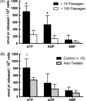
Previous studies raised the idea that ecto-nucleotidase activ-ity could be related with parasite virulence in vivo. To address this, we determined the enzymatic activity of short- or long-term cultured parasites. As shown in Fig. 2A, long-term maintenance of parasites in culture led to a significant decrease in the activ-ity of metacyclic promastigotes over extracellular ATP and ADP. This result reinforces the hypothesis that ecto-nucleotidase, in par-ticular, ecto-NTPDase expression inLeishmaniapromastigotes is a virulence factor for these parasites.
To further corroborate the correlation between
ecto-nucleotidasic activity and infectivity in L. amazonensisinfection, we added adenosine to the culture medium of short-term cultured parasites. This treatment led to a decreased activ-ity of metacyclic promastigotes over extracellular nucleotides, especially over ATP (Fig. 2B). This observation expands on the
results from Berredo-Pinho et al. (2001) where a decreased
Fig. 1.Long-term culture decreases the infectivity ofL. amazonensispromastigotes. C57BL/6J mice were inoculated in the left hind footpad with 1×10⁵L. amazonensis
metacyclic promastigotes obtained from parasites that were kept in culture for few (<15) or many (>100) passages. Lesion development was followed weekly (A). Tissue parasitism was evaluated by limiting dilution at the 8th week of infection (B). IFN-␥ production by draining lymph node cells from mice infected for 8 weeks. Cells were stimulated for 72 h with particulate Ag (C). Results are expressed as mean + 1 SD of three independent experiments with four animals per group. *p< 0.05.
contributed to a down modulation of the enzymatic activ-ity.
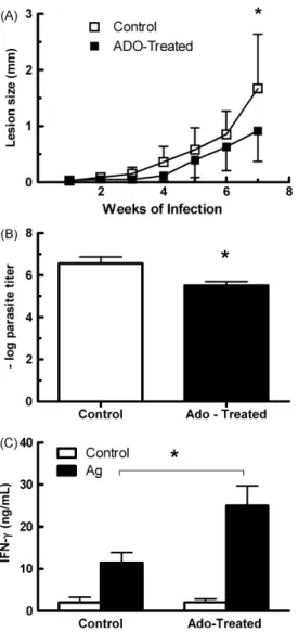
In addition, animals inoculated with adenosine-treated para-sites developed smaller lesions and presented a 10-fold decrease in tissue parasitism (Fig. 3A and B). Interestingly, this decreased infectivity of adenosine-treated parasites was also correlated with an increased IFN-␥production by antigen-stimulated lymph node cells from infected mice (Fig. 3C).
Parasite isolates are considered a mixture of several subpop-ulations of the microorganism. To evaluate the possibility that subpopulations of parasites might display different levels of ecto-nucleotidasic activity we took advantage of the existence in our laboratory of clones of the PH8L. amazonensisstrain. These clones were obtained several years prior to the present study and have always shown decreased infectiveness in the mouse model (data not shown).
Mice inoculated with metacyclic promastigotes from each of the two clones developed only small lesions that contained fewer par-asites than those from animals inoculated with the parental strain (Fig. 4A and B). Interestingly, the ability of these two clones to hydrolyze extracellular ATP was significantly reduced as compared
Fig. 2. Ecto-nucleotidasic activity ofL. amazonensismetacyclic promastigotes. (A) Metacyclic promastigotes were isolated on the 5th day of culture and incubated with each nucleotide for 1 h at 30◦C. Enzymatic activity was evaluated by the
measurement of inorganic phosphate released. (B) Adenosine treatment decreases ecto-nucleotidasic activity of metacyclic promastigotes. Adenosine (5 mM) was added to culture medium of short-term (<15 passages) culturedL. amazonensis pro-mastigotes. Metacyclic promastigotes were isolated on the 5th day of culture and incubated with each nucleotide for 1 h at 30◦C. Enzymatic activity was evaluated by
the measurement of inorganic phosphate released. Results are expressed as mean + 1 SD of four (A) or five (B) independent experiments. *p< 0.05 between short- and long-term cultured parasites (A) or between control and treated groups (B).
to the original strain while AMP hydrolysis was increased (Fig. 4C). These results further corroborate our hypothesis that lesion devel-opment and parasite multiplication within the host are associated with the level of ecto-NTPDase activity by the parasite.
3.3. Alterations in immune response
3.3.1. In vivo experiments
To investigate the mechanisms by which the decreased expres-sion of ecto-NTPDase activity contributes to leexpres-sion development in the host, mice were inoculated with short- or long-term cultured parasites and the presence of regulatory T cells as well as cytokine and chemokine production was evaluated. In this regard, evalua-tion of IL-10, TNF and IL-12 producevalua-tion by stimulated cells from the draining lymph nodes also did not reveal differences between the two groups (data not shown).
Regulatory T cells (CD4+CD25+Foxp3+) modulate the response
of the host duringLeishmaniainfection (Belkaid et al., 2002). In the case ofL. amazonensisinfection it has been demonstrated that these cells rapidly disappear from the lesion site thus allowing for lesion development (Ji et al., 2005). Analysis of the presence of CD4+CD25+Foxp3+ at the draining lymph node from infected
cul-Fig. 3.Adenosine treatment decreases the infectivity ofL. amazonensis promastig-otes. C57BL/6J mice were inoculated in the left hind footpad with 1×10⁵ L. amazonensismetacyclic promastigotes obtained from short-term cultured parasites treated or not with 5 mM of adenosine in the culture medium. Lesion development was followed weekly (A). Tissue parasitism was evaluated by limiting dilution at the 8th week of infection (B). IFN-␥production by draining lymph node cells from mice infected for 8 weeks. Cells were stimulated for 72 h with particulate Ag (C). Results are expressed as mean + 1 SD of two independent experiments with four or five animals per group. *p< 0.05 between treated and control groups.
tured parasites presented reduced levels of CCL2 as compared to lesions from animals infected with short-term cultured parasites (Fig. 5B).
3.3.2. In vitro experiments
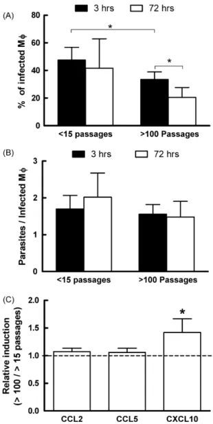
To further characterize the interactions between the parasites and the host, we evaluated the infectivity and cytokine produc-tion by infected thioglycolate-elicited peritoneal macrophages. A different pattern of infection was observed between short- and long-term cultured parasites. A decreased number of macrophages was found associated with parasites in cultures incubated with long-term cultured promastigotes early after incubation (Fig. 6A—3 h). Furthermore, after 72 h of incubation, the percentage of infected cells was reduced in these cultures, a fact not observed in cells incubated with short-term cultured parasites (Fig. 6A—72 h). Interestingly, the number of parasites per infected cell was simi-lar in both groups at all times (Fig. 6B). These results indicate that short-term cultured promastigotes are capable of not only interact-ing and infectinteract-ing a larger number of cells but also, somehow, these
Fig. 4. Avirulent clones ofL. amazonensisPH8 strain show decreased parasitism and ecto-NTPDase activity. (A) C57BL/6J mice were inoculated in the left hind footpad with 1×10⁵L. amazonensismetacyclic promastigotes obtained from short-term
cul-tured PH8 strain or its clones. Lesion development was followed weekly. (B) Tissue parasitism was evaluated by limiting dilution at the 10th week of infection. Results are expressed as mean + 1 SD of two independent experiments with five animals per group. *p< 0.05 between treated and control groups. (C) Ecto-nucleotidasic activity of metacyclic promastigotes was evaluated as described inFig. 2A. Results repre-sent mean + 1 SD for 3 (PH8) or 2 (clones) independent measurements in triplicate.
#p< 0.05 as compared to the parental strain (PH8).
parasites interfere with the host cells and inhibit their microbicidal mechanisms.
While we did not observe differences in the production of IL-10 or TNF by infected macrophages (data not shown), analysis of chemokine production by these cells demonstrated a significant increase in CXCL-10 message levels by peritoneal cells infected with long-term cultured promastigotes (Fig. 6C). These in vitro experiments clearly show that not only short-term cultured pro-mastigotes are able to more efficiently infect macrophages but also are less prone to activate this cell, favoring parasite establishment within the host cell.
4. Discussion
Fig. 5.Regulatory T cells and chemokine production in vivo. (A) The presence of regulatory T cells was evaluated at the draining lymph node 1 and 4 weeks after infection with short- and long-term cultured parasites. (B) Relative levels of message for different chemokines at 1 week after infection. Message levels of each chemokine were evaluated relatively to the housekeeping gene – HPRT and the ratio between animals infected with long- and short-term cultured parasites calculated. *p< 0.05 as compared to an equal level of message between the two groups.
and Sacks, 1987; Peres-Sampaio et al., 2008). One of the possible reasons for the decreased infectivity of parasites maintained for long periods in culture would be the decreased number of meta-cyclic parasites found in these cultures. Although we have also observed a decrease of almost 50% in the number of metacyclic promastigotes in our long-term cultures, this result cannot explain the differences in infectivity observed in our study given the fact that animals were inoculated with purified metacyclic promastig-otes. Thus, we decided to evaluate possible causes for the altered infectivity of long-term culturedL. amazonensis.
We, initially, investigated nucleotide hydrolysis by these par-asites, in order to verify the role of these hydrolytic enzymes in parasite virulence. We found that long-term cultured parasites were less virulent in vivo and showed a reduced hydrolytic activ-ity over ATP and ADP (Fig. 2A). Importantly, when adenosine was added to the culture media, parasites showed a reduction in their ability to hydrolyze nucleotides and became less virulent in vivo (Fig. 2B andFig. 3). The same relationship was found with distinct clones of parasites (Fig. 4), showing a straight correlation between parasite virulence in the mouse model and hydrolytic activity over nucleotides.
These data corroborate earlier data from our laboratory (Maioli et al., 2004; Marques-da-Silva et al., 2008) and reinforce the hypothesis that the enzymes involved in the extracellular metabolism of nucleotides are important virulence factors for Leish-mania(Berredo-Pinho et al., 2001; Pinheiro et al., 2006) andT. cruzi (Santos et al., 2009).
Next, we evaluated the immune response developed by mice infected with short- or long-term cultured parasites. A reduction in the population of regulatory T cells in the lymph nodes of infected mice from 1 to 4 weeks of infection was observed (Fig. 5A), which is consistent with the increase in lesion development and
Fig. 6.Long-term culture reduces infectivity ofLeishmaniaparasites to peritoneal macrophages. Thyoglicolate-elicited peritoneal macrophages were infected with metacyclicL. amazonensispromastigotes (5 parasites/cell) for 3 or 72 h. (A) Percent-age of infected cells *p< 0.05 Student’sttest as indicated. (B) Number of associated parasites per infected macrophage. (C) Relative levels of message for different chemokines. Message levels of each chemokine were evaluated relatively to the housekeeping gene – HPRT and the ratio between cultures infected with long- and short-term cultured parasites calculated. *p< 0.05 as compared to an equal level of message between the two cultures.
corroborates previous findings byJi et al. (2005). No differences, however, were observed between animals infected with long- or short-term cultured parasites. On the other hand, we observed that the chemokine CCL2 was preferentially induced by more virulent parasites (Fig. 5B) indicating that this chemokine could be involved in animal susceptibility, in accordance with previous studies that show the involvement of this chemokine with lesion develop-ment inLeishmaniainfection (Gu et al., 2000; Santiago et al., 2004; Teixeira et al., 2005).
facilitate parasite infection (Berredo-Pinho et al., 2001; Pinheiro et al., 2006; Santos et al., 2009).
When the production of chemokines by infected peritoneal macrophages was evaluated, a higher production of CXCL10 by cells infected with long-term cultured parasites was observed (Fig. 6C). It has already been demonstrated that CXCL10 is important in the generation and migration of effector T cells (Dufour et al., 2002). In regard to leishmaniasis, a previous study indicates that the suscep-tibility of BALB/c mice toL.major infection could be related to low expression of the receptor for CXCL10 by activated cells from these animals in comparison to C57BL/6 mice (Barbi et al., 2008). Fur-thermore, treatment of bone marrow derived macrophages with CXCL10 before infection byL. amazonensisresulted in a smaller cellular parasitism (Vasquez and Soong, 2006). In agreement with these data, treatment with CXCL10 enhances the production of IL-12p40 by dendritic cells infected withL. amazonensis(Vasquez et al., 2008).
In view of the results described here, we propose that the reduction in extracellular ATP levels and consequent increase in adenosine production by virulent parasites would decrease the ability of the host to mount a protective IFN-␥response. Further-more, these parasites may also bind to phagocytes more easily, due to their higher expression of ecto-NTPDase. In addition, they would stimulate CCL2 synthesis by the host cell, resulting in an increased recruitment of cells, especially monocytes, to the infection site. In an environment with decreased IFN-␥production, these recruited monocytes would be more suitable to parasite proliferation. On the other hand, in the presence of less virulent parasites, the increased availability of extracellular ATP would favor the differentiation of IFN-␥-producing T cells by activated DC thus increasing the host ability to control the infection. Also, due to the decreased expression of ecto-NTPDase, their adhesion and further uptake by macrophages would be decreased. Furthermore, cells infected by long-term cultured parasites synthesize CXCL10, resulting in phagocyte activation and parasite control.
Taken together, our results support the hypothesis that ecto-enzymes involved in nucleotide metabolism may be considered virulence factors to Leishmania parasites, at least in the mouse model. The correlation of the expression of these enzymes with the clinical manifestation of the disease in leishmaniasis patients is currently under investigation in our laboratory.
Acknowledgments
The authors wish to thank Ms. Adriana Lúcia Ferreira for animal care, Mr. Leandro Henrique dos Santos and Mr. Marcorelio Divino de Souza for technical support in the laboratory and Dr. Leda Q. Vieira for critical reading of the manuscript. This study received financial support from FAPEMIG, CNPq and Rede Mineira de Bio-terismo/FAPEMIG. L.C.C. Afonso and M.N. Melo are CNPq research fellows. E.A. Marques da Silva received fellowships from FAPEMIG and CNPq. M.C. Souza received a fellowship from FAPEMIG. R.S. Gomes received a fellowship from CAPES.
References
Afonso, L.C.C., Scott, P., 1993. Immune responses associated with suscepti-bility of C57BL/10 mice to Leishmania amazonensis. Infect. Immun. 61, 2952–2959.
Alexander, J., Satoskar, A.R., Russell, D.G., 1999.Leishmaniaspecies: models of intra-cellular parasitism. J. Cell Sci. 112, 2993–3002.
Barbi, J., Brombacher, F., Satoskar, A.R., 2008. T cells from Leishmania major-susceptible BALB/c mice have a defect in efficiently up-regulating CXCR3 upon activation. J. Immunol. 181, 4613–4620.
Belkaid, Y., Mendez, S., Lira, R., Kadambi, N., Milon, G., Sacks, D., 2000. A natural model ofLeishmaniamajor infection reveals a prolonged “silent” phase of para-site amplification in the skin before the onset of lesion formation and immunity. J. Immunol. 165, 969–977.
Belkaid, Y., Piccirillo, C.A., Mendez, S., Shevach, E.M., Sacks, D.L., 2002. CD4+CD25+
regulatory T cells controlLeishmaniamajor persistence and immunity. Nature 420, 502–507.
Berredo-Pinho, M., Peres-Sampaio, C.E., Chrispim, P.P., Belmont-Firpo, R., Lemos, A.P., Martiny, A., Vannier-Santos, M.A., Meyer-Fernandes, J.R., 2001. A Mg-dependent ecto-ATPase inLeishmania amazonensisand its possible role in adenosine acquisition and virulence. Arch. Biochem. Biophys. 391, 16–24. Bisaggio, D.F., Peres-Sampaio, C.E., Meyer-Fernandes, J.R., Souto-Padron, T., 2003.
Ecto-ATPase activity on the surface ofTrypanosoma cruziand its possible role in the parasite–host cell interaction. Parasitol. Res. 91, 273–282.
Bours, M.J., Swennen, E.L., Di Virgilio, F., Cronstein, B.N., Dagnelie, P.C., 2006. Adenosine 5′-triphosphate and adenosine as endogenous signaling molecules
in immunity and inflammation. Pharmacol. Ther. 112, 358–404.
Cohn, C.S., Gottlieb, M., 1997. The acquisition of purines by trypanosomatids. Para-sitol. Today 13, 231–235.
Convit, J., Pinardi, M.E., Rondón, A.J., 1972. Diffuse cutaneous leishmaniasis: a disease due to an immunological defect in the host. Trans. R. Soc. Trop. Med. Hyg. 66, 603–610.
Coutinho-Silva, R., da Cruz, C.M., Persechini, P.M., Ojcius, D.M., 2007. The role of P2 receptors in controlling infections by intracellular pathogens. Purinergic Signal 3, 83–90.
Cysne-Finkelstein, L., Temporal, R.M., Alves, F.A., Leon, L.L., 1998. Leishmania amazonensis: long-term cultivation of axenic amastigotes is associated to meta-cyclogenesis of promastigotes. Exp. Parasitol. 89, 58–62.
da Silva, R., Sacks, D.L., 1987. Metacyclogenesis is a major determinant ofLeishmania
promastigote virulence and attenuation. Infect. Immun. 55, 2802–2806. de Sa Pinheiro, A.A., Cosentino-Gomes, D., Lanfredi-Rangel, A., Ferraro, R.B., De, S.W.,
Meyer-Fernandes, J.R., 2008. Giardia lamblia: biochemical characterization of an ecto-ATPase activity. Exp. Parasitol. 119, 279–284.
Desjeux, P., 2004. Leishmaniasis. Nat. Rev. Microbiol. 2, 692.
Di Virgilio, F., 2007. Purinergic signalling in the immune system. A brief update. Purinergic Signal 3, 1–3.
Dufour, J.H., Dziejman, M., Liu, M.T., Leung, J.H., Lane, T.E., Luster, A.D., 2002. IFN-␥-inducible protein 10 (IP-10; CXCL10)-deficient mice reveal a role for IP-10 in effector T cell generation and trafficking. J. Immunol. 168, 3195–3204. Fietto, J.L., DeMarco, R., Nascimento, I.P., Castro, I.M., Carvalho, T.M., De Souza,
W., Bahia, M.T., Alves, M.J., Verjovski-Almeida, S., 2004. Characterization and immunolocalization of an NTP diphosphohydrolase of Trypanosoma cruzi. Biochem. Biophys. Res. Commun. 316, 454–460.
Fonseca, F.V., Fonseca De Souza, A.L., Mariano, A.C., Entringer, P.F., Gondim, K.C., Meyer-Fernandes, J.R., 2006.Trypanosoma rangeli: characterization of a Mg-dependent ecto ATP-diphosphohydrolase activity. Exp. Parasitol. 112, 76–84. Gomes, M.L., Araujo, S.M., Chiari, E., 1991.Trypanosoma cruzi: growth of clones
on solid medium using culture and blood forms. Mem. Inst. Oswaldo Cruz 86, 131–132.
Gu, L., Tseng, S., Horner, R.M., Tam, C., Loda, M., Rollins, B.J., 2000. Control of Th2 polarization by the chemokine monocyte chemoattractant protein-1. Nature 404, 407–411.
Jesus, J.B., Lopes, A.H., Meyer-Fernandes, J.R., 2002. Characterization of an ecto-ATPase ofTritrichomonas foetus. Vet. Parasitol. 103, 29–42.
Ji, J., Masterson, J., Sun, J., Soong, L., 2005. CD4+CD25+regulatory T cells restrain
pathogenic responses duringLeishmania amazonensisinfection. J. Immunol. 174, 7147–7153.
Komoszynski, M., Wojtczak, A., 1996. Apyrases (ATP diphosphohydrolases, EC 3.6.1.5): function and relationship to ATPases. Biochim. Biophys. Acta 1310, 233–241.
Lattime, S.C., Stopacciaro, A., Stutman, O., 1988. Limiting dilution analysis of TNF producing cells in C3H/HeJ mice. J. Immunol. 141, 3422–3428.
Leite, M.S., Thomaz, R., Fonseca, F.V., Panizzutti, R., Vercesi, A.E., Meyer-Fernandes, J.R., 2007.Trypanosoma brucei brucei: biochemical characterization of ecto-nucleoside triphosphate diphosphohydrolase activities. Exp. Parasitol. 115, 315–323.
Lowry, O.H., Rosebrough, N.J., Farr, A.L., Randall, R.J., 1951. Protein measurement with the folin phenol reagent. J. Biol. Chem. 193, 265–275.
Maioli, T.U., Takane, E., Arantes, R.M., Fietto, J.L., Afonso, L.C., 2004. Immune response induced by New WorldLeishmaniaspecies in C57BL/6 mice. Parasitol. Res. 94, 207–212.
Marques-da-Silva, E.A., Oliveira, J.C., Figueiredo, A.B., Lima Jr., D.S., Carneiro, C.M., Fietto, J.L., Afonso, L.C.C., 2008. Extracellular nucleotide metabolism in Leishma-nia: Influence of adenosine in the establishment of infection. Microbes Infect. 10, 850–857.
Matin, A., Khan, N.A., 2008. Demonstration and partial characterization of ecto-ATPase in Balamuthia mandrillaris and its possible role in the host–cell interactions. Lett. Appl. Microbiol. 47, 348–354.
McMahon-Pratt, D., Alexander, J., 2004. Does theLeishmaniamajor paradigm of pathogenesis and protection hold for New World cutaneous leishmaniases or the visceral disease? Immunol. Rev. 201, 206–224.
Meyer-Fernandes, J.R., Dutra, P.M., Rodrigues, C.O., Saad-Nehme, J., Lopes, A.H., 1997. Mg-dependent ecto-ATPase activity inLeishmania tropica. Arch. Biochem. Bio-phys. 341, 40–46.
Pereira, B.A., Alves, C.R., 2008. Immunological characteristics of experimental murine infection withLeishmania (Leishmania) amazonensis. Vet. Parasitol. 158, 239–255.
Pinheiro, C.M., Martins-Duarte, E.S., Ferraro, R.B., Fonseca De Souza, A.L., Gomes, M.T., Lopes, A.H., Vannier-Santos, M.A., Santos, A.L., Meyer-Fernandes, J.R., 2006.Leishmania amazonensis: biological and biochemical characterization of ecto-nucleoside triphosphate diphosphohydrolase activities. Exp. Parasitol. 114, 16–25.
Righetti, P.G., 2005. Electrophoresis: the march of pennies, the march of dimes. J. Chromatogr. A 1079, 24–40.
Robson, S.C., Sevigny, J., Zimmermann, H., 2006. The E-NTPDase family of ectonucleotidases: structure function relationships and pathophysiological sig-nificance. Purinergic Signal 2, 409–430.
Sacks, D., Noben-Trauth, N., 2002. The immunology of susceptibility and resistance toLeishmaniamajor in mice. Nat. Rev. Immunol. 2, 845–858.
Sansom, F.M., Robson, S.C., Hartland, E.L., 2008. Possible effects of microbial ecto-nucleoside triphosphate diphosphohydrolases on host-pathogen interactions. Microbiol. Mol. Biol. Rev. 72, 765–781.
Santiago, H.C., Oliveira, C.F., Santiago, L., Ferraz, F.O., de Souza, D.G., de Freitas, L.A., Afonso, L.C.C., Teixeira, M.M., Gazzinelli, R.T., Vieira, L.Q., 2004. Involvement of the chemokine RANTES (CCL5) in resistance to experimental infection with
Leishmaniamajor. Infect. Immun. 72, 4918–4923.
Santos, R.F., Possa, M.A., Bastos, M.S., Guedes, P.M., Almeida, M.R., DeMarco, R., Verjovski-Almeida, S., Bahia, M.T., Fietto, J.L., 2009. Influence of ecto-nucleoside triphosphate diphosphohydrolase activity onTrypanosoma cruziinfectivity and virulence. PLoS Negl. Trop. Dis. 3, e387.
Sissons, J., Alsam, S., Jayasekera, S., Khan, N.A., 2004. Ecto-ATPases of clin-ical and non-clinclin-ical isolates of Acanthamoeba. Microb. Pathog. 37, 231– 239.
Souza-Neto, S.M., Carneiro, C.M., Vieira, L.Q., Afonso, L.C.C., 2004. Leishmania braziliensis: partial control of experimental infection by IL-12p40 deficient mice. Mem. Inst. Oswaldo Cruz 99, 289–294.
Späth, G.F., Beverley, S.M., 2001. A lipophosphoglycan-independent method for isolation of infectiveleishmaniametacyclic promastigotes by density gradient centrifugation. Exp. Parasitol. 99, 97–103.
Tanuri, A., de Andrade, P.P., de Almeida, D.F., 1985.Trypanosoma cruzi: isolation of cloned strains and characterization of their infectivity. J. Parasitol. 71, 397–402. Taussky, H.H., Shorr, E., 1953. A microcolorimetric method for the determination of
inorganic phosphorus. J. Biol. Chem. 202, 675–685.
Teixeira, M.J., Fernandes, J.D., Teixeira, C.R., Andrade, B.B., Pompeu, M.L., San-tana, d.S., Brodskyn, C.I., Barral-Netto, M., Barral, A., 2005. DistinctLeishmania braziliensisisolates induce different paces of chemokine expression patterns. Infect. Immun. 73, 1191–1195.
Vasquez, R.E., Soong, L., 2006. CXCL10/gamma interferon-inducible protein 10-mediated protection againstLeishmania amazonensisinfection in mice. Infect. Immun. 74, 6769–6777.
Vasquez, R.E., Xin, L., Soong, L., 2008. Effects of CXCL10 on dendritic cell and CD4+
T-cell functions duringLeishmania amazonensisinfection. Infect. Immun. 76, 161–169.
Vieira, L.Q., Hondowicz, B.D., Afonso, L.C.C., Wysocka, M., Trinchieri, G., Scott, P., 1994. Infection withLeishmaniamajor induces interleukin-12 production in vivo. Immunol. Lett. 40, 157–161.